www.bjournal.com.br
www.bjournal.com.br
Institutional Sponsors
The Brazilian Journal of Medical and Biological Research is partially financed by
Volume 43 (5) 409-521 May 2010
Braz J Med Biol Res, May 2010, Volume 43(5) 431-436
Induction of a gloverin-like antimicrobial polypeptide in the
sugarcane borer Diatraea saccharalis challenged by septic
injury
J.L.C. Silva, J.F. Barbosa, J.P. Bravo, E.M. de Souza, L.F. Huergo, F.O. Pedrosa, E. Esteves,
Induction of a gloverin-like antimicrobial
polypeptide in the sugarcane borer
Diatraea saccharalis
challenged by septic injury
J.L.C. Silva
1, J.F. Barbosa
1, J.P. Bravo
1, E.M. de Souza
2, L.F. Huergo
2,
F.O. Pedrosa
2, E. Esteves
3, S. Daffre
3and M.A. Fernandez
11Departamento de Biologia Celular e Genética, Universidade Estadual de Maringá, Maringá, PR, Brasil 2Departamento de Bioquímica, Universidade Federal do Paraná, Curitiba, PR, Brasil 3Departamento de Parasitologia, Instituto de Ciências Biomédicas,
Universidade de São Paulo, São Paulo, SP, Brasil
Abstract
Diatraea saccharalis (Fabricius, 1794) (Lepidoptera: Crambidae) is an important pest for Brazilian sugarcane. In the present study, we detected two distinct spots in hemolymph from septic injured larvae (HDs1 and HDs2), which are separated by 2DE gel electrophoresis. Both spots were subjected to in-gel tryptic digestion and MALDI-TOF/TOF analysis, which revealed the sequence VFGTLGSDDSGLFGK present in both HDs1and HDs2. This sequence had homology and 80% identity with specific
Lepidoptera antimicrobial peptides called gloverins. Analyses using the ImageMaster 2D software showed pI 8.94 of the HDs1 spot, which is similar to that described to Hyalophora gloveri gloverin (pI 8.5). Moreover, the 14-kDa molecular mass of the spot HDs1 is compatible to that of gloverins isolated from the hemolymph of Trichoplusia ni, Helicoverpa armigera and H. gloveri.
Antimicrobial assays with partially purified fractions containing the HDs1 and HDs2 polypeptides demonstrated activity against
Escherichia coli. This is the first report of antimicrobial polypeptides in D. saccharalis, and the identification of these peptides
may help in the generation of new strategies to control this pest.
Key words: Diatraea saccharalis; Lepidoptera; Immune response; Gloverin; Sugarcane borer; Hemolymph
Introduction
Correspondence: M.A. Fernandez, Departamento de Biologia Celular e Genética, Universidade Estadual de Maringá, Av. Colombo, 5790, 87020-900 Maringá, PR, Brasil. Fax: +55-44-3261-4893. E-mail: [email protected]
Received July 5, 2009. Accepted February 19, 2010. Available online May 7, 2010. Published May 14, 2010.
Insects are evolutionarily successful organisms that occupy almost all habitats in nature. They are continuously exposed to potentially pathogenic microorganisms and
eu-karyotic parasites and they are able to react with an efficient
immune response against these invaders (1). Over the last decades, innate immunity has been extensively studied in insects, with emphasis on insect pest management in agriculture, the discovery of bioactive molecules and the revelation of the evolutionary roots of innate immunity (2). One of the most studied mechanisms of innate immunity in insects is the production of antimicrobial peptides (AMPs), which contain less than 150-200 amino acids (3).
Since the first report of the innate immune response of
Hyalophora cecropia (4) (Lepidoptera: Saturniidae), many taxa of insects have been the subject of studies of innate immune response to septic injury. Insects can produce a variety of AMPs in response to microbial infection or body
injury, and most of these peptides are synthesized by the fat body, which is a major insect tissue involved in the immune response (5). AMPs are secreted into the hemolymph and are responsible for the growth arrest of the microbe at an early stage in the immune response (6).
The molecular mechanisms of the insect’s innate im-mune system have been studied using both molecular and proteomic methods (7,8). Drosophila melanogaster
432 J.L.C. Silva et al.
to investigate insect innate immunity (11-14).
In Brazil, the sugarcane borer Diatraea saccharalis
(Fabricius, 1794; Lepidoptera: Crambridae) is a pest with great economic impact on the sugarcane crop, causing considerable damage to the sugar and alcohol industries (15). Several studies intended to develop new strategies for biological control have been conducted to identify the physiological changes resulting from host-parasitoid as-sociations (16). Micro-hymenopteran parasitoids were
first used for the biological control of D. saccharalis in the 1980’s and they have become one of the most valuable biopesticides currently used in commercial sugarcane crop management in Brazil (17). Although biological agents can control D. saccharalis infestation, further research on new approaches to insect management and the knowledge of
D. saccharalis antimicrobial defenses should improve the biological control of this important pest (18).
In this study, we have detected two distinct peptides in the hemolymph from D.saccharalis 5th-instar larvae after septic injury. To identify these polypeptides we performed antimicrobial activity assay, electrophoretic analysis, in-gel tryptic digestion, matrix-assisted laser
desorption/ionization-time of flight mass spectrometry (MALDI-TOF/TOF), and
bioinformatic analysis.
Material and Methods
Experimental D. saccharalis challenge and sample
preparation
Sugarcane borer larvae were reared on an artificial diet
(19) under controlled environmental conditions (12-h light/ dark, 25 ± 1°C, 70% humidity). For immune challenge, the 5th-instar larvae were pierced with a long 27G dental needle dipped into a pellet (1:1) of viable Escherichia coli (ATCC strain 11229) and Bacillus subtilis (ATCC strain 6623). Prior to hemolymph collection, the insects were chilled for 15 min at 4°C, and surface sterilized with a 70% (v/v) ethanol solution. Fifty larvae of each experimental group (naive and septic injured) were used in the experiments. The hemolymph (about 20 µL from each larva) was collected 14 h after the immune challenge for polypeptide extraction. The samples were obtained by cutting off an abdominal proleg with sterile
scissors. The outflowing hemolymph was collected with a
micropipette and immediately transferred to a sterile chilled Eppendorf tube containing a few crystals of phenylthiourea to prevent melanization. Hemocyte-free hemolymph was obtained by centrifugation at 300 g for 10 min to pellet hemo-cytes and the supernatant was subsequently centrifuged at 10,000 g for 10 min at 4°C to pellet any cell debris (20).The cell-free hemolymph was boiled for 10 min, centrifuged at 10,000 g for 20 min at 4°C and the resulting supernatant was subjected to electrophoretic analysis (21).
Isoelectric focusing and 2DE analysis
Total protein in the sample was quantified by the method
of Bradford (22) and 5 µg total protein was loaded onto 16% Tricine SDS-PAGE. Alternatively, the samples were
concentrated through filter systems YM-50 and YM-10
(Microcon Millipore, Ireland). Samples that displayed an
induced immune response expression profile were then
used for isoelectric focusing (IEF) and two-dimensional gel electrophoresis (2DE). For IEF, 50 µg concentrated protein was mixed with 250 µL rehydration buffer (GE Healthcare, Sweden) and loaded onto an IPG strip (13 cm, pH 3-10, linear; GE Healthcare, Sweden) following manufacturer instructions. Rehydration and IEF were carried out on the IPGphor platform (GE Healthcare, USA) at 20°C and 50 µA/strip, according to the following protocol: Step 1: 200 V-200 Vhr; Step 2: 500 V-500 Vhr; Step 3: Gradient 1000 V-800 Vhr; Step 4: Gradient 8000 V-11,300 Vhr; Step 5: 8000 V-12,000 Vhr; with a total of 24,800 Vhr accumulated in the IEF. After IEF separation, the gels were incubated for 30 min in equilibration buffer (50 mM Tris-base, 6 M urea, 30% glycerol, 2% SDS, and 0.002% bromophenol blue) containing 1% DTT, followed by the equilibration buffer containing 2.5% iodoacetamide for an additional 30 min. The gel strip was subjected to 15% SDS-PAGE sealed with 0.5% agarose. SDS-PAGE was performed at a constant power of 5 W/gel for 45 min and switched to 15 W/gel until the bromophenol blue frontier reached the bottom of the gel. The 2DE gels were visualized by Coomassie colloidal staining. The gels were scanned at an optical resolution of 300 dpi on a high-resolution image scanner (GE Healthcare, USA), and the images were analyzed using the ImageMaster Platinum 2D v6.0 software (GE Healthcare, USA). For both naive and challenged samples of larva hemolymph, three 2DE gels were prepared and analyzed in order to determine the reproducibility of the data.
Mass spectrometry
The HDs1 and HDs2 spots detected in the induced hemolymph samples were excised from the 2DE gels and subjected to in-gel digestion with sequencing-grade trypsin (Promega, USA) as described by Shevchenko et al. (23)
with minor modifications. After incubation at 37°C for 16
h, aliquots of each hydrolyzed sample were mixed with a saturated matrix solution of a-cyano-4-hydroxycinnamic acid, spotted onto MALDI target plates and allowed to air dry. Mass spectra (MS) were acquired using a MALDI-TOF/
TOF Autoflex II spectrometer (Bruker Daltonics, Germany)
that was operated at a laser frequency of 50 Hz. MS analysis
was performed in a positive ion reflection mode. The voltage
parameters were set at IS1 19 kV, IS2 16.8 kV, lens 8 kV,
reflector 1 20 kV, and reflector 2 9.54 kV. The delay time
was 70 ns, and the acquisition mass range was 800-3200 Da. External calibration was performed using a peptide mixture containing ACTH (residues 1-24), ACTH (residues 18-39), Somatostatin, angiotensin I and angiotensin II (all from Sigma, USA).MS/MS analyses were performed in a
were set at IS1 6 kV, IS2 5.3 kV, lens 3.15 kV, reflector 1 23.5 kV, reflector 2 9.7 kV, LIFT 1 19 kV, and LIFT 2 4 kV.
The acquisition mass range was 40-2400 Da. Peak lists were created using the FlexAnalysis 3.0 software (Bruker Dal-tonics). The sophisticated numerical annotation procedure algorithm was used to detect the monoisotopic peak values, with a quality factor threshold of 30. The S/N threshold was set at 6 for MS analysis and at 3 for MS/MS analysis. Automated de novo sequencing was performed using the Peaks Studio software 4.5 Demo version (Bioinformatics Solutions, Canada). The parent ion mass tolerance was 50
ppm, carbamidomethylation of cysteines was set at fixed modification, fragment error was set at 0.3 Da, and all ion
charge states were set as +1.
Antimicrobial assay
Antimicrobial activity was monitored by a liquid growth inhibition assay against Micrococcus luteus, Bacillus subti-lis, Escherichia coli SBS363, and Candida albicans MDM8 using a procedure described by Silva Jr. et al. (24), and the microorganisms tested were obtained as previously described (24,25).
Results and Discussion
Bacterial challenge induced a 14-kDa polypeptide in
D. saccharalis
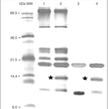
The 1DE analysis with hemolymph from bacterial-challenged larvae showed the presence of an approximately 14-kDa polypeptide that was not observed in naive larvae
(Figure 1, lanes 1 and 2). After the pre-purification processes
with the filter systems YM-50 and YM-10 (Microcon Millipore;
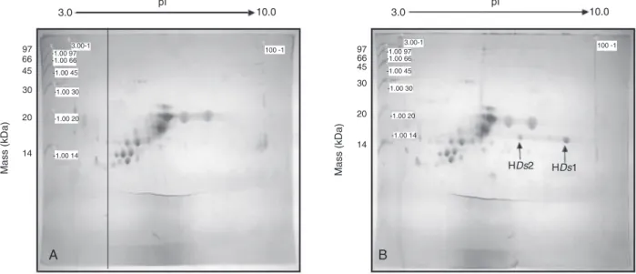
Figure 1, lanes 3 and 4), the polypeptide of approximately 14 kDa was detected in challenged larva hemolymph samples. When these samples were separated on 2DE gels (Figure 2A and B), two spots of different pIs at approximately 14 kDa were present only in hemolymph samples from challenged insects and were denoted HDs1 and HDs2 (Figure 2B). Both HDs1 and HDs2 were subjected to in-gel tryptic digestion and MALDI-TOF analysis, and the MS spectra were com-posed of 11 and 8 m/z monoisotopic peaks, respectively (Table 1). All peaks found in the HDs2 spectra were also found in the HDs1 spectra (molecular mass error <0.1 Da). Thus, the HDs1 and HDs2 fractions probably represent the same polypeptide and the differences observed in pI
values suggest a putative post-translational modification.
Recently, Mrinal and Nagaraju (26) reported that the evo-lutionary genetic mechanisms originated four isoforms of gloverins in Bombyx mori, Bmglv1, Bmglv2, Bmglv3, and
Bmglv4. These different isoforms have a molecular mass of 19 kDa; however, no reports on biochemical aspects, such as the isoelectric point, are available. In contrast to
being the result of post-translational modification, the two
proteins described here could be the result of gene duplica-tion events in which different alleles produce isoforms of gloverin-like proteins.
The sequencing and antimicrobial assay showed a gloverin-like polypeptide
In order to identify the HDs1/HDs2 polypeptides, the tryptic peptides of HDs1 and HDs2 were subjected to MS/ MS analysis (Table 1). No positive hits were observed on
Figure 1. One-dimensional gel electrophoretic (1DE) protein
analysis of hemolymph from Diatraea saccharalis larva. Lane 1, Naive hemolymph; lane2, induced hemolymph collected 14 h after septic injury; lanes 3 and 4, naive and induced hemolymph
concentrated by Microcon YM-50 and YM-10 filters. Black stars
434 J.L.C. Silva et al.
MS/MS spectra submitted to Mascot ion search using the NCBInr database (12/03/2009). The MS/MS data were subjected to automated de novo sequencing using the Peaks 4.5 software. Only one peptide (m/z of 1499.682) generated an MS/MS spectrum that allowed de novo
se-quencing with confidence higher than 50%, yielding the
sequence VFGTLGSDDSGLFGK for both HDs1and HDs2
(Table 1, Figure 3). These sequences were confirmed by
manual inspection of the MS/MS data. A protein homology search of the NCBI database using the BLASTP 2.2.18+ algorithm adjusted to search for a short input sequence showed homology of the peptide with gloverins from diverse species of Lepidoptera. Gloverins are antibacterial proteins
ranging in sizefrom 8 to 30 kDa and along with other small peptides, such as cecropins and defensins, are components of the immune response in Lepidoptera (27). The approxi-mately 14-kDa molecular mass of the HDs1 polypeptide is similar to that of gloverins isolated from the hemolymph of Trichoplusia ni, Helicoverpa armigera and Hyalophora gloveri (27-29). The pI of the HDs1 polypeptide (pI 8.94) is also similar to that described for H. gloveri gloverin (pI 8.5) (29). Gloverins are generally basic, heat-stable, glycine-rich (16-20%) proteins that act by inhibiting the synthesis of the outer membrane proteins, which would result in increased membrane permeability to Gram-negative bacteria (29).
Antimicrobial activity assays were carried out using
Figure 2. Two-dimensional gel electrophoretic (2DE) analysis of proteins from Diatraea saccharalis larva hemolymph. Partially purified
samples, as shown in Figure 1, lanes 3 and 4, were resolved by 2DE gels. A, Sample from naive larva hemolymph; B, sample from septic injury-induced larva hemolymph. The polypeptide spots with molecular mass of approximately 14 kDa, HDs1 and HDs2, are indicated by arrows. The HDs1 and HDs2 spots were excised, submitted to tryptic digestion and analyzed by MALDI-TOF/TOF (Table 1). 15% Tris-glycine SDS-PAGE visualized by colloidal Coomassie staining; pI 3-10; 14 to 97 kDa low molecular weight calibration marker (GE Heathcare, USA).
Table 1. Hemolymph polypeptides from induced Diatraea saccharalis larvae.
Spot Mass (kDa) pI m/z of tryptic peptides in MS spectraa
HDs1 14 8.94 892.306; 979.422; 1302.687*; 1362.678*; 1427.661*; 1499.759* (VFGTLGSDDSGLFGK) - 99%; 1513.769; 1521.741*; 1612.806; 2288.310; 3058.829
HDs2 14 7.27 892.256; 979.394; 1302.638*; 1362.620*; 1427.601*; 1499.682* (VFGTLGSDDSGLFGK) - 77%; 1612.741; 3058.809
Molecular mass (kDa) was obtained by comparison of mobility with globular proteins(14 to 97 kDa low molecular weight calibration marker, GE Heathcare, USA)and pI by assuming IEF strips are linear(pI 3-10)- both with the ImageMaster Platinum 2D v6.0 software.
aMonoisotopic m/z list was generated using FlexAnalysis 3.0 (Bruker Daltonics)but sequences were not obtained, except those shown
in the Table.*Peptides subjected to MALDI-TOF/TOF MS/MS analysis. The sequence of the peptide of m/z 1499 indicated in the table was obtained after MS/MS and automatic de novo sequencing using the Peaks Studio Demo version software (Bioinformatics
samples that were partially purified through the filter systems
(Figure 1, lanes 3 and 4). The antimicrobial activity of the
induced sample was efficient against E. coli (Gram-negative) growth. However, both the naive and induced samples were inactive against the Gram-positive bacterium M. luteus and the yeast Candida albicans (Table 2). Although in this study we could not obtain data on minimum inhibitory concentration, the activity against a Gram-negative bacterium, amino acid se-quence and biochemical data (pI and molecular mass) support
the classification of HDs1 as a gloverin-like polypeptide. These results indicate that the polypeptide HDs1 may be involved in the innate immune response of D. saccharalis. Although several AMPs have been described in Lepidoptera,
this is the first report of an immune polypeptide in this im -portant sugarcane culture pest. Recent studies have shown that control strategies combined with fungi and nematodes are effective in controlling D. saccharalis (30,31). However, the action of these biological agents in the expression of antimicrobial peptides, as described for the Lepidoptera
Spodoptera exigua when infected with Xenorhabdus nematophila, is unknown in D. saccharalis (32).
Therefore, a better understanding of the immune re-sponse of D. saccharalis will help with the development of new strategies to control this pest. Future RT-PCR experiments will be carried out using the sequence of the gloverin-like polypeptide in order to clone the gene. There-after, we willanalyze gene expression in D. saccharalis
and its possible applications in methods to control this sugarcane pest.
Acknowledgments
We thank Valmir Peron and Marli Licero Schuete Silva for dedicated technical assistance, and Universidade Es-tadual de Maringá for the use of its facilities (COMCAP laboratories).
References
1. Gillespie JP, Kanost MR, Trenczek T. Biological mediators of insect immunity. Annu Rev Entomol 1997; 42: 611-643.
2. Lee YS, Yun EK, Jang WS, Kim I, Lee JH, Park SY, et al. Purification, cDNA cloning and expression of an insect de -fensin from the great wax moth, Galleria mellonella. Insect Mol Biol 2004; 13: 65-72.
3. Bulet P, Stocklin R, Menin L. Anti-microbial peptides: from invertebrates to vertebrates. Immunol Rev 2004; 198:
169-184.
4. Steiner H, Hultmark D, Engstrom A, Bennich H, Boman
HG. Sequence and specificity of two antibacterial proteins
involved in insect immunity. Nature 1981; 292: 246-248. 5. Bulet P, Hetru C, Dimarcq JL, Hoffmann D. Antimicrobial
peptides in insects; structure and function. Dev Comp Im-munol 1999; 23: 329-344.
6. Haine ER, Moret Y, Siva-Jothy MT, Rolff J. Antimicrobial
Figure 3. MS/MS mass spectrum of the parental peptide 1499.682 (m/z) derived from trypsin digestion of spot HDs1. The mass
spec-trum and the de novo interpretation of the data were generated using the Peak 4.5 software. The y and b ion series shown confirm the
VFGTLGSDDSGLFGK peptide sequence.
Table 2. Antimicrobial assay with Diatraea saccharalis polypeptides
partially purified from naive and induced larvae hemolymph.
Microorganisms Naive
hemolymph
Induced hemolymph
Escherichia coli SBS363 +
-Micrococcus luteus + +
Bacillus subtilis + +
Candida albicansMDM8 + +
The hemolymph samples were concentrated through filter sys
436 J.L.C. Silva et al.
defense and persistent infection in insects. Science 2008; 322: 1257-1259.
7. Kaneko Y, Tanaka H, Ishibashi J, Iwasaki T, Yamakawa M.
Gene expression of a novel defensin antimicrobial peptide in the silkworm, Bombyx mori. Biosci Biotechnol Biochem
2008; 72: 2353-2361.
8. Vierstraete E, Verleyen P, Baggerman G, D’Hertog W, Van den Bergh G, Arckens L, et al. A proteomic approach for the analysis of instantly released wound and immune proteins in Drosophila melanogaster hemolymph. Proc Natl Acad Sci U S A 2004; 101: 470-475.
9. Lemaitre B, Hoffmann J. The host defense of Drosophila melanogaster. Annu Rev Immunol 2007; 25: 697-743. 10. Gandhe AS, Arunkumar KP, John SH, Nagaraju J. Analysis
of bacteria-challenged wild silkmoth, Antheraea mylitta
(Lepidoptera) transcriptome reveals potential immune genes. BMC Genomics 2006; 7: 184.
11. International Silkworm Genome Consortium. The genome of a lepidopteran model insect, the silkworm Bombyx mori.
Insect Biochem Mol Biol 2008; 38: 1036-1045.
12. Mita K, Kasahara M, Sasaki S, Nagayasu Y, Yamada T, Ka -namori H, et al. The genome sequence of silkworm, Bombyx mori. DNA Res 2004; 11: 27-35.
13. Wang Y, Zhang P, Fujii H, Banno Y, Yamamoto K, Aso Y. Pro -teomic studies of lipopolysaccharide-induced polypeptides in the silkworm, Bombyx mori. Biosci Biotechnol Biochem
2004; 68: 1821-1823.
14. Li XH, Wu XF, Yue WF, Liu JM, Li GL, Miao YG. Proteomic
analysis of the silkworm (Bombyx mori L.) hemolymph dur-ing developmental stage. J Proteome Res 2006; 5: 2809-2814.
15. Huang F, Leonard BR, Andow DA. Sugarcane borer (Lepidoptera: Crambidae) resistance to transgenic Bacillus thuringiensis maize. J Econ Entomol 2007; 100: 164-171.
16. Alleyne M, Wiedenmann RN, Diaz RR. Quantification and
development of teratocytes in novel-association host-para-sitoid combinations. J Insect Physiol 2001; 47: 1419-1427. 17. Degaspari N, Macedo N, Botelho PSM, Araújo JR, Almeida
LC. Predação e parasitismo de ovos da Diatraea saccharalis
em cana-de-açúcar. Pesqui Agropecu Bras 1987; 22: 785-792.
18. Falco MC, Marbach PAS, Pompermayer P, Lopes FCC, Silva-Filho MC. Mechanisms of sugarcane response to herbivory. Genet Mol Biol 2001; 24: 113-122.
19. Hensley SD, Hammond AM. Laboratory techniques for
rear-ing the sugarcane borer on an artificial diet. J Econ Entomol Geneva 1968; 61: 1742-1743.
20. Cytrynska M, Mak P, Zdybicka-Barabas A, Suder P, Jakubo
-wicz T. Purification and characterization of eight peptides
from Galleria mellonella immune hemolymph. Peptides
2007; 28: 533-546.
21. Bae S, Kim Y. Lysozyme of the beet armyworm, Spodoptera exigua: activity induction and cDNA structure. Comp Bio-chem Physiol B BioBio-chem Mol Biol 2003; 135: 511-519. 22. Bradford MM. A rapid and sensitive method for the
quantita-tion of microgram quantities of protein utilizing the principle of protein-dye binding. Anal Biochem 1976; 72: 248-254. 23. Shevchenko A, Wilm M, Vorm O, Mann M. Mass
spectro-metric sequencing of proteins silver-stained polyacrylamide gels. Anal Chem 1996; 68: 850-858.
24. Silva PI Jr, Daffre S, Bulet P. Isolation and characterization of gomesin, an 18-residue cysteine-rich defense peptide from the spider Acanthoscurria gomesiana hemocytes with sequence similarities to horseshoe crab antimicrobial peptides of the tachyplesin family. J Biol Chem 2000; 275: 33464-33470.
25. Fogaca AC, da Silva PI Jr, Miranda MT, Bianchi AG, Miranda A, Ribolla PE, et al. Antimicrobial activity of a bovine hemo-globin fragment in the tick Boophilus microplus. J Biol Chem
1999; 274: 25330-25334.
26. Mrinal N, Nagaraju J. Intron loss is associated with gain of function in the evolution of the gloverin family of antibacte-rial genes in Bombyx mori. J Biol Chem 2008; 283: 23376-23387.
27. Lundstrom A, Liu G, Kang D, Berzins K, Steiner H. Tricho-plusia ni gloverin, an inducible immune gene encoding an antibacterial insect protein. Insect Biochem Mol Biol 2002; 32: 795-801.
28. Mackintosh JA, Gooley AA, Karuso PH, Beattie AJ, Jardine DR, Veal DA. Gloverin-like antibacterial protein is synthe-sized in Helicoverpa armigera following bacterial challenge.
Dev Comp Immunol 1998; 11: 276-288.
29. Axen A, Carlsson A, Engstrom A, Bennich H. Gloverin, an antibacterial protein from the immune hemolymph of Hyalo-phora pupae. Eur J Biochem 1997; 247: 614-619.
30. Acevedo JP, Samuels RI, Machado IR, Dolinski C. Interac-tions between isolates of the entomopathogenic fungus
Metarhizium anisopliae and the entomopathogenic nema-tode Heterorhabditis bacteriophora JPM4 during infection of the sugar cane borer Diatraea saccharalis (Lepidoptera: Pyralidae). J Invertebr Pathol 2007; 96: 187-192.
31. Carneiro CN, Damatta RA, Samuels RI, Silva CP. Effects of entomopathogenic bacterium Photorhabdus temperata
infection on the intestinal microbiota of the sugarcane stalk borer Diatraea saccharalis (Lepidoptera: Crambidae). J Invertebr Pathol 2008; 99: 87-91.