Detection of
Metarhizium anisopliae
var.
anisopliae
within infected sugarcane
borer
Diatraea saccharalis
(Lepidoptera, Pyralidae) using specific primers
Ricardo Henri Rodrigues Destéfano1, Suzete A. Lanza Destéfano2and Claudio Luiz Messias1
1
Universidade Estadual de Campinas, Instituto de Biologia, Departamento de Genética e Evolução, Campinas, São Paulo, Brazil.
2
Instituto Biológico, Laboratório de Bacteriologia Vegetal, Campinas, São Paulo, Brazil.
Abstract
In order to construct specific primers for the detection and identification of the entomopathogenic fungusMetarhizium within infected sugarcane borer (Diatraea saccharalis) larvae we analyzed the ITS1 -5.8S- ITS2 rDNA regions of strains and varieties ofM. anisopliae, M. album and M. flavoviride. The PCR amplification of these regions yielded a unique fragment of approximately 540 bp forM. anisopliae variety anisopliae strains E9, B/Vi and C (isolated in Brazil), 600 pb forM. a. anisopliae strain 14 (isolated in Australia), 650 bp for the M. album and 600 bp for M. flavoviride strains. The PCR products were digested with different restriction endonucleases (Afa I, Alu I, Dde I, Hae III,Hpa II and Sau 3A) and the PCR-RFLP profiles showed clear differences between the species. Sequencing of the ITS-5.8S rDNA regions allowed us to design one specific primer (ITSMet: 5’ TCTGAATTTTTTATAAGTAT 3’) for the BrazilianM. a. anisopliae strains (E9, B/Vi and C) and another specific primer (ITSMet14: 5’ GAAACCGGGAC TAGGCGC 3’) for the Australian strain (strain 14). Amplification was not observed with M. album, M flavoviride and Beauveria bassiana strains. DNA extracted from larvae infected with the Brazilian or Australian strains were tested using the specific primers designed by us to identify the fungal strains with which the larva had been infected. The correct fungal strain was successfully detected within 48 h of the insect having been infected, showing that this molecular technique allows rapid and secure detection and identification ofM. anisopliae.
Key words:Metarhizium anisopliae, entomopathogenic fungi, PCR-RFLP, ITS region. Received: April 1, 2003; Accepted: October 24, 2003.
Introduction
About 80% of the etiologic agents involved in insect diseases are fungi, encompassing 90 genera and more than 700 species. Several research groups have verified the entomopathogenicity of the Deuteromycete fungi Metarhizium anisopliae, which has become an important biocontrol agent used in the microbial control of insect pests.
The sugarcane spittlebug (Mahanarva posticata) (Homoptera, Cercopidae) causes serious losses in sugar-cane crops but has been successfully controlled in north-eastern Brazil using biocontrol byM. anisopliae, which has also been used in the biocontrol of other spittlebug genera (Aenolamia, Deois and Zulia) infesting pasture grasses (Onofreet al., 2002). Biological control of the sugarcane borer (Diatraea saccharalis) (Lepdoptera, Pyralidae) by
variousM. anisopliaeandBeauveria bassianaisolates has been achieved by Alveset al.(1984; 1985).
The production of mycoinsecticides is very simple, but their use as biocontrol agents depends critically on the standardization of the production runs and the stability of the mycoinsecticide under field conditions where the for-mulation must allow the fungus to maintain its virulence. It is also important to monitor how mycoinsecticides dissemi-nate and survive in the environment after application (Hegedus and Khachatourians, 1996a).
There is a need for specific and sensitive systems for the detection and identification of fungi which can be used to evaluate the dispersion and environmental persistence of mycoinsecticides. Commercial implications such as the identification of existing or new fungal isolates, quality control and patent protection must also be considered be-cause several researcher groups and commercial compa-nies are developing production, formulation and application methods involving entomopathogens for use in the control of insect pests of agricultural and public health importance.
www.sbg.org.br
Send correspondence to Ricardo Henri Rodrigues Destéfano. Uni-versidade Estadual de Campinas, Departamento de Genética e Evolução, Caixa Postal 6109, 13083-970 Campinas, SP, Brazil. E-mail: stefano@unicamp.br.
The classical methods for the identification of entomopathogenic fungi are based on spore morphology, biochemical characteristics and immunological properties, but PCR and DNA sequence analysis are also being inten-sively used as standard tools for the detection, identifica-tion and phylogenetic analysis of many fungal species. These techniques are particularly valuable for the large number of species which are incapable of growth in the lab-oratory under artificial conditions and from which pure DNA is not easily obtained (Bindslevet al.,2002).
Several molecular genetic methods have been sug-gested as potential systems for the identification and moni-toring of entomopathogenic fungi. Techniques which use DNA probes to create restriction fragment length polymor-phism (RFLP) fingerprints have been used to distinguish between species (Hegedus and Khachatourians, 1993a) or individual isolates (Hegedus and Khachatourians, 1993b), although direct DNA probing methods may not exhibit the desired degree of sensitivity required for detection under field conditions (Hegedus and Khachatourians, 1996b). Other methods based on the polymerase chain reaction (PCR), such as the random amplified polymorphic DNA (RAPD) method, also exhibit the ability to discriminate be-tween entomopathogenic fungal isolates (Jensen et al., 2001; Freireet al., 2001; Alveset al., 2001; Urtz and Rice, 1997; Fungaroet al., 1996; Lealet al., 1994; Bidochkaet al, 1994) but the use of random oligonucleotide primers in the design of these systems is not useful in the detection of fungi within environmental samples which can contain a significant amount of DNA from indigenous biotic materi-als.
An alternative approach is to use ribosomal DNA (rDNA), an important molecular marker widely used in the identification and differentiation of species. The rDNA
operon of eukaryotes can be present in multiple copies per genome, with each unit consisting of regions coding for rRNA, 18S, 5.8S and 28S genes as well as the two internal spaces (ITS 1 and ITS 2) between these regions, each rDNA unit being separated by one intergenic space (IGS). The rDNA unit presents sequence variations which may be used in systematic studies at different taxonomic levels (Foulyet al.,1997; Argentina, 1999). The rDNA 18S and 28S re-gions are the most conserved units, and may be used in dif-ferentiating genera and species, while the ITS and IGS spacer regions have accumulated more variability and are better used to differentiate species or strains within the same species (Esteve-Zarzosoet al., 1999).
In the study reported in this paper we sequenced the ITS1 –5.8S- ITS2 region of various Metarhiziumstrains and designed specific primers for the detection and identifi-cation ofM. anisopliaewithin infectedD. saccharalis lar-vae.
Material and Methods
Fungal strains
TheMetarhizium albumstrain was supplied by Dr. Myrian Tigano-Milani (Cenargen-EMBRAPA, Brasília, DF, Brazil) while the other Metarhizium and Beauveria bassianastrains were obtained from the Germplasm Bank of the Laboratório de Genética de Microrganismos Ento-mopatogênicos (Departamento de Genética e Evolução, Instituto de Biologia, Universidade Estadual de Campinas -UNICAMP, Campinas, SP, Brazil. All strains are listed in Table 1.
Each fungal strain was individually grown on rice medium (50g of food grade rice plus 40 mL distilled water, sterilized at 121 °C for 20 min) for ten days at 28 °C.
Table 1- Fungal strains used in this study and their insect hosts.
Fungal Strain Host insect Source GenBank accession n.
Metarhizium anisopliaevarietyanisopliae
E9 Deois flavopicta(Homoptera: Cercopidae) Brazil AY 373632
B/Vi (E9auxotrophic mutant tya-) Brazil AY 373633
C Mahanarva posticata(Homoptera: Cercopidae) Brazil AY 373634
14 unknown Australia AY 375445
Metarhizium album
201 (ARSEF 2082) Cofana spectra(Homoptera: Cicadellidae) Indonesia AY 375446
Metarhizium flavoviride
204 (ARSEF 2024) Otiorhynchus sulcatus(Coleoptera: Curculionidae) France AY 375447
209 (ARSEF 2133) Ceutorhynchusmacula alba(Coleoptera: Curculionidae) Czechoslovakia AY 375449
Beauveria bassiana
ARSEF 959 Spodoptera frugiperda(Lepdoptera: Noctuidae) Brazil
ARSEF 2253 Autographa gamma(Lepdoptera: Noctuidae) France
ARSEF 2629 Diatraea saccharalis(Lepdoptera: Pyralidae) Brazil
Host insects and infection procedure
Third instar sugarcane borer (Diatraea saccharalis) larvae (produced from stock cultures maintained in CTC/ COPERSUCAR, Piracicaba, SP, Brazil) were infected by allowing them to walk over the sporulating fungi for 10 min so that each larva received a potential inoculum of about 3.5 x 10⁶conidia. Each infected larva was placed in a plas-tic plate containing a small piece of sugarcane stalk and the plates kept at 28 °C and 80-100% relative humidity. Twenty-four larvae per treatment were collected 48 h post-infection, sacrificed immediately by freezing at -20 °C for 4 h and transferred to -80 °C. This procedure was repeated for each fungal strain.
To remove external fungi, the surface of the dead lar-vae were carefully washed, with gentle agitation, three times in 2.5% (v/v) sodium hypochloride solution for 10 min, three times in 0.1% (v/v) Tween 80 solution and a further three times with 0.85% (w/v) sodium chloride solu-tion and the individual larva placed on sterilized filter paper and dried in a laminar flow chamber. To confirm the effec-tiveness of the surface disinfection process 0.1 mL of the effluent from the last wash was inoculated onto agar plates of the complete medium of Pontecorvoet al.(1953) and the plates incubated for up to ten days at 28 °C. Infection of the larvae was confirmed by placing some washed larvae onto the surface of the same medium and incubating the plate under the same conditions.
The dead, washed, cleaned and dried larvae were stored in Petri dishes in an ultra-freezer at -80 °C. Non-infected sugarcane larvae were washed and stored in the same way and the extracted DNA used as a control.
DNA extraction and amplification
Genomic DNA was extracted from fungal mycelia, non-infected larvae and infected larvae (48 h post-infection) as described by Garber and Yoder (1983). The ITS regions were amplified using the ITS1 and ITS4 prim-ers (Whiteet al.,1990). The PCR reactions were carried out using a PTC 200 DNA Thermal Cycler (MJ Research) us-ing 25µL reaction volumes, each containing 100 ng of tem-plate DNA, 1XTaqbuffer, 0.4 mM of each primer ITS1 and ITS4, 200 mM of dNTP mix, 0.1% bovine serum albu-min (BSA) and 1U of TaqDNA polymerase (Genotaq-GENON) using one cycle of 3 min at 95 °C followed by 32 cycles of 1 min at 94 °C, 1 min at 50 °C and 1 min at 72 °C with a final extension of 3 min at 72 °C.
RFLP analysis of PCR amplified DNA
The PCR products (3µL) were individually digested
with restriction endonucleasesAfaI,AluI,DdeI,HaeIII, HpaII andSau3A I under conditions specified by the man-ufacturer (Amersham Biosciences) and the restriction frag-ments separated by electrophoresis in 3% agarose gels using 1X TAE buffer (Sambrooket al., 1989). The gels
were stained with ethidium bromide and then visualized under UV. The molecular weights of the fragments were determined by comparison with a 100 bp DNA ladder (Amersham Biosciences).
Sequence analysis
The ITS1 – 5.8S – ITS2 amplified products (about 540 bp) were purified with the GFX PCR DNA and Gel Band Purification kit (Amersham Biosciences) and se-quenced in an automated system (ABI 377, Perkin Elmer, Foster City, CA, USA). The sequences were aligned using the ClustalW (http://www.ebi.ac.uk/clustalw) program and compared with those available in the GenBank data base for M. a. anisopliae(accession numbers AF516295 and AF134150), M. album (AF137067), M. flavoviride (AF138269) andBeauveria bassiana(BBA345090) using the Genetic Data Environment (GDE) software version 2.2 and phylogenetic trees constructed using the Neigh-bor-joining method (gopher://megasun.bch. umontreal.ca: 70/11/GDE).
Primer design and PCR conditions forMetarhizium anisopliae
We found that the sequenced regions of the products from the BrazilianM. a.anisopliaestrains E9, B/Vi and C
differed from those of the AustralianM. a. anisopliaestrain 14 and this allowed us to design a specific primer for each of these distinct groups based on the location of the differ-ences in the ITS1 – 5.8 S – ITS2 sequdiffer-ences. Two Oligonu-cleotide primers were designed: a 20 mers oligonuOligonu-cleotide primer (5’ TCTGAATTTTTTATAAGTAT 3’), which we named ITSMet, for the E9, B/Vi and C strains and an 18
mers primer (5’ GAAACCGGGACTAGGCGC 3’), which we named ITSMet14, for strain 14. These primers were used in PCR reactions with DNAs from the different Metarhiziumstrains, the ITS4 oligonucleotide being used as the reverse primer and primer specificity checked by comparison with available GenBank/EMBL/DDBJ data base sequences.
The DNAs fromM. a. anisopliaestrains E9, B/Vi and
C and 14 were used as targets in PCRs performed in 25µL
Detection ofMetarhizium anisopliae within infected insects
The ability of the PCR-based system to identifyM. anisopliaewithin infected larvae was determined using to-tal DNA extracted from infected larvae 48 h after inocula-tion withM. a.anisopliae.PCR was conducted either with the ITSMet/ITS4 or ITSMet14/ITS4 primer sets according to the strain involved, DNAs from pure cultures of the dif-ferentMetarhiziumstrains were used as controls and DNA from non-infected larvae was also tested. The presence of Metarhiziumstrains within infected larvae was confirmed by restriction profiling (using the endonucleasesAfaI,Alu I,DdeI,HaeIII,Sau3A I andTaqI) of the PCR products obtained from infected larvae and pure cultures of the fungi.
Results
PCR-RFLP of the ITS1 – 5.8S – ITS2 sequence
The amplification of the ITS region resulted in a sin-gle product for all isolates. The size of the product was about 540 bp forM. a. anisopliaestrains E9, B/Vi and C,
600 bp forM. a. anisopliaestrain 14, 650 bp for theM. al-bumstrain and 600 bp forM. flavoviridestrains. The PCR products digested with the restriction enzymesAfaI,AluI, DdeI, HaeIII,HpaII andSau3A I (Figure 1, Table 2) showed distinct RFLP patterns for different strains. Diges-tions withAfaI generated fragments of from 100 to 600 bp whileAluI yielded fragments of from 100 to 650 bp. Diges-tions withHaeIII produced fragments ranging from 100 to 450 bp andDdeI fragments of from 150 to 450 bp, but with slightly different profiles for different strains. TheHpaII digestions revealed fragments ranging from 100 to 370 bp, showing clear differences betweenM. anisopliae,M. al-bum and M. flavoviride.Fragments from 100 to 410 bp were obtained in theSau3A I digestions and differentiation between the threeMetarhiziumspecies was again evident. The Australian strain ofM. a. anisopliae(strain 14) showed a distinct profile in comparison with the other strains of the same species with all restriction enzymes tested, represent-ing a separate group.
Phylogenetic analysis of the ITS sequence
The alignments of the nucleotide sequences of the ITS1 –5.8S– ITS2 regions of the strains investigated by us with GenBank ITS sequences for the same region and spe-cies of fungi are shown in Figure 2. The alignments and phylogenetic analysis (Figure 3) confirmed the taxonomic identity of the strains used in our study, with M. album strain 201 grouping with the GenBankM. albumsequence AF137067 while M. flavoviride strains 204 and 209 grouped with the GenBank M. flavoviride sequence AF138269, although strain 204 was phylogenetically more distantly related. TheM. a. anisopliaeITS sequences from strains E9, B/Vi and C were phylogenetically identical to
Figure 1- PCR-RFLP of the ITS1 – 5.8S – ITS4 region digested with (A) AfaI,AluI,DdeI andHaeIII, and (B)HpaII eSau3A I. 1 =Metarhizium anisopliaevar.anisopliaestrain E9; 2 =M. a. anisopliaestrain C; 3 =M. a.
anisopliaestrain 14; 4 =Metarhizium albumstrain 201; (5)Metarhizium flavoviridestrain 204. M = 100 bp marker.
Table 2- RFLP profiles produced by digestions of the ITS-PCR products with different restriction enzymes.
Restriction enzymes Fragments (bp) Fungal strains
AfaI 540
100, 500 180, 450 600
M. anisopliae: E9, B/Vi, C M. anisopliae: 14 M. album: 201 M. flavoviride: 209
AluI 540
200, 400 650 100, 450
M. anisopliae: E9, B/Vi, C M. anisopliae: 14 M. album: 201 M. flavoviride: 204 and 209
DdeI 150, 400
180, 430 190, 450 160, 450
M. anisopliae: E9, B/Vi, C M. anisopliae: 14 M. album: 201 M. flavoviride: 204 and 209
HaeIII 120, 410
130, 450 100, 410 100, 390
M. anisopliae: E9, B/Vi, C M. anisopliae: 14 M. album: 201 M. flavoviride: 204 and 209
HpaII 100, 360
100, 370 100, 120, 250 100, 160, 340
M. anisopliae: E9, B/Vi, C M. anisopliae: 14 M. album: 201 M. flavoviride: 209
Sau3A I 170, 190, 210
100,120, 190, 2 10
210 180, 410
each other and to GenBank M. a. anisopliae sequences AF516295 and AF134150, whileM. a. anisopliaestrain 14 from Australia formed a separate group, supporting the high level of polymorphy detected in the PCR-RFLP exper-iments. TheB. bassianastrains grouped with the GenBank sequences (Figure 3).
Design of newM. anisopliae primers
As pointed out in the Material and Methods section, the presence of sequence differences in the PCR products of the ITS-5.8 S rDNA regions meant that the specificity of the system to generate primers capable of recognizing re-gional differences in the ITS sequences of the strains could be increased by sequencing the PCR products from the M. a. anisopliae,M. albumandM. flavoviridestrains and comparing the sequences to each other. We observed some differences in the sequenced regions of the products from the BrazilianM. a. anisopliae(strains E9, B/Vi and C) and
the AustralianM. a. anisopliaestrain 14 which allowed us to develop a specific primer for each of these distinct
groups, ITSMet for the Brazilian strains and ITSMet14 for Australian strain.
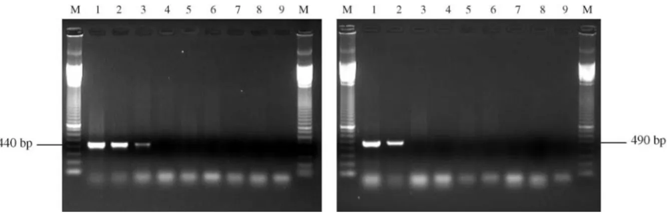
With all theM. a. anisopliaestrains studied by us the ITSMet/ITS4 primer set yielded a specific 440 bp PCR product. Although this set has been developed only to Bra-zilian strains, the Australian strain 14 manifested a low-intensity band in the gels. The ITSMet14/ITS4 primer set produced a 490 bp fragment only from strain 14. Neither of these primers produced amplified products with any of the M. album,M. flavoviride or Beauveria bassiana strains (Figure 4).
Detection ofMetarhizium anisopliae within infected insects
The presence ofMetarhiziumstrains within infected larvae was confirmed by the restriction profiles of the PCR
Figure 2- Alignment of the nucleotide sequences of the ITS1 – 5.8S – ITS2 region of the strains investigated by us (Metarhizium anisopliaevar. anisopliaestrains E9, B/Vi, C and 14;Metarhizium albumstrain 201;Metarhizium flavoviridestrains 204 and 209;Beauveria bassianastrains 959, 2253
products obtained from infected larvae and pure cultures of the fungi (Figures 5 and 6).
Discussion
Our PCR-RFLP data showed that the variation in length and restriction sites in the ITS regions could be used to differentiateM. a. anisopliae from M. album and M. flavoviride. The six distinct restriction enzymes tested in this study were able to clearly distinguish the different Metarhizium species investigated. Ribosomal genes and their ITS and IGS spacer regions have been widely used for the identification and differentiation of species (Foulyet al., 1997) as well as in taxonomic (Driveret al., 2000), phylogenetic (Rakotonirainyet al., 1994) and genetic di-versity (Andersonet al.,2001; Uetakeet al., 2002) studies, with ITS sequences having been reported as being useful for discriminating between different species of fungi (Neuvegliseet al., 1994; Foulyet al., 1997; Jensenet al., 2001; Andersonet al., 2001; Thomsen and Jensen, 2002).
Figure 2 (Cont).
Figure 3- Phylogenetic analysis of ITS1 – 5.8S – ITS4 rDNA sequences from Metarhizium anisopliae, Metarhizium album, Metarhizium flavovirideandBeauveria bassianastrains based on the Neighbor-joining method. GenBank accession numbers are listed after species names.
Figure 4- Amplification of DNA from pure cultures and infected larvae using the primer sets ITSMet/ITS4 (A) and ITS14/ITS4 (B). Primer set A: 1 = strain E9; 2 = larvae infected by strain E9; 3 = strain 14; 4 = larvae infected by strain 14; 5 = strain 201; 6 = strain 204; 7 = strain 959; 8 = non-infected
lar-vae; 9 =control reaction without DNA. Primer set B: 1 = strain 14; 2 = larvae infected by strain 14; 3 = strain E9; 4 = larvae infected by strain E9; 5 = strain
201; 6 = strain 204; 7 = strain 959; 8 = non-infected larvae; 9 = control reaction without DNA. M = 100 bp DNA marker.
Figure 5- PCR-RFLP profiles ofMetarhizium anisopliaevar.anisopliae strain E9using the ITSMet/ITS4 primer set with the restriction enzymes
AfaI,AluI,DdeI,HaeIII,Sau3A I andTaqI. 1 = DNA from a pure cul-ture of strain E9; 2 = DNA from larvae infected with strain E9. M = 100 bp
Our ITS1 – 5.8S – ITS2 sequencing data showed vari-ations which allowed us to design specific primers which could not only detect and identifyM. a. anisopliaebut also to differentiate between our Brazilian strains (E9, B/Vi and
C) and the Australian strain (strain 14) because the se-quence variation of strain 14 was very different from that of the other members of the same species, these sequence vari-ations allowing us to develop a specific primer for strain 14. Isolation of PCR-amplifiable DNA from environ-mental samples is often difficult due to the co-isolation of compounds with the ability to inhibit the PCR (Hegedus and Khachatourians, 1996b). Samples of DNA from plants (Do and Adams, 1991) and fungi (Pfeiferet al., 1993) may retain polysaccharides which can cause problems during amplification and insect cuticles may contain a number of compounds that can act as potent PCR inhibitors (Hack-man, 1974). Our extraction protocol produced DNA from infected larva with sufficient purity for direct PCR amplifi-cation and although this DNA produced a reduced amount of product in comparison with that produced when DNA from pure cultures was used the amount of product was still sufficient for us to be able to identify the fungal species with which the larva had been infected.
Several fungi have been cited as being potential mycoinsecticides (Samsonet al., 1988), although only a few have been intensively investigated and examined at the molecular level (Hegedus and Khachatourians, 1996b). Commercial mycoinsecticides (mycopesticides) require specific and sensitive methods for the identification of the specific fungal strain used during production, such methods being needed not only to ensure strain stability and protect patents but also for use under field conditions for such pur-poses as making environmental impact assessments.
Specific primers have been developed for the detec-tion and identificadetec-tion of some entomopathogenic fungi such asBeauveria bassiana(Hegedus and Khachatourians, 1996b) andGliocladium catenulatum(Paavanen-Huhtala
et al., 2000). In this study we developed a PCR-based primer set system specific forMetarhizium anisopliaevar. anisopliaewhich can simply, rapidly and securely detect this fungus inDiatraea saccharalislarvae 48 h after infec-tion without the need to use techniques such as probe hy-bridization or DNA sequencing.
Our study also showed thatM. a. anisopliae,M. al-bumandM. flavoviridecan be clearly differentiated using PCR-RFLP of the ITS 1-5.8S-ITS 2 region, supporting the view of Curranet al.(1994) who used amplification and se-quencing of the 5.8 S gene, intergenic regions ITS 1 and ITS 2 and phylogenetic analysis to show thatM. anisopliae andM. flavoviride+M. albumrepresent two separate evo-lutionary lines.
The fact that DNA extracted from infected insects can be rapidly analyzed using our set of primers means that the identification of the entomopathogen fungus M. a. anisopliaein laboratory and field studies can be used not only to evaluate the efficacy of mycopesticides and indicate when re-application is necessary but also to study the envi-ronmental persistence of entomopathogens, such evalua-tions normally taking a long time because entomopathogenic fungi must be re-isolated and cultured under specific conditions in order for them to be identified by morphological and physiological tests. Our method of-fers an alternative approach for typing M. a. anisopliae strains within infected insects and reduces the need for time-consuming conventional methods. This technique also opens up the possibility of monitoring the environmen-tal impact of M. a. anisopliae mycoinsecticides on pollinators and other non-target insects.
Acknowledgments
The authors are very grateful to Dr. Valéria Maia de Oliveira (CPQBA/UNICAMP, Campinas, SP, Brazil) for help in the phylogenetic analysis, Dr. Enrico De Beni Arri-goni (CTC/COPERSUCAR, Piracicaba, SP, Brazil) for supplying the insects, Irene M. G. Almeida, M.Sc. (Insti-tuto Biológico, Campinas, SP, Brazil) for revision of the manuscript, Cintia Losano, M.Sc., for help in the alignment of sequences, and the undergraduate students Denise M. Balani and Mariana Ferreira for their technical assistance.
References
Alves SB, Pádua LEM, Azevedo EMVM and Almeida LC (1985) Controle da broca da cana-de-açúcar pelo uso deBeauveria bassiana. Pesq Agrop Bras 20(4):403-406.
Alves SB, Risco SH, Silveira Neto S and Machado Neto R (1984) Pathogenicity of nine isolates of Metarhizium anisopliae
(Metsch.) Sorok. to Diatraea saccharalis (Fabr.). Z ang Entom 97:403-406.
Alves SB (2001) RAPD-PCR na distinção de linhagens de
Metarhizium anisopliae. VII Simpósio de Controle Bio-lógico, Foz do Iguaçú, Brazil.
Anderson IC, Chambers SM and Cairney WGJ (2001) ITS-RFLP and ITS sequence diversity inPisolithusfrom central and eastern Australian sclerophyll forests. Mycol Res 11:1304-1312.
Argentina SC (1999) Taxonomia polifásica deNeurospora produ-toras de aromas. PhD Thesis, UNICAMP, Campinas, Brazil. Bidochka MJ, McDonald MA, St. Leger RJ and Roberts DW (1994) Differentiation of species and strains of entomo-pathogenic by random amplification of polymorphic DNA (RAPD). Curr Gen 25:107-113.
Bindslev L, Oliver RP and Johansen B (2002)In situPCR for de-tection and identification of fungal species. Mycol Res 106:277-279.
Curran J, Driver F, Ballard JWO and Milner RJ (1994) Phylogeny ofMetarhizium: Analysis of ribosomal DNA sequence data. Mycol Res 98:547-552.
Do N and Adams, RP (1991) A simple technique for removing plant polysaccharide contaminants from DNA. BioTech 10:162-166.
Driver F, Milner RJ and Trueman JWH (2000) A taxonomic revi-sion ofMetarhiziumbased on a phylogenetic analysis of rDNA sequence data. Mycol Res 2:134-150.
Esteve-Zarzoso B, Belloch C, Uruburu F and Querol A (1999) Identification of yeasts by RFLP analysis of the 5.8S rRNA gene and the two ribosomal internal transcribed spacers. Int J Syst Bact 49:329-337.
Fouly H, Wilkinson HT and Chen W (1997) Restriction analysis of internal transcribed spacers and the small subunit gene of ribosomal DNA among four Gaeumannomyces species. Mycologia 89:590-597.
Freire LLC, Costa ABL, Góes LB and Oliveira NT (2001) DNA polymorphism and total protein in mutants ofMetarhizium anisopliaevar.anisopliae(Metsch.) Sorokin strain E9.Braz
J Microb 32:93-97.
Fungaro MHP, Vieira MLC, Pizzirani-Kleiner AA and Azevedo JL (1996) Diversity among soil and insect isolates of
Metarhizium anisopliaevar.anisopliaedetected by RAPD. Lett Appl Microb 22:389-392.
Garber RC and Yoder OC (1983) Isolation of DNA from filamen-tous fungi and separation into nuclear, mitochondrial, ribo-somal, and plasmid components. Anal Bioch 135:416-422. Hackman RH (1974) Chemistry of the insect cuticle. In:
Rockstein M (ed) The Physiology of Insecta, v. VI. Aca-demic Press, New York, pp 215-270.
Hegedus DD and Khachatourians GG (1993a) Construction of cloned DNA probes for the specific detection of the ento-mopathogenic fungusBeauveria bassianain grasshoppers. J Invert Path 62: 33-240.
Hegedus DD and Khachatourians GG (1993b) Identification of molecular variants in mitochondrial DNAs of members of the genera Beauveria, Verticillium, Paecilomyces, Tolypocladium, and Metarhizium. Appl Env Microb 59:4283-4288.
Hegedus DD and Khachatourians GG (1996a) Identification and differentiation of the fungusBeauveria bassianausing merase chain reaction and single-strand conformation poly-morphism analysis. J Invert Path 67:289-299.
Hegedus DD and Khachatourians GG (1996b) Detection of the entomopathogenic fungus Beauveria bassiana within in-fected migratory grasshoppers (Melanoplus sanguinpes)
us-ing polymerase chain reaction and DNA probe. J Invert Path 67:21-27.
Jensen AB, Thomsen L and Eilenberg J (2001) Intraespecific vari-ation and host specificity ofEntomophtora muscae sensu strictoisolates revealed by random amplified polimorphic DNA, universal primed PCR, PCR-restriction fragment lenght polymorphism, and conidial morphology. J Invert Path 78:251-259.
Leal SCM, Bertioli DJ, Butt TM and Peberdy JF (1994) Charac-terization of isolates of the entomopathogenic fungus
Metarhizium anisopliae by RAPD-PCR. Mycol Res 98:1077-1081.
Neuveglise C, Brygoo Y, Vercambre B and Riba G (1994) Com-parative analysis of molecular and biological characteristics of strains ofBeauveria brongniartiiisolated from insects. Mycol Res 98:322-328.
Onofre SB, Vargas LRB, Rossato M, Barros NM, Boldo JT, Nunes ARF and Azevedo JL (2002) Controle biológico de pragas na agropecuária, por meio de fungos entomopato-gênicos. In: Serafini LA, Barros NM and Azevedo JL (eds) Biotecnologia: Avanços na Agricultura e na Agroindústria, EDUCS, Caxias do Sul, pp 295-317.
Paavanen-Huhtala S, Akinainen H and Yli-Mattila T (2000) De-velopment of strain-specific primers for a strain of
Gliocladium catenulatumused in biological control. Eur J Plant Path 106:187-198.
Pfeifer TA and Khachatourians GG (1993) Isolation of DNA from entomopathogenic fungi grown in liquid cultures. J Invert Path 61:113-116.
Pontecorvo G, Roper JA, Hemons JM, Mac Donald KD and But-ton AWJ (1953) The genetics ofAspergillus nidulans. Adv Gen 5:141-238.
Rakotonirainy MS, Cariou ML, Brygoo Y and Riba G (1994) Phylogenetic relationships within the genus Metarhizium
based on 28S rRNA sequences and isozyme comparison. Mycol Res 98:225-230.
Sambrook J, Fritsch EF and Maniatis T (1989) Molecular Clon-ing: A Laboratory Manual. Cold Spring Harbor Laboratory Press, Cold Spring Harbor, New York, 545 pp.
Samson RA, Evans HC and Latge JP (1988). Atlas of Entomo-pathogenic Fungi. Springer-Verlag, Netherlands, 187 pp. Thomsen L and Jensen AB (2002) Application of nested-PCR
technique to resting spores from theEntomophthora muscae
species complex: implications for analyses of host-pathogen population interactions. Mycologia 94:794-802.
Uetake Y, Arakawa M, Nakamura H, Akahira T, Sayama A, Cheah L-HO, Okabe IO and Matsumoto N (2002) Genetic relationship among violet root rot fungi as revealed by hyphal anastomosis and sequencing of the rDNA ITS re-gions. Mycol Res 106:156-163.
Urtz BE and Rice WC (1997) RAPD-PCR characterization of
Beauveria bassiana isolates from the rice water weevil
Lissorhoptrus oryzophilus. Lett Appl Microb 25:405-409. White TJ, Bruns TD, Lee SB and Taylor JW (1990) Analysis of
phylogenetic relationships by amplification and direct se-quencing of ribosomal DNA genes. In: Innis DH, Sninsky JJ and White TJ (eds) PCR PROTOCOLS: A Guide to Methods and Applications. Academic Press, New York, pp 315-322.