The origin of cortical neurons
Department of Anatomy and Developmental Biology, University College London, London, UK
J.G. Parnavelas
Abstract
Neurons of the mammalian cerebral cortex comprise two broad classes: pyramidal neurons, which project to distant targets, and the inhibitory nonpyramidal cells, the cortical interneurons. Pyramidal neurons are generated in the germinal ventricular zone, which lines the lateral ventricles, and migrate along the processes of radial glial cells to their positions in the developing cortex in an inside-out sequence. The GABA-containing nonpyramidal cells originate for the most part in the ganglionic eminence, the primordium of the basal ganglia in the ventral telencephalon. These cells follow tangential migratory routes to enter the cortex and are in close association with the corticofugal axonal system. Once they enter the cortex, they move towards the ventricular zone, possibly to obtain positional information, before they migrate radially in the direction of the pial surface to take up their positions in the developing cortex. The mechanisms that guide inter-neurons throughout these long and complex migratory routes are currently under investigation.
Correspondence
J.G. Parnavelas
Department of Anatomy and Developmental Biology University College London Gower Street
London WC1E 6BT UK
Fax: +44-20-7679-7349 E-mail: [email protected]
Presented at the IV International UNESCO Course on “What the Developing Cerebral Cortex Tells About the Adult Cortex (and Vice Versa)”, Rio de Janeiro, RJ, Brazil, December 3-7, 2001.
Received July 12, 2002 Accepted October 16, 2002
Key words
·Neurogenesis ·Migration
·Cortical development
Introduction
The cerebral cortex is by far the largest part of the brain of mammals containing about half of the total number of neurons. Microscopic examination of a coronal sec-tion through the cortex shows that the neu-ronal cell bodies are arranged in six layers. The layering is produced by variation in the types and density of cell bodies through the cortical depth. Each layer contains a comple-ment of pyramidal, so-called for the shape of their somata, and nonpyramidal neurons. Pyramidal neurons, which make up approxi-mately 75% of all neurons in the cortex, are the projection cells that send axons to other areas of the cortex and to distant parts of the brain. They utilize the excitatory amino acid glutamate as a neurotransmitter. Nonpyrami-dal cells are interneurons, that is, their con-nections are all made locally. There are many
varieties of nonpyramidal neurons based on differences in size and shape of their den-drites and patterns of axonal branching. They all contain the inhibitory neurotransmitter GABA and also one or more neuropeptides and/or calcium-binding proteins (for a re-view, see Ref. 1).
de-picted in Figure 1 (2,3), shows that early post-mitotic neurons migrate away from the VZ towards the surface of the cerebral vesicles to form the primordial plexiform layer or pre-plate (PP). The later-generated neurons mi-grate to form a layer within the PP, the so-called cortical plate (CP), thus splitting it into a superficial marginal zone (MZ; layer I) and a deep subplate (SP). The neurons of the CP assemble into layers II-VI in an inside-out sequence, that is, the deepest cellular layers are assembled first and those closest to the surface last. Both the MZ and SP contain the earliest-generated neurons of the cerebral cor-tex. Those in layer I differentiate as Cajal-Retzius cells and other types of neurons that have not yet been fully characterized (4,5). The SP is separated from the VZ by the inter-mediate zone (IZ), a layer that will eventually contain the afferent and efferent axonal tracts of the cortex (future white matter). As the CP emerges, another layer of proliferating cells appears between the VZ and IZ, the so-called subventricular zone (SVZ). This germinal zone contains cells, produced in the VZ, that mainly give rise to glia (6-8). The SVZ expands greatly in late gestation and in early postnatal life as the VZ disappears. According to this conven-tional scheme of cortical development, radial
glial cells serve as a scaffold to support and direct the migration of young neurons from their origin in the VZ to their positions in the CP (9). However, this view is now changing in the light of emerging evidence which shows that radial glia produce cortical neurons and glia, in addition to guiding migration (10-13).
Development of neuronal lineages
At what point do undifferentiated cells in the VZ commit themselves to become pyra-midal or nonpyrapyra-midal neurons? Knowledge of the genealogical (lineage) relationships of cells in the cortex would facilitate our efforts to understand the mechanisms that underlie their developmental choices. The study of cell lineages in the cerebral cortex has be-come possible with the use of recombinant retroviruses (14,15). The advantage of this technique is that, by infecting single pro-genitor cells with a virus, a marker gene is introduced into them that can be inherited undiluted by all their progeny.
Earlier lineage studies, using predomi-nantly the BAG retrovirus, observed single-labeled cells or discrete clusters of two or more neurons in the cerebral cortex follow-ing the placement of intraventricular
injec-VZ
VZ IZ PP
MZ
CP
SP
IZ
SV
VZ
tions at any day of gestation during cortical neurogenesis in rats and mice (16,17). The clones of two or more cells contained for the most part either neurons or glial cells, a finding that was confirmed in dissociated cell cultures (16,18). When neuronal clones were examined further by electron micros-copy or immunohistochemistry, it was found that they were nearly all homogeneous, com-posed of all pyramidal or all nonpyramidal cells (19-21). These early observations sug-gested that the VZ, the germinal layer of the embryonic cortex, might be viewed as a mosaic of progenitor cells each with a differ-ent restricted potdiffer-ential.
Further examination of clonally related neurons from brains injected with retrovirus at different stages of corticogenesis revealed remarkable differences in the number and laminar distribution of pyramidal and non-pyramidal neurons. Nonnon-pyramidal cells ap-peared either as single cells or as clones of two neurons present in the same or in adja-cent layers. Clones of pyramidal neurons were larger, and they were either dispersed in one layer or in several layers following earlier injections. Their size and laminar distribution was progressively reduced fol-lowing later injections (20). This finding suggested the existence of different mechan-isms that generate the two main neuronal types of the cerebral cortex. Other lineage analyses have utilized libraries of retroviral vectors carrying a large number of DNA tags, each of which can be distinguished by the polymerase chain reaction (22). These studies have shown that not all clonally re-lated cells maintain the spatial relationship that they had before migration (23), but it is noteworthy that those that do invariably show the same phenotype (24).
The more recent use of a retroviral marker in combination with bromodeoxyuridine (BrdU) has shown that only pyramidal neu-rons maintain a close spatial relationship with their clonal relatives in the developing cortex, which can be achieved through radial
migration (25). In contrast, labeled nonpy-ramidal cells were found, in agreement with earlier lineage analyses, as isolated cells or pairs of clonally neurons. Their low content of BrdU indicated that these cells were part of larger clones, and suggested that their isolation was the result of non-radial (tan-gential) migration. These findings pose two alternative interpretations: either clonally related cells, instructed to develop a particu-lar phenotype, are also endowed with the ability to use a specific migratory pathway, or cues encountered during radial and tan-gential migration are responsible for the py-ramidal and nonpypy-ramidal phenotypes. Ex-periments by Tan et al. (26) have provided evidence for the former possibility with re-gards to the generation and migration of pyramidal neurons. Using highly unbalanced mouse embryonic stem cell chimeras, these authors found that radially dispersed neu-rons contained glutamate, the marker of py-ramidal neurons, whereas tangentially dis-persed cells were predominantly GABAergic. Their study has also demonstrated that spec-ification of the pyramidal lineage occurs at the level of the progenitor, before the onset of neurogenesis.
were the neurons found to migrate tangen-tially in the developing cortex after they were labeled with a fluorescent marker in the VZ (27). The presence in the cortex of a relatively small number of nonpyramidal neurons compared with the often very large pyramidal clones suggests that there are other potential sources of nonpyramidal neurons.
Nonpyramidal neurons: origin and tangential migration
Sources of neurons destined for the cere-bral cortex have been discovered in the gan-glionic eminence, the primordium of the ba-sal ganglia in the ventral telencephalon or subpallium. Porteus et al. (28) first reported that cells expressing Dlx2 appear to migrate from the ventral telencephalon to the neo-cortex. Subsequently, De Carlos et al. (29) and Tamamaki et al. (30) provided unequivo-cal evidence that cells in the lateral gangli-onic eminence (LGE), the primordium of the striatum, migrate into the developing neo-cortex, and are distributed predominantly in the IZ. More recently, a number of tracing studies (31-33) have shown that the cells that
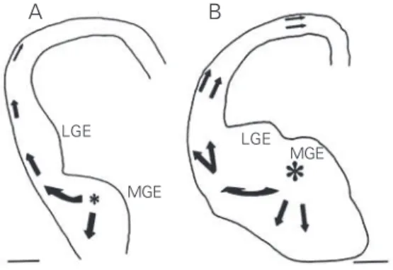
migrate from the ventral telencephalon to the cortex are indeed GABAergic. Three different streams of neurons originating in the ventral telencephalon were observed to round the corticostriatal notch and follow tangentially oriented paths to enter the cor-tex. An early cohort (E12 in mouse; E14 in rat), originating in the medial ganglionic eminence (MGE), invades mainly the PP. These cells are tangentially oriented and show features typical of Cajal-Retzius cells (Fig-ure 2A). A second and more prominent co-hort, composed also of MGE cells, has been observed to migrate predominantly through the IZ slightly later in development (E13-15 mouse; E15-17 rat) (Figure 2B). At the late stages of corticogenesis, cells originating in the LGE and MGE appear in the lower IZ and SVZ (34). Genetic manipulations have provided further evidence for the origin of the cortical GABAergic interneurons in the subpallium. Thus, mice lacking transcrip-tion factors that regulate regionalizatranscrip-tion (Nkx2.1) or differentiation (Dlx1/2, Mash1) in the ventral telencephalon have signifi-cantly reduced numbers of GABAergic neu-rons in the cortex (31,35,36). Specifically, Nkx2.1 mutants that lack a functional MGE
show approximately a 50% reduction in GABA-containing neurons in the cortex, but the olfactory bulbs of these mutants contain nearly a normal number of GABAergic cells. Further, tracing studies in slice cultures of these mutants showed a significantly reduced migration of labeled cells in the cortex from the LGE or from the abnormal MGE. In contrast, the Dlx1/2 mutants that lack migra-tion of cells out of the LGE and MGE in slice cultures, have about a 75% reduction of GABA cells in the cortex and virtually no GABAergic cells in the olfactory bulb. These observations suggest that the MGE is the main source of interneurons to the cerebral cortex, while the LGE provides some cells to the cortex and many to the olfactory bulb. This is supported by the finding that dissoci-ated MGE cells injected into the lateral
ven-Figure 2. Camera lucida drawings of coronal sections through part of the rat forebrain after placement of DiI
crystals in the medial ganglionic eminence (MGE) at E14 (A) and E16 (B). Arrows indicate the direction of migration of labeled cells in the cortical primordium 2 days (A) and 3 days (B) after DiI application. In A, fluorescent cells appeared as a stream rounding the corticostriatal sulcus and heading toward the preplate. In B, migrating cells were seen in the most superficial and the deeper aspects of the cortical primordium. LGE, lateral ganglionic eminence.
LGE
MGE
LGE MGE
tricle tend to migrate to the striatum, pallidum and neocortex, while LGE cells migrate pre-dominantly into the striatum and olfactory bulb (33). In a more recent study, Stühmer et al. (37) have shown that all cortical GABA-ergic neurons in the adult mouse brain are derived from cells that express the Dlx genes. This finding suggests that the vast majority, if not all, GABA-containing neurons in the mammalian cortex are derived from pro-genitors in the ventral telencephalon.
Neurons of the ganglionic eminence des-tined for the developing cerebral cortex ex-press the LIM homeobox gene Lhx6 (32,38). In the later stages of corticogenesis and in postnatal life, Lhx6 has been found to be expressed in cells scattered throughout the cortical thickness. Although double-label-ing experiments have not yet been conducted, the distribution of these cells resembles that of the GABA-containing neurons (Alifragis P and Parnavelas JG, unpublished observa-tions), suggesting that cortical interneurons express Lhx6 during corticogenesis and in later life. LIM homeodomain transcription factors have been previously implicated in neurotransmitter expression (39), and it may be that the expression of GABA in cortical interneurons is under the control of Lhx6.
The molecular mechanisms that guide the migration of interneurons from the gan-glionic eminence, around the corticostriatal notch and into the neocortex are unknown. What signals trigger the migration of these neurons? A number of factors have been shown to stimulate motogenic activity in neural and non-neural tissues. One of these molecules, hepatocyte growth factor/scatter factor (HGF/SF), and its receptor MET have recently been shown to be important in the migration of cortical interneurons. Disrup-tion of the normal expression of HGF/SF appears to result in undirected scattering of cells from the ganglionic eminence and in a significant reduction of interneurons in the cortex at the time of birth (40). Recent stud-ies have demonstrated that interneurons
des-tined to populate the cortex express neuro-pilin1 and neuropilin2, which enable them to respond to chemorepulsion produced by class 3 semaphorins in the striatal mantle (41). Further, it appears that the repulsive activity of semaphorins in the developing striatum would create an exclusion zone for migrating cortical interneurons and channel them into adjacent paths, leading to the for-mation of MZ- and IZ-migratory routes.
developing CP (Figure 3).
Earlier birthdating studies (47) and more recent transplantation studies (48) have dem-onstrated that, similar to pyramidal cells, cortical interneurons are disposed in an in-side-out pattern within the CP. How is it that two neuronal types generated in two distinct regions of the developing brain, the pallium and subpallium, and shown to fol-low different migratory paths, canassemble in the cortex in a way that they are linked temporally and spatially? Nadarajah and col-leagues (46) have proposed that cortical
in-terneurons follow ventricle-directed migra-tion actively seeking the VZ in order to obtain the type of positional information that the pyramidal cells acquire prior to becom-ing postmitotic. These cues may be obtained from the local environment or from the py-ramidal cells themselves through neural-neu-ral interactions. The mechanism that guides interneurons to the VZ before they move to the positions in the developing cortex is not known, but a combination of chemoattrac-tant (e.g., semaphorins) and chemorepellent (e.g., Slit) molecules may be involved.
References
1. Parnavelas JG, Dinopoulos A & Davies SW (1989). The central visual pathways. In: Björklund A, Hökfelt T & Swanson LW (Editors), Handbook of Chemical Neu-roanatomy.Vol. 7. Integrated Systems of the CNS, Part II. Elsevier, Amsterdam, The Netherlands, 1-164.
2. Boulder Committee (1970). Embryonic vertebrate central nervous system: re-vised terminology. Anatomical Record, 166: 257-262.
3. Uylings HBM, Van Eden CG, Parnavelas JG & Kalsbeek A (1990). The prenatal and postnatal development of rat cerebral cor-tex. In: Kolb B & Tees RC (Editors), The Cerebral Cortex of the Rat. MIT Press, Cambridge, MA, USA, 35-76.
4. Frotscher M (1997). Dual role of
Cajal-Retzius cells and reelin in cortical devel-opment. Cell and Tissue Research, 290: 315-322.
5. Meyer G, Goffinet AM & Fairén A (1999). What is a Cajal-Retzius cell? A reassess-ment of a classical cell type based on recent observations in the developing cor-tex. Cerebral Cortex, 9: 765-775. 6. Levison SW & Goldman JE (1993). Both
oligodendrocytes and astrocytes develop from progenitors in the subventricular zone of postnatal rat forebrain. Neuron, 10: 201-212.
7. Luskin MB & McDermott KA (1994). Di-vergent lineages for oligodendrocytes and astrocytes originating in the neonatal fore-brain subventricular zone. Glia, 11: 211-226.
8. Parnavelas JG (1999). Glial cell lineages in the rat cerebral cortex. Experimental Neu-rology, 156: 418-429.
9. Rakic P (1972). Mode of cell migration to the superficial layers of fetal monkey neo-cortex. Journal of Comparative Neurology, 145: 61-83.
10. Hartfuss E, Galli R, Heins N & Gotz M (2001). Characterization of CNS precursor subtypes and radial glia. Developmental Biology, 229: 15-30.
11. Miyata T, Kawaguchi A, Okano H & Ogawa M (2001). Asymmetric inheritance of radial glial fibers by cortical neurons. Neuron, 31: 727-741.
lish radial units in neocortex. Nature, 409: 714-720.
13. Parnavelas JG & Nadarajah B (2001). Ra-dial glial cells: are they really glia? Neuron, 31: 881-884.
14. Sanes JR, Rubenstein JLR & Nicolas J-F (1986). Use of recombinant retrovirus to study post-implantation cell lineage in mouse embryos. EMBO Journal, 5: 3133-3142.
15. Price J, Turner D & Cepko C (1987). Lin-eage analysis in the vertebrate nervous system by retrovirus-mediated gene transfer. Proceedings of the National Academy of Sciences, USA, 84: 156-160. 16. Luskin MB, Pearlman AL & Sanes JR (1988). Cell lineage in the cerebral cortex of the mouse studied in vivo and in vitro with a recombinant retrovirus. Neuron, 1: 635-647.
17. Price J & Thurlow L (1988). Cell lineage in the rat cerebral cortex: a study using ret-roviral-mediated gene transfer. Develop-ment, 104: 473-482.
18. Price J, Williams B & Grove E (1992). The generation of cellular diversity in the cere-bral cortex. Brain Pathology,2: 23-29. 19. Parnavelas JG, Barfield JA, Franke E &
Luskin MB (1991). Separate progenitor cells give rise to pyramidal and nonpy-ramidal neurons in the rat telencephalon. Cerebral Cortex,1: 463-468.
20. Luskin MB, Parnavelas JG & Barfield JA (1993). Neurons, astrocytes, and oligo-dendrocytes of the rat cerebral cortex originate from separate progenitor cells: an ultrastructural analysis of clonally re-lated cells. Journal of Neuroscience, 13: 1730-1750.
21. Mione MC, Danevic C, Boardman P, Har-ris B & Parnavelas JG (1994). Lineage anal-ysis reveals neurotransmitter (GABA and glutamate) but not calcium-binding pro-tein homogeneity in clonally related corti-cal neurons. Journal of Neuroscience, 14: 107-123.
22. Walsh C & Cepko CL (1992). Widespread dispersion of neuronal clones across func-tional regions of the cerebral cortex. Sci-ence, 255: 434-440.
23. Walsh C & Cepko CL (1993). Clonal dis-persion in proliferative layers of develop-ing cerebral cortex. Nature, 362: 632-635. 24. Reid CB, Liang I & Walsh C (1995). Sys-tematic widespread clonal organization in cerebral cortex. Neuron, 15: 299-310. 25. Mione MC, Cavanagh JFR, Harris B &
Par-navelas JG (1997). Cell fate specification and symmetrical/asymmetrical divisions in the developing cerebral cortex. Journal of Neuroscience, 17: 2018-2029.
26. Tan SS, Kalloniatis M, Sturm K, Tam PPL, Reese BE & Faulkner-Jones B (1998). Separate progenitors for radial and tan-gential cell dispersion during develop-ment of the cerebral cortex. Neuron, 21: 295-304.
27. O’Rourke NA, Sullivan DP, Kaznowski CE, Jacobs AA & McConnell SK (1995). Tan-gential migration of neurons in the devel-oping cerebral cortex. Development, 121: 2165-2176.
28. Porteus MH, Bulfone A, Liu JK, Puelles L, Lo LC & Rubenstein JLR (1994). DLX-2, MASH-1, and MAP-2 expression and bro-modeoxyuridine incorporation define mo-lecularly distinct cell populations in the embryonic mouse forebrain. Journal of Neuroscience, 14: 6370-6383.
29. De Carlos JA, Lopez-Mascaraque L & Val-verde F (1996). Dynamics of cell migra-tion from the lateral ganglionic eminence in the rat. Journal of Neuroscience, 16: 6146-6156.
30. Tamamaki N, Fugimori KE & Takauji R (1997). Origin and route of tangentially migrating neurons in the developing neo-cortical intermediate zone. Journal of Neu-roscience, 17: 8313-8323.
31. Anderson SA, Eisenstat DD, Shi L & Ru-benstein JLR (1997). Interneuron migra-tion from the basal forebrain to neocor-tex: dependence on Dlx genes. Science, 278: 474-476.
32. Lavdas AA, Grigoriou M, Pachnis V & Par-navelas JG (1999). The medial ganglionic eminence gives rise to a population of early neurons in the developing cerebral cortex. Journal of Neuroscience, 19: 7881-7888.
33. Wichterle H, Garcia-Verdugo JM, Herrera DG & Alvarez-Buylla A (1999). Young neu-rons from medial ganglionic eminence dis-perse in adult and embryonic brain. Na-ture Neuroscience, 2: 461-466.
34. Anderson SA, Marin O, Horn C, Jennings K & Rubenstein JLR (2001). Distinct corti-cal migrations from the medial and lateral ganglionic eminences. Development, 128: 353-363.
35. Casarosa S, Fode C & Guillemot F (1999). Mash1 regulates neurogenesis in the ven-tral telencephalon. Development, 126: 525-534.
36. Sussel L, Marin O, Kimura S & Ruben-stein JLR (1999). Loss of Nkx2.1 homeo-box gene function results in a ventral to dorsal molecular respecification within the basal telencephalon: evidence for a trans-formation of the pallidum into the stria-tum. Development, 126: 3359-3370. 37. Stühmer T, Puelles L, Ekker M &
Ruben-stein JLR (2002). Expression from a Dlx gene enhancer marks adult mouse corti-cal GABAergic neurons. Cerebral Cortex, 12: 75-85.
38. Grigoriou M, Tucker AS, Sharpe PT & Pachnis V (1998). Expression and regula-tion of Lhx6 and Lhx7, a novel subfamily of LIM homeodomain encoding genes, suggests a role in mammalian head devel-opment. Development, 125: 2063-2074. 39. Thor S & Thomas JB (1997). The
Droso-phila islet gene governs axon pathfinding and neurotransmitter identity. Neuron, 18: 397-409.
40. Powell EM, Mars WM & Levitt P (2001). Hepatocyte growth factor/scatter factor is a motogen for interneurons migrating from the ventral to the dorsal telencepha-lon. Neuron, 30: 79-89.
41. Marin O, Yaron A, Bagri A, Tessier-Lavigne M & Rubenstein JL (2001). Sort-ing of striatal and cortical interneurons regulated by semaphorin-neuropilin inter-actions. Science, 293: 872-875. 42. Mètin C & Godement P (1996). The
gan-glionic eminence may be an intermediate target for corticofugal and thalamocortical axons. Journal of Neuroscience, 16: 3219-3235.
43. Rakic P (1985). Contact regulation of neu-ronal migration. In: Edelman GM & Thiery JP (Editors), The Cell in Contact. Wiley-Liss, Inc., New York, NY, USA, 67-91. 44. Gray GE, Leber SM & Sanes JR (1990).
Migratory patterns of clonally related cells in the developing central nervous system. Experientia, 46: 929-940.
45. Denaxa M, Chan C-H, Schachner M, Par-navelas JG & Karagogeos D (2001). The adhesion molecule TAG-1 mediates the migration of cortical interneurons from the ganglionic eminence along the corticofu-gal fiber system. Development, 128: 4635-4644.
46. Nadarajah B, Alifragis P, Wong R & Parna-velas JG (2002). Ventricle-directed migra-tion in the developing cerebral cortex. Na-ture Neuroscience, 5: 218-224.
47. Miller MW (1985). Cogeneration of retro-gradely labeled corticocortical projection and GABA-immunoreactive local circuit neurons in cerebral cortex. Developmen-tal Brain Research,23: 187-192. 48. Wichterle H, Turnbull DH, Nery S, Fishell