Sotalia
(Gray, 1866) (Cetacea, Delphinidae)
Fettuccia, DC.
a* da Silva, VMF.
aand Simões-Lopes, PC.
b aLaboratório de Mamíferos Aquáticos – INPA,Av. André Araújo, 2936, Aleixo, CEP 69060-001, Manaus, AM, Brazil
bLaboratório de Mamíferos Aquáticos – LAMAQ, Departamento de Ecologia e Zoologia, CCB, Universidade Federal de Santa Catarina – UFSC,
CP 5102, CEP 88040-970, Florianópolis, SC, Brazil *e-mail: [email protected]
Received December 7, 2007 – Accepted August 27, 2008 – Distributed August 31, 2009 (With 12 figures)
Abstract
Analyses of non-metric characters of the skull and cervical vertebrae were performed among samples of dolphins of the genus Sotalia from the north, northeast and south Brazilian coast (S. guianensis) and also samples from the Amazon River Basin (S. fluviatilis) as part of an osteological descriptive study. The results demonstrated that there was a higher percentage of occurrence of fenestrae in the occipital region (66%) and cervical ribs in the cervical vertebrae (87%) in the riverine species. The vomer in wide shape was more frequent in the riverine species (57%), followed by the intermediate (32%) and narrow shape (11%), that was found to be more frequent in the marine spe-cies (66 to 76%). In relation to the lacerate anterior foramen, it was observed that an open/elongated shape is more common in the riverine species (88%). Most samples in the marine species present this foramen divided by a spike shaped projection (72 to 98%). The ventrally visible location of the hypoglossal foramen was more often observed externally displaced in S. guianensis (88 to 98%), while in S. fluviatilis, most samples (87%) presented this foramen internally displaced to the jugular notch, and not visible in ventral view. The fluvial species seems to present neoteny (or maintenance of juvenile characters in adults) in relation to the position of the pterygoids and in development of lacerate anterior foramen.
Keywords: Morphology, Sotalia, osteology, skull, cervical vertebrae.
Caracteres não métricos nas duas espécies de
Sotalia
(Gray, 1866)
Resumo
Este é um trabalho osteológico descritivo entre exemplares do gênero Sotalia da costa Norte, Nordeste e Sul do Brasil e exemplares fluviais da bacia amazônica, analisando caracteres não métricos no crânio e nas vértebras cervicais. A frequência de ocorrência de fenestras na região occipital (66%) e de costelas cervicais (87 %) foi maior na espécie fluvial (S. fluviatilis). Na espécie fluvial, a forma do vômer largo foi mais frequente (57%), seguida da forma interme-diária (32%) e estreita (11%). A forma do vômer estreito foi mais frequente na espécie marinha (S. guianensis) (66 a 76%). Em relação ao forâmen lacerado anterior, foi observado que a forma aberta/alongada é mais comum na espécie fluvial (88%). Na espécie marinha, a maioria dos exemplares apresenta este forâmen dividido por uma projeção em forma de espinho (72 a 98%). A localização do forâmen hipoglossal visível ventralmente foi mais observada em S. guianensis (88 a 98%), enquanto que em S. fluviatilis, a maioria dos exemplares (87%) apresentou este forâmen deslocado internamente à reentrância jugular, não podendo ser observado em vista ventral. A espécie fluvial parece apresentar neotenia (ou manutenção de caracteres juvenis no adulto) em relação ao posicionamento dos pterigoides e ao desenvolvimento do forâmen lacerado interior.
Palavras-chave: morfologia, Sotalia, osteologia, crânio, vértebra cervical.
1. Introduction
The genus Sotalia (Gray, 1866) was until recently considered monospecific, however recent molecular ge-netics (Cunha et al., 2005; Caballero et al., 2007), and geometric morphometric (Monteiro-Filho et al., 2002)
parison of forms or presence and absence of determined characters for this genus. Therefore, this study aims to compare the two species of the genus Sotalia using non-metric characters.
2. Material and Methods
A total of 149 specimens were analysed from five distinct Brazilian states: Mammal Collection of National Institute for Amazônia Research (INPA), Manaus, Amazonas (AM); Emilio Goeldi Museum (MPEG), Belém, Pará (PA); Aquatic Mammals Laboratory (LMA) of the Department of Ecology and Zoology, Federal University of Santa Catarina (UFSC), Florianópolis, Santa Catarina (SC); as well as the Osteological Archive of the Association of Research and Preservation of Aquatic Ecosystems (AQUASIS), Caucaia, Ceará (CE) (Tables 1-4). The INPA collection contains riverine spec-imens (S. fluviatilis) from the Amazonas State (AM), ma-rine specimens from the coast of the Amapá (AP) and es-The marine species is broadly distributed along the
tropical and subtropical Atlantic coast of South and Central America, having been recorded from Florianópolis, Santa Catarina, Brazil (27° 35’ S and 48° 34’ W) (Simões-Lopes, 1988; Borobia, 1989) to Honduras (15° 58’ N and 85° 42’ W) (da Silva and Best, 1996). The riverine species is endemic to the Amazon Basin, occurring from Belém (at the mouth of the Amazon River), in Brazil, to the rivers of Peru, Colombia and Ecuador (Borobia et al., 1991, da Silva and Best, 1996).
Osteological descriptions for the genus Sotalia have been published by Miranda-Ribeiro (1936); Casinos et al. (1981); Borobia (1989); Menezes and Simões-Lopes (1996); da Silva and Best (1996); Alves Júnior and Monteiro-Neto (1999); Avila et al. (2002); Fettuccia and Simões-Lopes (2004), Simões-Lopes (2006). With the exception of Simões-Lopes (2006), these studies were mainly based on traditional studies of morphomet-rics. There are no previous comparative studies using non-metric characteristics such as, for example, the
com-Table 1. List of Sotalia fluviatilis analysed from the northern region (Amazon State, Brazil), deposited at INPA’s mam-mals collection. F - female, M - male, I - indeterminate gender. N = 44.
Collection number
Sex Age
class
Locality Collection
number
Sex Age
class
Locality
INPA 005 I adult Japurá River INPA 054 M calf Amazonas River INPA 007 I adult Tefé Lake INPA 055 I adult Amazonas River INPA 008 I adult Tefé Lake INPA 056 F adult Amazonas River INPA 009 M juvenile Negro River INPA 057 F adult Amazonas River INPA 015 F calf Japurá River INPA 059 M juvenile Marchantaria,
Solimões River INPA 016 I adult Japurá River INPA 060 M juvenile Marchantaria,
Solimões River INPA 017 M adult Japurá River INPA 062 F adult Tefé Lake INPA 018 I calf Japurá River INPA 065 M calf Japurá River INPA 020 I adult Japurá River INPA 067 I adult Japurá River INPA 024 I adult Japurá River INPA 069 F adult Negro River/
Anavilhanas INPA 026 M juvenile Purus River INPA 071 F adult Japurá River INPA 029 F juvenile Tefé Lake INPA 072 I adult Pará River INPA 038 F calf Japurá River INPA 073 F adult Purus River INPA 039 M immature Japurá River INPA 074 M adult Purus River INPA 040 F adult Amanã Lake INPA 080 M calf Catalão, Negro
River INPA 041 M adult Negro River/
Anavilhanas
INPA 081 M juvenile Cabaliana Lake, Manacapuru INPA 043 M juvenile Japurá River INPA 082 F calf Tapajós River INPA 047 M adult Juruá River INPA 093 M adult Tocantis River/
analyzed (AP = 24; PA = 02; CE = 41; SC = 38). The marine specimens were divided into 3 large areas: NOR (including specimens from AP and PA), for the speci-mens from the coast of Amapá and Pará States, CE for tuarine specimens (S. guianensis) from Pará State (PA).
The other collections only possess marine specimens from their respective localities. Forty-four specimens of S. fluviatilis and 105 specimens of S. guianensis were
Table 2. List of Sotalia guianensis analyzed from northern region (Amapá and Pará States, Brazil), deposited at INPA and MPEG mammals collections, respectively. F - female, M - male, I - indefinite gender. N = 26.
Collection number
Sex Age
class
Locality Collection
number
Sex Age
class
Locality
INPA 120 M juvenile Amazonas Estuary INPA 133 M juvenile North of Amapá INPA 121 M juvenile Amazonas Estuary INPA 134 F adult North of Amapá INPA 122 M juvenile Amazonas Estuary INPA 135 F calf North of Amapá INPA 123 M adult North of Amapá INPA 136 M calf North of Amapá INPA 124 F juvenile North of Amapá INPA 137 M calf North of Amapá INPA 125 F juvenile Amazonas Estuary INPA 138 F juvenile North of Amapá INPA 126 M juvenile North of Amapá INPA 139 F juvenile North of Amapá INPA 127 M calf North of Amapá INPA 140 M juvenile North of Amapá INPA 128 M calf North of Amapá INPA 141 F adult North of Amapá INPA 129 M adult North of Amapá INPA 142 F adult North of Amapá INPA 130 F juvenile North of Amapá INPA 143 F adult North of Amapá INPA 131 F calf Amazonas Estuary MPEG 24548 I adult Marajó Island INPA 132 F calf Amazonas Estuary MPEG 10945 F juvenile Marajó Island
Table 3. List of Sotalia guianensis analysed from the Northeast region (Ceará state, Brazil), deposited at AQUASIS’s collec-tion. F - female, M - male, I - indeterminate gender. N = 41.
Collection number
Sex Age class Locality Collection
number
Sex Age
class
Locality
Aq 002 M adult Caucaia Aq 210 F adult Paracuru
Aq 004 I juvenile Fortaleza Aq 212 F adult Fortaleza
Aq 012 I adult Fortaleza Aq 213 M calf Fortaleza
Aq 013 M adult Fortaleza Aq 214 F juvenile Caucaia
Aq 023 M adult Caucaia Aq 215 I adult São G. Amarante
Aq 026 I adult Caucaia Aq 218 M juvenile São G. Amarante
Aq 036 F adult Fortaleza Aq 222 F adult Fortaleza
Aq 038 M adult Caucaia Aq 231 M adult Caucaia
Aq 039 I adult São G. Amarante Aq 232 M adult Fortaleza Aq 040 I calf São G. Amarante Aq 234 M adult Caucaia Aq 041 I immature Fortaleza Aq 236 I adult São G. Amarante
Aq 042 M adult Fortaleza Aq 239 M adult Fortaleza
Aq 058 F adult Fortaleza Aq 240 I immature Paraipaba
Aq 071 I adult Cascavel Aq 241 I adult Caucaia
Aq 084 M adult Fortaleza Aq 247 F adult Fortaleza
Aq 132 M juvenile São G. Amarante Aq 250 M calf Paraipaba
Aq 134 M adult Fortaleza Aq 251 I adult Trairé
Aq 139 I adult Itapipoca Aq 253 M adult Fortaleza
Aq 159 I adult Fortaleza Aq 259 M adult Fortaleza
Aq 184 F adult Fortaleza Aq 262 I adult Bitupitá
-the specimens off -the Ceará coast and SC for specimens from the Santa Catarina coast.
The skulls were compared using non-metric char-acters, in accordance with Perrin et al. (1982) and two newly proposed characters (Table 5; Figures 1-4). The
Table 4. List of Sotalia guianensis analysed from the Southern region (Santa Catarina state, Brazil), deposited at LAMAq’s col-lection (UFSC). F - female, M - male, I - indeterminate gender. N = 38.
Collection number
Sex Age
class
Locality Collection
number
Sex Age
class
Locality
UFSC1010 I juvenile Daniela Beach UFSC1222 M immature Estreito
UFSC1073 I adult Biguaçu UFSC1223 M calf Anhatomirim Island UFSC1079 F adult Beira Mar Norte UFSC1226 M adult G. Celso Ramos UFSC1082 I adult Beira Mar Norte UFSC1236 F calf Sambaqui UFSC1083 M adult Beira Mar Norte UFSC1245 I juvenile São F. do Sul UFSC1104 F immature Beira Mar Norte UFSC1247 I adult Cacupé UFSC1108 M adult Beira Mar Norte UFSC1253 M juvenile Beira Mar Norte UFSC1117 I adult Biguaçu UFSC1266 M immature Biguaçu UFSC1130 F adult G. Celso Ramos UFSC1268 F adult Biguaçu UFSC1174 M calf Sambaqui UFSC1289 F adult G. Celso Ramos UFSC1175 M adult São F. do Sul UFSC1291 M adult G. Celso Ramos UFSC1176 I adult Costeira UFSC1296 M juvenile Anhatomirim UFSC1178 M juvenile Sambaqui UFSC1297 M adult G. Celso Ramos UFSC1179 M juvenile Beira Mar Norte UFSC1302 I adult Itapoá
UFSC1180 F adult Cacupé UFSC1307 M juvenile Daniela Beach UFSC1203 F adult Anhatomirim UFSC1311 I immature São F. do Sul UFSC1208 F adult Estreito UFSC1312 M adult Estreito UFSC1218 F adult Curtume Beach UFSC1321 F adult Itaguaçu Beach UFSC1219 F adult Costeira UFSC1327 M calf Beira Mar Norte
Figure 1. Scheme of Delphinidae skull in dorsal view, show-ing non-metric characters: a) Asymmetry of position of the two anterior most large maxillary foramina; b) Number of small foramina in the maxillary; c) Number of foramina in the maxil-lary behind a line at the level of the anterior edge of the external nares; d) Contact between maxillary and occipital; e) Contact between premaxillary and nasal, on right side; f) Asymmetry of position of the two premaxillary foramina; and g) Devel-opment of dorsal mesethmoid spine at the anterior margin of external nares. Adapted from Perrin et al. (1982).
Figure 2. Composition of the anteorbital process, in lateral view, of the left side: 1) formed by lacrimal; 2) formed by lacrimal and frontal; 3) formed by lacrimal and maxillary. la: lacrimal; ma: maxilar; fr: frontal. Adapted from Perrin et al. (1982).
3.1. Fenestrae
A high frequency of fenestrae in the occipital region was observed, near the foramen magnum in S. fluviatilis (66%; n = 44). In S. guianensis the percentage of oc-currence of fenestrae varied between 31% (specimens from NOR, n = 26), 10 % (CE, n = 41) and 13% (SC, n = 39).
The number of fenestrae near the condyle was also larger in the riverine species, varying from one to four per individual. In both species, these structures were found in all age classes.
In the marine species, the specimens from CE and NOR exhibited one to three fenestrae, while those from SC exhibited only one to two. Fenestrae can occur only on one side (more common in the marine species) or on cervical vertebrae were analyzed according to the
pres-ence or abspres-ence of cervical ribs.
In order to observe ontogenetic variation, the char-acters were evaluated by age classes (calf, juvenile, im-mature and adult) (adapted from Dawbin et al., 1970). Variations between the sexes were not analysed in this study due to the small sample size.
The terminology used for skeletal bones followed Flower (1885), Kraglievich (1937), Lessertieur and Saban (1967), Rommel (1990) and Simões-Lopes (2006).
3. Results
Of the 22 non-metric characters analysed, only six were informative for the distinction between the two spe-cies.
Table 5. List of non-metric characters analysed in skulls and cervical vertebrae from specimens of the genus Sotalia. Adapt-ed from Perrin et al. (1982) and two new proposAdapt-ed characters (4a e 4g). NI: not illustratAdapt-ed.
Figure character and character condition
1 a. Asymmetry of position of the two anterior most large maxillary foramina: 1- symmetrical; 2- left foramen more anteriorly placed; right foramen more anterior.
b. Number of small foramina in the maxillary anterior to the anterior-most of the three large foramina. c. Number of foramina in the maxillary behind a line at the level of the anterior edge of the external nares and perpendicular to the long axis of the skull.
d. Contact between maxillary and occipital, at point where occipital crest intersects margin of temporal fossa: 1- contact (or space of <1 mm); 2- no contact.
e. Contact between premaxillary and nasal, on right side: 1- contact (or space of <1 mm); 2- no contact. f. Asymmetry of position of the two premaxillary foramina: 1- symmetrical; 2- left foramen more anteriorly placed; right foramen more anterior
g. Development of dorsal mesethmoid spine at the anterior margin of external nares, between angles of premaxillaries: 1- elevation of the ossified portion of the mesethmoid to, or near to the level of the dorsal surfaces of the premaxillaries; 2- no such elevation.
2 Composition of the anteorbital process, in lateral view, of the left side: 1- formed by lacrimal; 2- formed by lacrimal and frontal; 3- formed lacrimal and maxillary.
3 a. Medial occipital ridge, projecting above level of the occipital swellings at mid-height: 1- present; 2- absent, or not projecting above swellings.
b. Accessory foramen above foramen magnum: 1- present; 2- absent. c. Clear notch in upper margin of foramen magnum: 1- present; 2- absent.
d. Number of fenestrations in occipital, near foramen magnum and in exoccipital region. e. Number of fenestrations in region of occipital swellings.
4 a. Shape of the vomer among posterior process of the pterygoids: 1- wide; 2- intermediate; 3- narrow. b. Anterior contact between pterygoid hamuli: 1- open (gap > 1 mm); 2- closed.
b. Posterior contact between pterygoid hamuli: 1- open (gap > 1 mm); 2- closed.
d. Shape of posterior projection of left pterygoid hamulus: 1- longer than wide (y > x); 2- wider than long, or equal (x > y).
e- Vomer’s posterior alignment in relation to pterygoids’s lamellar process: 1- anterior; 2- aligned; 3- posterior.
f- Number of fenestrations in region of basoccipital.
g- Shape of the anterior lacerate foramen (right): 1- open; 2- with projection spine form; 3- narrow. h- Visibility, in ventro-occipital view of mesially directed hypogossal foramen between basoccipital and exoccipital process (jugular notch): 1- visible; 2- not visible.
a
b
c
d
Figure 4. a) Skull of Sotalia fluviatilis in ventral view, show-ing non-metric characters (adapted from Perrin et al., 1982): a’) shape of the vomer among posterior process of the ptery-goids: 1) wide; 2) intermediate; 3) narrow; b’) anterior con-tact between pterygoid hamuli; c’) posterior concon-tact between pterygoid hamuli; d’) shape of posterior projection of left pterygoid hamulus: 1) longer than wide (y > x); 2) wider than long, or equal (x > y); e’) vomer’s posterior alignment in rela-tion to pterygoids’s lamellar process: 1) anterior; 2) aligned; 3) posterior; f’) number of fenestrations in basoccipital re-gion; g’) Shape of the anterior lacerate foramen (right); h’) visibility, in ventro-occipital view of mesially directed hypogossal foramen between basoccipital and exoccipital process (jugular notch). b) Detail of vomer shape (character a’); c) Detail of hamular process shape from left pterygoide (character d’); d) Detail of alignment from posterior border of vomer (character e’). Skull design from Izeni P. Farias.
both sides of the occipital condyle (more common in the riverine species). In newborn and young specimens, other openings in the occipital region (located above the fenestrae) were observed, corresponding to the fonta-nelles, formed by the union of the exoccipital, parietal and supraoccipital bones (Figure 5).
In the region of occipital protuberance, in general, the occurrence of fenestrae was less frequent: 4.5% in S. fluviatilis (n = 44), 10% for S. guianensis of CE (n = 41), 5% for specimens from SC (n = 38) and no occurrence for marine specimens from NOR (n = 26). In some specimens of the two species, small fenestrae of irregular contour in the basioccipital were observed. In the marine species, such fenestrae occurred in 2% (n = 41) and 8% (n = 39) of the specimens from CE and
a
b
SC, respectively. In the riverine species, these fenestrae occurred in 9% (n = 43) of the specimens. These open-ings were not observed in the marine specimens from NOR.
3.2. Vomer
There was a prevalence of one form of vomer for the marine species, and another form for the riverine species (Figure 6). The wide form (wider in the posterior region) was more frequent in the riverine species, occurring in 57% (n = 37) of specimens, followed by the intermediate form (32%) and the narrow form (11%) (Figure 7). In the marine species, the narrow form was most frequent, oc-curring in 76% (n = 21) of the specimens from NOR and in 66% (n = 38) of the specimens from CE. The interme-diate form of the vomer occurred in 9.5% (n = 21) of the specimens from NOR and in 31.5% (n = 38) of the speci-mens from CE. This characteristic was added later to the analysis, thus the specimens from SC were not included in the analyses. This characteristic seems to be related with individual variation and not with development.
3.3. Pterygoids
The contact, anterior and posterior, between ptery-goids exhibited no variation, being separate (gap >1 mm)
a b c
a
b
Figure 6. Shapes of the vomer observed in the genus Sotalia: a) wide; b) intermediate; and c) narrow. vo: vomer, pt: posterior process of pterygoide.
Figure 7. Variation of the vomer shapes in the genus Sotalia: 1- wide; 2- intermediate; 3- narrow. AM- Amazonas, CE- Ceará, NOR (samples from AP and PA together).
Figure 8. Shape of the anterior lacerate foramen (fo) ob-served in the genus Sotalia: a) open; and b) with spine form projection (black arrow).
greater distance in the posterior portion. This could in-dicate neoteny in the riverine species, but in order to be able to interpret this variation a more thorough morpho-metric study is needed.
3.4. Anterior lacerate foramen
The predominant form of anterior lacerate foramen was distinct in the two species (Figures 8 and 9). In S. fluviatilis, the majority of specimens (88%, n = 43) exhibited open/elongated anterior lacerate foramen. In S. guianensis, the majority of the specimens analysed ex-hibited foramen divided by a projection in the form of a spine: 72% (NOR, n = 25), 77.5% (CE, n = 40) and 98%, (SC, n = 38). This spine projection, which rarely devel-ops in adults of the riverine species, seems to be associat-ed with ontogenetic development in the marine species, leading to the assumption that it is related to neoteny. A more detailed study with a larger number of young and juveniles of both species could provide an answer to this question. In the three marine samples analysed, it was observed that some individual adults exhibited a forma-tion of fusion points between the projecforma-tion and one side of the lacerate foramen.
3.5. Hypoglossal foramen
The location of hypoglossal foramen next to the jugular notch was considerably higher in S. guianensis: 88% (NOR, n = 25), 95% (CE, n = 41) and 98% (SC, n = 39). In this species, this foramen is generally visible in the ventral view, externally displaced to the jugular notch. Conversely, in the riverine species, the majority of specimens (87%, n = 45) exhibited this foramen inter-nally displaced to the jugular notch, not easily observed in the ventral view (Figures 10 and 11). It is worth not-ing, however, that variation exists in the proximity of this
foramen to the jugular notch in both species, where some individuals exhibit a displaced foramen, being either separated or together with the notch. This characteristic doesn’t seem to be related with development.
3.6. Cervical vertebrae
In the cervical vertebrae, projections on the seventh cervical vertebrae (Ce7) associated with the transver-sal canal (= vertebrarterial canals), are called pleuroa-pophiseal plates or cervical ribs (Figures 12). The oc-currence of these structures was higher in S. fluviatilis (87%; n = 31) than in S. guianensis from CE (9%, n = 23)
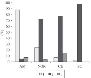
Figure 9. Variation of the anterior lacerate foramen shapes (fo) observed in the specimens of genus Sotalia: 1- open; 2- with projection spine form; 3- narrow or partially closed. AM- Amazonas, CE- Ceará, NOR (samples of AP and PA together), SC- Santa Catarina.
a
b
and from SC (19%, n = 32). Furthermore, the size of the ribs is proportionally smaller in the riverine species than in the marine species. In S. guianensis, the cervical ribs were generally observed only on one side (right or left), while in S. fluviatilis they were observed on both sides. The UFSC specimen 1117 exhibited this structure on the on the right side of the third cervical vertebra (Ce3). The cervical ribs were observed in all age classes.
4. Discussion
4.1. Fenestrae
The presence of fenestrae commonly observed in the occipital region, near the condyles of Sotalia fluviatilis was also recorded in juveniles and adults of Pontoporia blainvillei (Gervais and d’Orbigny, 1844) (Pinedo,1991). However, in Pontoporia, the number of fenestrae was greater than that observed for the genus Sotalia.
In the genus Sotalia, the occurrence of fenestrae next to protuberances of the occipital region was low com-pared with the region next to the condyles. In the ba-sioccipital, the presence of fenestrae was less frequent in immature specimens than in mature specimens. In Pontoporia blainvillei and in Stenella the occurrence of this structure was rare and occasional, respectively (Pinedo, 1991; Perrin et al., 1982).
Considering that the fenestrae observed next to the condyles in S. fluviatilis do not have the same origin as the fontanelles present in newborn individuals, given their different location, the origin and function of these fenestrae are not clear.
4.2. Vomer
The cranial floor is not generally included in mor-phologic studies and revealed interesting results. Simões-Lopes (2006), analysing specimens of S. guianensis in the southern region, observed that the laminar posterior process of the vomer is narrower than the lamelar ptery-goid processes.
In the same study, the author found the converse in S. fluviatilis – where the vomer is broader and the lamelar pterygoid processes narrower. In the present work we verified that this characteristic described by Simões-Lopes (2006) was present in the majority of the marine specimens analysed. However, in the samples from CE and AP, we observed that some specimens also possessed a broad vomer, similar to the riverine species, herein called the “wide form”. Apparently, this charac-teristic can be used to distinguish the two species, but it is important to note that a small percentage of marine specimens possess a broad vomer just as some riverine specimens possess a narrow vomer. Thus, the use of this characteristic should be considered in combination to others when separating the species.
In relation to the posterior alignment of the vomer, the arrangement anterior to the lamelars pterygoid proc-esses was observed in 100% of the cases for the marine species. In the riverine species (n = 39), 18% were ob-served with the posterior edge of the vomer aligned with the pterygoids, while in another 18% the posterior edge of the vomer extended beyond the pterygoides. Dawbin et al. (1970) attribute the alignment of the vomer in rela-tion to the posterior pterygoid processes to age. For these authors, in sub-adults Peponocephala electra (Gray, 1846) the vomer extends to the level of the sutures be-tween the pterygoides and basioccipital and in adults, the vomer extends beyond the adjacent pterygoides. This
Figure 11. Percentage of the occurrence of hipoglossal
foramen: 1- Hipoglossal foramen visible in ventral view; 2- Hipoglossal foramen not visible in ventral view. AM- Amazonas, CE- Ceará, NOR (samples of AP and PA togeth-er), SC- Santa Catarina.
a b
c
pattern was not observed in the current study, since all adults of S. guianensis were observed with the posterior suture of the vomer anterior to the pterygoides. Moreover, the variation observed in the riverine species does not ap-pear to be related to ontogenetic development, but rather to individual variation.
4.3. Pterygoides
The pterygoides were found to be medially separated by a projection in the tip of the palatines in all speci-mens, corroborating the data found in the literature (van Bénéden, 1875; Flower, 1885; Miranda-Ribeiro, 1936, da Silva and Best, 1994; 1996; Avila et al., 2002; Simões-Lopes, 2006). The variation in the form of the posterior projection of the left pterygoide was very subtle between the two species.
4.4. Anterior lacerate foramen
The anterior lacerate foramen is formed by two ramens (the optic foramen and the orbitorotundum fo-ramen). These two structures are divided by a “wall” (Yamagiwa, et al., 1999) here called a spike-shaped pro-jection. In S. guianensis, the majority of the adult speci-mens (between 72 and 98%) exhibited this projection be-tween the optic and the orbitorotundum foramen. On the other hand, the narrow form was only observed in adults, suggesting that with time, this spiny projection fused with one side of the lacerated foramen (as observed in some marine specimens). In S. fluviatilis, in contrast, the absence of this projection was more common (88%), and could indicate neoteny.
4.5. Hypoglossal foramen
The location of the hypoglossal foramen varied be-tween the two species. In S. guianensis the hypoglossal foramen generally meets between the crest of the basioc-cipital and the paraocbasioc-cipital process, more precisely in the jugular notch as it occurs in other species of marine Delphinidae, for example, Grampus griseus (Cuvier, 1812) (Yamagiwa et al., 1999) and Tursiops truncatus (Montagu, 1821) (Rommel, 1990). In contrast, the hypoglossal fo-ramen in S. fluviatilis, in the majority of cases, was found to be internally displaced.
4.6. Cervical vertebrae
The occurrence of cervical ribs was higher (87%) in the riverine species. The presence of pleuroapo-phiseal plates was initially suggested for some groups of mammals, especially monotremes and marsupials (Lessertisseur and Saban, 1967). Unilateral or bilateral processes in the cervical vertebrae are common in some groups of mammals (including Homo), and the costal ru-diments are associated with the vertebral or vertebrarte-rial foramens (Lessertisseur and Saban, 1967). These au-thors also mention their presence in marine mammals as Orcinus orca (Linnaeus, 1758), Tursiops truncatus and Balaenoptera sp. Such processes have been considered serially homologous to the cervical ribs, also appearing in the literature as costal plates or pleuroapophiseal plates
(Flower and Lyddeker, 1891). The presence of cervical ribs is an intriguing characteristic in comparative anato-my, as these structures are typically observed in reptiles (Paula Couto, 1979; Ferigolo, 1987). In S. guianensis, these structures have already been reported in about 22.5% (n = 31) of the specimens of the southern region (Fettuccia and Simões-Lopes, 2004). In this work, con-sidering a larger sample, the observed frequency of cer-vical ribs in the specimens from SC was 19% (n = 33), and of 9% (n = 23) from CE. Cervical ribs in mam-mals are examples of atavism (reappearance of an extinct character, common to ancestral lineages that rarely oc-cur in oc-current populations). Other cases of atavism are described in the literature as the occurrence of vestigial posterior members in whales and extra-numeric teeth in bats and sea lions (Bejder and Hall, 2002; Rui and Drehmer, 2004 and Drehmer et al., 2004). According to Hall (1984), there are four basic criteria for the recogni-tion of an atavism: 1) persistence of the characteristic in adult life; 2) absence of this characteristic in the parents or recent ancestors; 3) occurrence in one or a few indi-viduals within a population; and 4) similarity or identity with the same character exhibited by all the members of the ancestral population. Moreover, if the incidence of the character is still relatively high in a population, it is considered a polymorphism (Hall, 1984). Thus, consid-ering the high occurrence of cervical ribs in specimens of the riverine species, these structures do not appear to be atavistic, suggesting the need for a more detailed study with a more representative sample number for a more consistent conclusion.
Acknowledgements — The authors would like to thank Angel Enzo Crovetto, César Drehmer, Fernando Rosas, Lena Geise and two anonymous referees for their helpful suggestions on improving the manuscript. We would also like to thank Nina Best for the English review. Special thanks to the Museu Paraense Emilio Goeldi (MPEG) and Associação de Pesquisa e Preservação de Ecossistemas Aquáticos (AQUASIS). This work was supported by grants from CNPq and IEB.
References
ALVES-JÚNIOR, TT. and MONTEIRO-NETO, C., 1999. Caracterização morfológica e morfométrica craniana do boto-cinza, Sotalia fluviatilis Gervais, 1953, da Costa do Ceará, Brasil. Arquivos de Ciências do Mar, no. 32, p. 89-101. ÁVILA, FJC., ALVES-JÚNIOR, TT., PARENTE, CL., VAZ, LAL. and MONTEIRO-NETO, C., 2002. Osteologia do boto-cinza, Sotalia fluviatilis Gervais, 1853, da Costa do Estado do Ceará, Brasil. Arquivos de Ciências do Mar, no. 35, p. 145-155.
BEJDER, L. and HALL, BK., 2002. Limbs in whales and limblessness in other vertebrates: mechanisms of evolutionary and developmental transformation and loss. Evolution and Development, vol. 4, no. 6, p. 445-458.
BOROBIA, M., 1989. Distribution and morphometrics of South American dolphins of the genus Sotalia. Montreal: McDonald College of McGill University. 81 p. [Master’s thesis].
BOROBIA, M., SICILIANO, S., LODI, L. and HOEK, W., 1991. Distribution of the South American dolphin Sotalia fluviatilis. Canadian Journal Zoology, no.69, p. 1025-1039. CASINOS, A., BISBAL, F. and BOHER, S., 1981. Sobre três exemplares de Sotalia fluviatilis del Lago Maracaibo (Venezuela) (Cetacea, Delphinidae). Proceedings of the Departament of Zoology, no. 7, p. 93-96.
CUNHA, HA., DA SILVA, VMF., LAILSON-BRITO Jr, J., SANTOS, MCO., FLORES, PAC., MARTIN, AR. et al., 2005. Riverine and marine ecotypes of Sotalia dolphins are different species. Marine Biology, vol. 148, no. 2, p. 449-457.
CABALLERO, S., TRUJILLO, F., VIANNA, JA., BARRIOS-GARRIDO, H., MONTIEL, MG., BELTRÁN-PEDREROS, S. et al., 2007. Taxonomic status of the genus Sotalia: species level ranking for “tucuxi” (Sotalia fluviatilis) and “costero” (Sotalia guianensis) dolphins. Marine Mammal Science, vol. 23, no. 2, p. 358-386.
DA SILVA, VMF. and BEST, RC., 1994. Tucuxi Sotalia fluviatilis (Gervais, 1853). In RIDGWAY, SH., HARRISON, SR. (Eds). Handbook of Marine Mammals. London: Academic Press. p. 43-69.
______, 1996. Sotalia fluviatilis. Mammalian Species, no. 527, p. 1-7.
DAWBIN, WH., NOBLE, BA. and FRASER, FC., 1970. Observations on the electra dolphin, Peponocephala electra. Bulletin of the British Museum (Natural History), vol. 20, no. 6, p. 173-201.
DREHMER, CJ., FABIÁN, ME. and MENEGHETI, JO., 2004. Dental anomalies in the Atantic population of South American sea lion, Otaria byronia (Pinnipedia, Otariidae): evolutionary implications and ecological approach. LAJAM, vol. 3, no. 1, p. 7-18.
FERIGOLO, J., 1987. Anatomia comparada, paleontologia e paleopatologia de vertebrados. Paula-Coutiana, no. 1, p. 105-127.
FETTUCCIA, DC. and SIMÕES-LOPES, PC., 2004. Morfologia da coluna vertebral do boto-cinza, Sotalia guianensis (Cetacea, Delphinidae). Biotemas, vol. 17, no. 2, p. 125-148.
FLOWER, WH., 1885. An Introduction to the Osteology of the Mammalia. 3 ed. London: Macmillan & Co. 344 p.
FLOWER, WH. and LIDEKKER, R., 1891. An introdution to the study of Mammals living and extinct. New York: Arno Press. 763 p. (Reprint edition 1978).
HALL, BK., 1984. Developmental echanisms underlying the formation of atavisms. Biological Reviews of the Cambridge Philosophical Society, no. 78, p. 409-433.
KRAGLIEVICH, L., 1937. Manual de Paleontologia Rio Platense Comparada de los Mamíferos. Montevideo: Ed. Siglo Ilustrado. 190 p.
LESSERTIEUR, J. and SABAN, R., 1967. Squelette Axial. In GRASSÉ, PP. (Ed.). Traité de Zoologie, Mammifères: Tèguments et Squelettes. Paris: Masson et Cie. Èditeurs. p. 585-708.
MENEZES, ME. and SIMÕES-LOPES, PC., 1996. Osteologia e morfologia da aleta peitoral da forma marinha de Sotalia fluviatilis (Cetacea-Delphinidae) no litoral do Brasil. Estudos de Biologia, vol. 4, no. 40, p. 23-31.
MIRANDA-RIBEIRO, A., 1936. Notas cetológicas dos gêneros Steno, Sotalia eStenopontistes. Boletim do Museu Nacional do Rio de Janeiro, no. 12, p. 3-23.
MONTEIRO-FILHO, ELA., MONTEIRO, LR. and REIS, SF., 2002. Skull shape and divergence in dolphins of the genus Sotalia: a tridimensional morphometric analysis. Journal of Mammalogy, vol. 83, no. 1, p. 125-134.
PAULA COUTO, C., 1979. Tratado de Paleomastozoologia. Rio de Janeiro: Academia Brasileira de Ciências. 590 p.
PERRIN, WF., YABLOKOV, AV. and CASS, VL., 1982. Preliminary report on the use of non-metrical skull characters to discriminate populations of pelagic dolphins. E.U.A.: National Marine Fisheries Services.32 p.
PINEDO, MC., 1991. Development and variation of the franciscana, (Pontoporia blainvillei), Santa Cruz. California: University of California. 406 p. [PhD. Thesis].
ROMMEL, SA., 1990. Osteology of the Bottlenose Dolphin. In LEATHERWOOD, S.; REEVES, R. (Eds.). The Bottlenose Dolphin. San Diego: Academic Press. p. 29-49.
RUI, AM. and DREHMER, CJ., 2004. Anomalias e variações na fórmula dentária em morcegos do gênero Artibeus Leach (Chiroptera, Phyllostomidae). Revista Brasileira de Zoologia, vol. 21, no. 3, p. 639-648.
SIMÕES-LOPES, PC., 1988. Ocorrência de uma população de Sotalia fluviatilis (Gervais, 1853) (Cetacea, Delphinidae) no limite sul de sua distribuição, Santa Catarina, Brasil. Biotemas, vol. 1, no. 1, p. 57-62.