___________________________
Corresponding author: Nevenka Velickova, Faculty of medical science, University “ Goce
Delcev” - Krste Misirkov 10-A, 2000 –Stip, R.Macedonia, [email protected] UDC 575 DOI: 10.2298/GENSR1503927V
Original scientific paper
CYTOGENETIC ABNORMALITIES IN LYMPHOCYTES EVALUATED WITH MICRONUCLEUS ASSAY IN MEDICAL PERSONNEL OCCUPATIONALLY
EXPOSED TO IONIZING RADIATION
Nevenka VELICKOVA, Misko MILEV, Tatjana RUSKOVSKA, Biljana PETROVA, Bojana NEDELJKOVIK, Pale GORGIEVA
Faculty of medical science, University “Goce Delcev” - Stip,R. of Macedonia
Velickova N., M. Milev, T. Ruskovska, B. Petrova, B. Nedeljkovik, P. Gorgieva (2015): Cytogenetic abnormalities in lymphocytes evaluated with micronucleus assay in medical personnel occupationally exposed to ionizing radiation.--Genetika, Vol 47, No. 3, 927-939.
The aim of this study was to evaluate the genotoxicity of ionizing radiation on medical personnel using the micronucleus assay and to determine the human health
risk. Paired Student’s t-test shows significant statistical difference between the total number of binucleated (BN) cells with micronuclei within the two groups (exposed and control) (t=6,812; p<0,05). The mean of MN frequencies in the exposed group increased in comparison with the mean of MN frequencies in the control group. The formation of small and large micronuclei indicates that medical personnel who are exposed on radiation in their work place, have a chromosomal instability and a risk of cancer.
Key words:micronucleus, lymphocytes, genotoxicity, ionizing radiation, cancer
INTRODUCTION
level and genetic monitoring is becoming an integral part of preventive medical surveillance, because chromosomal mutation is the main reason for carcinogenesis. In this point of view, the MN assay is one of the methods for cytogenetic evaluation for both, chromosome loss and chromosome breakage (FENECH et al., 1999; FENECH, 2000). Also, the Cytokinesis-Blocked Micronucleus (CBMN) assay provides an analysis for the other abnormal nuclear shapes (ANS), like micronuclei (MNi), nucleoplasmic bridges (NPBs) and nuclear buds (NBUDs). These types of formations have been associated with mitotic and chromosomal instability (CAMPS et al., 2005). CBMN is suitable and standardized test in genotoxicology used for assessment of genotoxicological effects of different chemical or physical agents and it is the most commonly used method for measuring DNA damage in human lymphocytes (FENECH, 1998; FENECH et al., 1999; HESSEL et al., 2001). MNi originates from acentric fragments, whole chromatids or chromosomes and it is a result of chromosome breaks or spindle disruption (KIRSCH-VOLDERS et al., 1997, 2001, 2003; FENECH, 2005). The biological response of human tissues to radiation could be unpredictable and it is very complex, because many, even more complex chemical interactions can occur if two chemicals act in different, but related ways. In extreme cases, there may be synergistic effects, in which case the effects of two substances together are greater than the sum of either effect alone. 60Co is one of the radiation sources, which can cause various illnesses and deaths worldwide (CAO et al., 2013). Blood, like hematological tissue and principally lymphocytes are the most sensitive cells to ionizing radiation (BONASSIet al., 2003, 2007, 2011).
The aim of this research was to evaluate and confirm the genotoxicity of ionizing radiation using the CBMN assay and to determine the human health risk. We analyzed the results of lymphocyte MNi, NPBs, NBUDs as biomarker of DNA miss repair complexes (FENECH, 2007) on the medical personnel occupationally exposed to ionizing radiation via control group.
MATERIALS AND METHODS
Study groups
The study was approved by the Ethical Committee of the Faculty of medical science in “Goce Delcev” University in Stip, R. of Macedonia, and all subjects of the exposed and the control group provided their written consents, according to the Declaration of Helsinki (2013). The study population included (Table 1):
- 20 individuals in the exposed group, medical personnel exposed to ionizing radiation (radiologist, technicians and nurses) and
- 20 individuals in the control group, healthy people, (who have never been exposed to ionizing rays and other chemical or physical agents).
All participants were informed about the aim of the study and they completed a comprehensive questionnaire about demographic information such as age, lifestyle factors (tobacco usage), profession, medical history and exposure to chemical, physical agents or radiotherapy. Blood samples were obtained during a routine annual checkup of health.
(Nunc), the tubes were mixed gently by invertingfor a few minutes and incubated (CO2 incubator for 24 hours) for 44h at 37 °C in a slant position. Cytochalasin B was then added to each culture at a concentration of 3 μg/ml to block cell cytokinesis and cultures were reincubated at 37 °C for further 28 h. Cells were then harvested by centrifugation at 1,000 rpm for 10 min. Supernatant was discarded by pipetting the media, leaving as little medium as possible over the cell pellet. Cell pellet was resuspended in the supernatant remains and 10 ml of 0.56% KCl warm hypotonic solution was added gently to each tube. Fixation was carried out during 2x15 minutes by fixative (glacial acetic acid: methanol = 1:3). Fixation steps were repeated for three more times. After that, the fixed lymphocyte cells were dropped from about 30 cm height using a Pasteur pipette onto microscopic slides. The slides were stained by 2% alkaline Giemsa for 8 minutes, then washed in distilled water and examined by light microscope Leica DM4500 P (×40 and×100).
Table 1. Demographic characteristics of the study population (the exposed and the control group)
RESULTS AND DISCUSSION
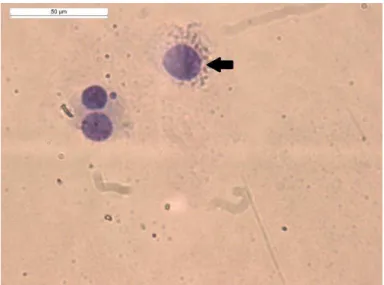
The analyses of MN were carried out on 1000 BN lymphocites per patient. Additionally with the findings of these studies (FENECH, 2007; KIRSCH-VOLDERS et al., 2001) the authors classified the cells as mononucleates (Fig.1), binucleates (Fig.1 and Fig.2 a and b), or multinucleate (Fig.3 a and b).
Fig. 1 Photomicrograph of mononucleated cell scored in CBMN assay
Exposed group
Control group
Number of men
13
10
Number of women
7
10
Age range
45
±
15
18
±22
Range of years of
professional exposure
15-35
/
a) b)
Fig. 2 Photomicrographs of BN cells scored in CBMN assay a) and b)
a) b)
Fig. 3 Photomicrographs of multinucleated cells scored in CBMN assay a) and b)
a) b)
c)
d)Fig. 4 Photomicrographs of BN cells containing MN scored in CBMN assay a), b), c) and d)
a)
b)
a)
b)
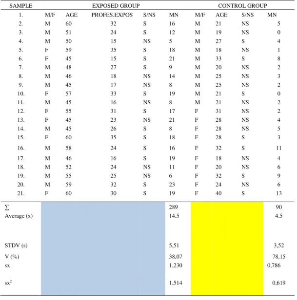
Fig. 6 Photomicrographs of BN cells containing NBUDs scored in CBMN assay a) and b)Cytogenetic analysis of peripheral blood lymphocytes showed that the number of BN cells, with one MN ranged from 5 to 23 in the exposed, and from 1 to 13 in the control group (Table 2). In both groups the most frequent micronucleated cells were the ones with one MN, the
cells with two or three MNi were very rare. Student’s t-test showed significant statistical differences between the total number of BN cells with MNi within the two groups (the exposed and the control) (t=6,812; p<0,05).
The observed MN frequencies were compared with the criteria for spontaneous MNi
(4.4±2.6 per 500 BN cells) given by FENECH and MORLEY (1985, 1986). The authors confirme than in the exposed group, 12 individual samples (60 %) showed an increase in the MN frequency while in the control group, the increased frequency of MNi was found in 1 of the blood samples (5%). 10 subjects with increased MN frequency in the exposed group and 1 in the control group smoked tobacco. In other words, this study revealed that the increased number of MNi in the exposed group was more common in smokers. Moreover, the clear evidence of increased number of MN formation was in a smoking woman in the control group. In addition to age and gender, smoking habits may change the MN frequency. The very important factors which explain the evidence of MN in the lymphocytes are the years of the exposure and the age of the subjects, especially in the exposed group. The samples with long time of exposure on ionizing radiation have much more MNi than the other samples (Table 2). In both analyzed samples of women in the exposed and the control group, age represents serious factor for the MNi values. BONASSI et al. (2003) confirmed a significant effect of smoking on MN frequency but, the study’s of HESSEL et al. (2001) and COSTA et al. (2006) observed no relation between smoking habit and MN frequency. EL-ZEIN, et al. (2008) and GARAJ-VRHOVAC et al. (2008) assessed that genotoxic hazards from cigarettes cause cell changes or cell death. Although men dominated in the exposed group, all the women from this exposed group showed increased MN frequency (Table 2). In the control group, one subject with increased MN frequency was a woman. The results of the other
Table 2. General characteristics, MNi frequency and MN distribution in male and female individuals in the exposed and the control group
The fact that the frequency of MNi is greater in female with reproductive disorders is confirmed in another study (FENECH, 1994), that also indicated greater chromosomal instability in
lymphocytes. Other study’s (THIERENS et al., 1999; 2000;JOSEPH et al., 2009) also assessed that the frequencies of MN are greater in females than in males. In this study the authors confirmed that two men in the exposed group, who have hypothyroidism (receive a therapy with euthyrox), have increased number of MNi (Table 2). In the research of AL FAISAL et al. (2012), there was an
SAMPLE EXPOSED GROUP CONTROL GROUP
1. M/F AGE PROFES.EXPOS S/NS MN M/F AGE S/NS MN
2. M 60 32 S 16 M 21 NS 5
3. M 51 24 S 12 M 19 NS 0
4. M 50 15 NS 5 M 27 S 4
5. F 59 35 S 18 M 18 NS 1
6. F 45 15 S 21 M 33 S 8
7. M 48 27 S 9 M 20 NS 2
8. M 46 18 NS 14 M 25 NS 3
9. M 45 17 NS 8 M 25 NS 2
10. F 57 33 S 19 M 21 S 0
11. M 45 16 NS 8 M 21 NS 2
12. F 55 31 S 17 F 31 NS 2
13. F 45 23 NS 21 F 28 NS 4
14. M 45 26 S 8 F 28 NS 5
15. F 60 35 S 18 F 28 S 3
16. M 58 24 S 16 F 32 S 11
17. M 46 16 S 19 F 18 NS 4
18. M 52 24 NS 11 F 20 NS 6
19. M 55 25 NS 6 F 32 S 9
20. M 59 32 S 23 F 24 NS 6
21. F 60 30 S 19 F 40 S 13
∑ 289 90
Average (x) 14.5 4.5
STDV (s) 5,51 3,52
V (%) 38,07 78,15
sx 1,230 0,786
evidence of increased MN frequencies in patients with thyroid disorder, compared to healthy population. So many factors like age, gender, smoking etc. can cause MN frequency variation, but gender is one of the main factor estimated in many studies, which confirmed that MN frequencies are higher in women than in men (FENECH, 1998; BOLOGNESI, 2002) generally by 1.2 to 1.6 times (BOLOGNESI, 2002). This was explained by over-prevalence of X chromosome in the female organism (CATALANet al., 1998; HANDO, 1997).
The observed MN frequencies were compared with the criteria for spontaneous MNi
(4.4±2.6 per 500 BN cells) given by FENECH and MORLEY (1985, 1986). The authors confirme than in the exposed group, 12 individual samples (60 %) showed an increase in the MN frequency while in the control group, the increased frequency of MNi was found in 1 of the blood samples (5%). 10 subjects with increased MN frequency in the exposed group and 1 in the control group smoked tobacco. In other words, this study revealed that the increased number of MNi in the exposed group was more common in smokers. Moreover, the clear evidence of increased number of MN formation was in a smoking woman in the control group. In addition to age and gender, smoking habits may change the MN frequency. The very important factors which explain the evidence of MN in the lymphocytes are the years of the exposure and the age of the subjects, especially in the exposed group. The samples with long time of exposure on ionizing radiation have much more MNi than the other samples (Table 2). In both analyzed samples of women in the exposed and the control group, age represents serious factor for the MNi values. BONASSI et al. (2003) confirmed a significant effect of smoking on MN frequency but, the study’s of HESSEL et al. (2001) and COSTA et al. (2006) observed no relation between smoking habit and MN frequency. EL-ZEIN, et al. (2008) and GARAJ-VRHOVAC et al. (2008) assessed that genotoxic hazards from cigarettes cause cell changes or cell death. Although men dominated in the exposed group, all the women from this exposed group showed increased MN frequency (Table 2). In the control group, one subject with increased MN frequency was a woman. The results of the other
study’s (FENECH, 1994) that also used CBMN assay, suggest that sex is an important factor for evaluation of the cytogenetic damage. The authors of the study, also found an enormously high frequency of MNi in control women in comparison with men in the same group. One woman in the control group (at age 40) used ordinary therapy on oral estrogen hormones for two years before the test and she had increased number of MN in the lymphocytes (Table 2). The fact that the frequency of MNi is greater in female with reproductive disorders is confirmed in another study (FENECH, 1994), that also indicated greater chromosomal instability in lymphocytes. Other
study’s (THIERENS et al., 1999; 2000; JOSEPH et al., 2009) also assessed that the frequencies of MN are greater in females than in males. In this study the authors confirmed that two men in the exposed group, who have hypothyroidism (receive a therapy with euthyrox), have increased number of MNi (Table 2). In the research of AL FAISAL et al. (2012), there was an evidence of increased MN frequencies in patients with thyroid disorder, compared to healthy population. So many factors like age, gender, smoking etc. can cause MN frequency variation, but gender is one of the main factor estimated in many studies, which confirmed that MN frequencies are higher in women than in men (FENECH, 1998; BOLOGNESI, 2002) generally by 1.2 to 1.6 times (BOLOGNESI, 2002). This was explained by over-prevalence of X chromosome in the female organism (CATALANet al., 1998; HANDO, 1997).
found when there is dense stippling in a specific region of the cytoplasm, or when a NBUD has a narrow connection to the nucleus, so it appears like an MN or when nuclear blebs have no obvious constriction or bridge among them and the nucleus (FENECH, 2000). They represent a constant connection between the two nuclei in a BN cell and their origin is from dicentric chromosomes whose centromeres were dragged to the opposite poles of separating cells. Micronucleus frequency in the BN cells is compatible (or well-matched) biomarker on mutagen or cytostatic effects of different agents on human cells (BONASSI et al., 2011; EL-ZEIN et al., 2011; KOCAMAN et al., 2008;). Also, this frequency can represent an indicator for chromosome damage, instability and can assess the risk of cancer, acquired mutations and genetic susceptibility (FENECH, 2000; JOSEPH et al., 2009). The difference of MN occurrence between males and females, smokers and non-smokers in both exposed and control group can be consequence to isolated exposure of some member of the group to non-specified genotoxic agent. Oxidative stress can also be a huge contributing factor for increased MN frequencies which can lead up to infertility. This was shown by other author by performing analysis of MN numbers in correlation to oxidative stress intensity (BONASSI, 2007).
In four blood samples of exposed subjects (men exposed on ionizing radiation more than 20 years) in their lymphocytes, the authors found NPBs (Fig.5 a and b), and NBUDs (Fig.6a and b), (17). NBUDS are considered to have same structure as an MN, but they differ from them with the fact that they are linked to the nucleus by a narrow or wide stalk of nucleoplasmic material. The size of the link is dependent by the stage of the budding process which occurs during the S phase of the cell cycle. Examples of cellular structures that resemble MNi, but should not be classified as MNi, originating from chromosome breakage or loss are illustrated in Fig. 6. Sometimes, amplification of genes can cause formation of NBUDS (FENECH, 2000), after that when they detach from the nucleus they start to form a MN, a process specific for the S phase (SHIMIZU, et al., 1998). The ‘‘cytome’’ concept includes that the cells are scored cytologically due to their possibility situation like necrosis, or apoptosis, its mitotic status (mononucleated, BN, multinucleated) and its genetic instability (presence of MNi, NPBs, NBUDs) (FENECH, 2007). The rate of chromosomal changes during the cell cycle often is described as chromosomal instability. When this chromosomal instability is evident in lymphocytes then it can be linked with various human health issues (FENECH, 1998). In all the studies on MN formation the authors recognize four mechanisms of MN formation, apoptosis like controlled cell death, disappearance of whole chromosomes during cell division, disappearance of acentric fragments during cell division and structural chromosomal changes. Different genotoxicological agents cause different MN formation, for example clastogenic agents usually induce the formation of smaller micronuclei while aneugenic agents induce the larger MN formation (HEDDLE, 1991; TUCKER, 1996).
CONCLUSIONS
Mutagenic substances are widely spread in the work environment, so from this point of view, cytogenetics provides numerous methods and assays for observation of the genotoxiclogical effects of these physical and chemical agents. One of the biomarkers in genotoxicology, for
assessing the chromosomal instability and damage is scoring of MN frequency’s in lymphocytes,
particularly for the observation of the chronic exposure of organisms to a potentially genotoxic
agents, like ionizing radiation. The author’s results show that the mean of MN frequencies in the
chromosomal instability in lymphocytes. These results suggest that chromosomal instability is in correlation with MN frequencies in medical workers exposed to ionizing radiation. The formation of small and large MNi, NPBs, NBUDs etc. indicates that medical workers are exposed on clastogenic and aneugenic agents, like ionizing radiation and have chromosomal instability and high risk of cancer. This study has a practical importance because it indicates the necessity of introducing a permanent genotoxicological and other monitoring on the vulnerable category of workers (in this study medical workers) who would contribute to better protection in this segment. The multidisciplinary elaboration of the topic, suggests the needs for a more serious approach to this important phenomenon (genotoxicological impact of ionizing radiation), application of other molecular cytogenetic techniques which allow easy detection of the rate of chromosome rearrangements and the origin of the chromosome instability.
List of abbreviations:
ANS Abnormal nuclear shapes CBMN Cytokinesis-blocked micronucleus DNA Deoxyribonucleic acid
BN Binucleated
MN Micronucleus
MNi Micronuclei NBUDs Nuclear buds
NPBs Nucleoplasmic bridges PBL Peripheral blood lymphocytes S/NS Smoker/Nonsmoker
M/F Male/Female
ACKNOWLEDGMENTS
This study like project was supported by the University “Goce Delcev” in Stip, R. of Macedonia.
Received June 02st, 2015 Accepted October 20th, 2015
REFERENCES
AL FAISAL, A.H.M., I.J.K., AL-RAMAHI, I.A.R., ABDUL-HASSAN (2012): Micronucleus frequency among Iraqi thyroid disorder patients, Comp. Clin. Pathol., DOI 2012;10.1007/s00580-012-1671-7.
BOLOGNESI, C., E., PERRONE, E. LANDINI (2002): Micronucleus monitoring of a floriculturist population from western Liguria, Italia. Mutagenesis; 17:391-7.
BONASSI, D., R., EL-ZEIN, C., BOLOGNESI, M., FENECH (2011): Micronuclei frequency in peripheral blood lymphocytes and cancer risk: evidence from human studies. Mutagenesis, 26(1):93–100.
BONASSI, S., M., NERI, C., LANDO, M., CEPPI, Y.P., LIN, W.P., CHANG, N., HOLLAND, M., KIRSCH-VOLDERS, E., ZEIGER, M., FENECH, HUMN COLLABORATIVE GROUP (2003): Effect of smoking habit on the frequency of micronuclei in human lymphocytes: results from the Human Micro Nucleus project. Mutat. Res.,543:155-66.
increased micronucleus frequency in peripheral blood lymphocytes predicts the risk of cancer in humans. Carcinogenesis, 28:625-31.
CAMPS, J., I., PONSA, M., RIBAS, E., PRAT, J., EGOZCUE, M.A., PEINADO, R. MIRO (2005): Comprehensive measurement of chromosomal instability in cancer cells: combination of fluorescence in situ hybridization and cytokinesis-block micronucleus assay, The Journal of the Federation of American Societies for Experimental Biology (The FASEB journal, Vol.19: 828-830.
CAO, J., J., ZHANG, Y., WANG, L.Q., DU, C., XU, Q., WANG, J.X., LIU, X., SU, FY., FAN, Q., LIU S.J., FAN (2013): Cytogenetic Abnormalities in Lymphocytes from Victims, Exposed to Cobalt-60 Radiation, International Journal of Molecular Sciences, 14: 17525-17535.
CATALAN, J., K., AUTIO, E., KUOSMA, H., NORPPA (1998): Age-dependent inclusion of sex chromosomes in lymphocyte micronuclei of man. Am. J. Hum. Genet., 63:1464-72.
COSTA, C., J.P., TEIXEIRA, S., SILVA, J., ROMA-TORRES, P., COELHO, J., GASPAR, M., ALVES, B., LAFFON, J., RUEFF, O., MAYAN (2006): Cytogenetic and molecular biomonitoring of a Portuguese population exposed to pesticides. Mutagenesis, 21:343-50.
COTTERILL, S.J., M.S., PEARCE, L., PARKE (2001): Thyroid cancer in children and young adults in the North of England. Is increasing incidence related to the Chernobyl accident? Eur. J. Cancer, 37(8):1020–1026.
EL-ZEIN, R.A., M., FENECH, M.S., LOPEZ, M.R., SPITZ, C.J., ETZEL (2008): Cytokinesis-Blocked Micronucleus Cytome assay biomarkers identify lung cancer cases amongst smokers, Cancer Epidemiol. Biomarkers Prev., 17:1111-1119.
EL-ZEIN, R.A., A., VRAL, C.J., ETZEL (2011): Cytokinesis-blocked micronucleus assay and cancer risk assessment. Mutagenesis, 26(1):101–106.
FENECH, M. and A.A., MORLEY (1985): Measurement of micronuclei in human lymphocytes. Mutat. Res., 147: 29–36. FENECH, M. (1998): Important variables that influence base-line micronucleus frequency in cytokinesis-blocked
lymphocytes a biomarker for DNA damage in human populations. Mutat. Res., 404:155-65.
FENECH, M.(2000): The in vitro micronucleus technique, Mutation Research, 455: 81–95.
FENECH, M. (2005): In vitro micronucleus technique to predict chemo sensitivity. Methods Mol. Med., 111:3-32. FENECH, M. (2007): Cytokinesis-block micronucleus cytome assay, Nature protocols,Vol.2, No.5: 1084-1104.
FENECH, M. (2011): Micronuclei and their association with sperm abnormalities, infertility, pregnancy loss, pre-eclampsia and intra-uterine growth restriction in humans. Mutagenesis, 26:63-7.
FENECH, M., J., CROTT, J., TURNER, S., BROWN (1999): Necrosis, apoptosis, cytostatic and DNA damage in human lymphocytes measured simultaneously within the cytokinesis-block micronucleus assay: description of the method and results for hydrogen peroxide. Mutagenesis, 14(6):605-12.
FENECH, M., N., HOLLAND, W.P., E., CHANG ZEIGER, S., BONASSI (1999): The Human Micronucleus project—an international collaborative study on the use of the micronucleus technique for measuring DNA damage in humans. Mutat. Res., 428: 271–283.
FENECH, M., A.A., MORLEY (1986): Cytokinesis-block micronucleus method in human lymphocytes: effect of in vivo ageing and low-dose x-irradiation Mutat. Res., 161: 193–198.
FENECH, M., S., NEVILLE, J., RINALDI (1994): Sex is an important variable affecting spontaneous micronucleus frequency in cytokinesis-blocked lymphocytes. Mutat. Res., 313:203-7.
GARAJ-VRHOVAC, V., M., DURINEC, N., KOPJAR, V.,OREŠČANIN (2008): A survey on the cytogenetic status of the Croatian general population by use of the cytokinesis-block micronucleus assay. Mutat. Res., 649:91-100.
GE, Y., H., NING, S., WANG, J., WANG (2005): Damage in thyroid gland cells of rats exposed to long term intake of high fluoride and low iodine. Fluoride, 38(4):318–323.
HEDDLE, J.A., M.C., CIMINO, M., HAYASHI, F., ROMAGNA, M.D., SHELBY, J.D., TUCKER, P.H., VANPARYS, J.T., MACGREGOR (1991): Micronuclei as an index of cytogenetic damage: past, present, and future. Environ. Mol. Mutagen., 18: 277-91.
HESSEL, H., K., RADON, A., PETHRAN, B., MAISCH, S., GROBMAIR, I., SAUTTER, G., FRUHMANN (2001): The genotoxic risk of hospital, pharmacy and medical personnel occupationally exposed to cytostatic drugs- evaluation by the micronucleus assay. Mutat. Res., 497:101-9.
JOSEPH, L.J., U.S., BHARTIYA, Y.S., RAUT, P., KAND, R.W., HAWALDAR, N., NAIR (2009): Micronuclei frequency in peripheral blood lymphocytes of thyroid cancer patients after radioiodine therapy and its relationship with metastasis. Mutat. Res., 675(1– 2):35–40, 30.
KIRSCH-VOLDERS, M., A., ELHAJOUJI, E., CUNDARI, P., VAN HUMMELEN (1997): The in vitro micronucleus test: a multi-endpoint assay to detect simultaneously mitotic delay, apoptosis, chromosome breakage, chromosome loss and non-disjunction. Mutat. Res., 392:19-30.
KIRSH-VOLDERS, M., M., FENECH (2001): Inclusion of micronuclei in non-divided mononuclear lymphocytes and necrosis/apoptosis may provide a more comprehensive cytokinesis block micronucleus assay for biomonitoring purposes. Mutagenesis, 16: 51 – 8.
KIRSCH-VOLDERS, M., T., SOFUNI, M., AARDEMA, et al. (2003): Report from the in vitro micronucleus assay working group. Mutat. Res., 540, 153–163.
KOCAMAN, A.Y., E., RENCUZOGULLARI, H., BASRIILA, M., TOPAKTAS (2008): The genotoxic effect of potassium metabisulfite using chromosome aberration, sister chromatid exchange, micronucleus tests in human lymphocytes and chromosome aberration test in bone marrow cells of rats. Environ. Mol. Mutagen., 49 (4):276–282.
SHIMIZU, N., N., ITOH, H., UTIYAMA, G.M., WAHL (1998): Selective entrapment of extra chromosomally amplified DNA by nuclear budding and micronucleation during S phase, J. Cell Biol., 140: 1307–1320.
THIERENS, H., A., VRAL, L., DE RIDDER, N., TOUIL, M., KIRSCH-VOLDERS, V., LAMBERT, C., LAURENT (1999): Inter-laboratory comparison of cytogenetic endpoints for the biomonitoring of radiological workers. Int. J. Radiat. Biol., 75: 23–34.
THIERENS H., A., VRAL, R., MORTHIER, B., AOUSALAH, L., DE RIDDER (2000): Cytogenetic monitoring of hospital workers occupationally exposed to ionizing radiation using the micronucleus centromere assay. Mutagenesis, 15: 245– 249.
CITOGENETIČKE ABNORMALNOST U LIMFOCITIMA MEDICINSKOG OSOBLJA IZLOŽENOG JONIZUJUĆEM ZRAČENJU
Nevenka VELICKOVA, Misko MILEV, Tatjana RUSKOVSKA, Biljana PETROVA, Bojana NEDELJKOVIK i Pale GORGIEVA
Fakultet medicinskih nauka, Univerzitet “ Goce Delčev” Štip, Republika Makedonija
Izvod
Cilj ovih istraživanja je bio izučavanje genotoksičnosti jonizujućeg zračenja kod medicinskog osoblja koristeći mikronukleus –analizu. Istraživanja su vršena radi utvrđivanja rizika zračenjem na ljudsko zdravlje. Student’s t-test pokazuje značajne statistčke razlike između ukupnog broja
binukleotidnih ćelija sa mikronukleusima unutar dve grupe (ozračene i kontrolna grupa) (t=6,812; p<0,05). Prosečna MN učestalost u ozračenoj grupi se povećava u poređenju sa prosečnom učestalošću u kontrolnoj grupi. Formiranje malih i velikih mikronukleusa ukazuje da medicinsko
osoblje izloženo zračenju na random mestu ima hromozomalnu nestabilnost i rizik od pojave
kancera.