Comparing the Effects of Isoflurane and
Alpha Chloralose upon Mouse Physiology
Lucie A. Low1*, Lucy C. Bauer1, Brenda A. Klaunberg2
1National Center for Complementary and Integrative Health, National Institutes of Health, Bethesda, Maryland, United States,2NIH Mouse Imaging Facility, National Institute of Neurological Disorders and Stroke, National Institutes of Health, Bethesda, Maryland, United States
Abstract
Functional magnetic resonance imaging of mice requires that the physiology of the mouse (body temperature, respiration and heart rates, blood pH level) be maintained in order to prevent changes affecting the outcomes of functional scanning, namely blood oxygenation level dependent (BOLD) measures and cerebral blood flow (CBF). The anesthetic used to sedate mice for scanning can have major effects on physiology. While alpha chloralose has been commonly used for functional imaging of rats, its effects on physiology are not well characterized in the literature for any species. In this study, we anesthetized or sedated mice with isoflurane or alpha chloralose for up to two hours, and monitored physiological parameters and arterial blood gasses. We found that, when normal body temperature is maintained, breathing rates for both drugs decrease over the course of two hours. In addi-tion, alpha chloralose causes a substantial drop in heart rate and blood pH with severe hypercapnia (elevated blood CO2) that is not seen in isoflurane-treated animals. We sug-gest that alpha chloralose does not maintain normal mouse physiology adequately for func-tional brain imaging outcome measures.
1. Introduction
Functional magnetic resonance imaging (fMRI) monitors blood flow to the brain as a surrogate measure of regional brain activation. In humans, physiological changes that can influence blood flow, such as heart rate, breathing rate, body temperature and blood oxygen saturation levels, can be continuously and non-invasively measured. However, when scanning rodents for basic pre-clinical research, these measures can be challenging to monitor, and less amenable to correction. In particular, blood oxygenation and carbon dioxide levels are particularly impor-tant to monitor, as these can directly affect cerebral blood flow (CBF), and hence the outcome measures used in functional scanning [1–4].
In the rat, there have been a number of successful studies of rat brain activation using fMRI in both anesthetized [5–7] and awake [8–11] rats. For accurate fMRI results it is imperative to maintain normal physiology to minimize any potential effects on regional changes in CBF and blood oxygen level dependent (BOLD) contrast. A decrease in blood gas partial pressures of
a11111
OPEN ACCESS
Citation:Low LA, Bauer LC, Klaunberg BA (2016) Comparing the Effects of Isoflurane and Alpha Chloralose upon Mouse Physiology. PLoS ONE 11 (5): e0154936. doi:10.1371/journal.pone.0154936
Editor:Kathleen R. Pritchett-Corning, Harvard University Faculty of Arts and Sciences, UNITED STATES
Received:November 24, 2015
Accepted:April 21, 2016
Published:May 5, 2016
Copyright:This is an open access article, free of all copyright, and may be freely reproduced, distributed, transmitted, modified, built upon, or otherwise used by anyone for any lawful purpose. The work is made available under theCreative Commons CC0public domain dedication.
Data Availability Statement:All relevant data are within the paper and its Supporting Information files.
Funding:This research was funded by the Division of Intramural Research at the National Center for Complementary and Integrative Health (NCCIH), and the NIH Mouse Imaging Facility at the National Institute for Neurological Disorders and Stroke (NINDS). The funders had no role in study design, data collection and analysis, decision to publish, or preparation of the manuscript.
oxygen (hypoxemia) [12] or an increase in blood gas partial pressures of carbon dioxide (hypercapnia) will both increase cerebral blood flow [13]. Blood gas levels can be indirectly monitored by end tidal carbon dioxide measurement (capnography) in the rat, or by insertion of an arterial catheter for periodic blood gas sampling in anesthetized animals. Variations in acid-base status can have severe consequences for functional scanning because sustained devia-tions in pH can cause a cascade of metabolic effects, including changes in oxygen binding capacities [14]. Systemically, wide divergence of pH can have severe to life-threatening conse-quences including lethargy and coma (if blood becomes too acidic), or muscle twitching, tet-any, and seizures (if blood becomes too basic).
The advent of genetic knockout technologies in mice has led to a window of opportunity in rodent scanning, where important information can be gleaned about brain processing in genetically modified mice. Functional MR scanning of these mice, combined with genetic and behavioral approaches, could powerfully combine to provide better translational animal models for basic and preclinical research. However, the physiological monitoring and homeostatic maintenance of mice under anesthesia is technically challenging. End tidal CO2
of the mouse cannot generally be measured accurately with standard available equipment, as the lung capacity of the mouse is too small (1.2–1.8 ml)[15]. Arterial catheters are invasive and challenging to implant in the mouse, and the volume of blood that can be taken safely is extremely limited. While volumes up to 150μl of blood can be taken from a 200g rat every 24
hours [16] (and NIH ARAC guidelines,http://oacu.od.nih.gov/ARAC/documents/Rodent_ Bleeding.pdf), the entire circulating blood volume of a 20g mouse may only be 1.4 ml, and therefore taking even a small sample can result in significant changes in blood pressure and, hence, functional outcome measures [17–19].
To minimize stress in the noisy scanner environment and reduce movement artifact, rodents are often anesthetized. A variety of anesthetics and sedatives have been used for in-vivo imaging, including inhalation anesthetics such as isoflurane [20–22], sedatives such as medetomidine (Domitor1
) or alpha chloralose [23–26] often combined with a paralytic such as pancuronium bromide [27,28], and dissociative agents such as ketamine in combination with muscle relaxants such as xylazine [29,30]. For functional scanning, alpha chloralose has been recommended as an ideal sedative for rodent studies as it preserves cortical blood flow signals and a good signal-to-noise ratio (SNR)[31]. However, alpha chloralose is associated with prolonged and poor recovery [32], so it is not generally allowed for longitudinal studies. Inhalant anesthetics such as isoflurane can depress cortical activity and can be potent vasodila-tors, potentially confounding blood flow signals [33–35]. Additionally, dissociative anesthetics such as ketamine may alter the function of neural signaling, inherently altering the desired out-come measure for functional imaging [36]. The use of anesthesia introduces many potential confounds into brain imaging in the rodent, so it is important for researchers to be aware of the effects of anesthesia regimen upon their experimental animal.
In this study, we investigated the physiological effects of isoflurane anesthesia and alpha chloralose sedation in the mouse up to 120 minutes post-induction, a time course similar to that used for functional scanning of the mouse brain. Breathing rate, heart rate and body tem-perature were monitored. Arterial blood pH, arterial blood oxygen and carbon dioxide partial pressures (PaO2and PaCO2) were measured during, and/or at the end of anesthesia. We found
2. Materials and Methods
2.1 Animals
C57Bl/6 male mice (Jackson), 28-46g, were used. All mice were housed on a 12-hour light/dark cycle in AAALAC-accredited NIH animal housing facilities withad libitumaccess to food and
water. Studies were approved by the National Institutes of Health National Institute of Neuro-logical Disorders and Stroke (NINDS) Animal Care and Use Committee (ACUC) under proto-col #1368–14. All efforts were taken to ameliorate animal suffering.
Anesthesia was induced by isoflurane and sedation by alpha chloralose. Animals were arbi-trarily chosen to receive isoflurane or alpha chloralose (AC) on experimental days, which were counterbalanced between groups. Animals undergoing isoflurane anesthesia were not subse-quently used for the alpha chloralose portion of the experiment. Euthanasia after alpha chloral-ose administration was performed by cervical dislocation.
2.2 Anesthetics
Mice were anesthetized with either isoflurane (n = 24–36, depending on outcome measure) or sedated with alpha chloralose (n = 15–17, depending on outcome measure). Isoflurane mice were induced in a chamber at 4–5% and maintained at 1.5–2% in 60% oxygen-supplemented air via nosecone [37]. Alpha chloralose mice were first induced with 4–5% isoflurane, and then given an 114mg/kg i.p. injection of alpha chloralose [38]. Mice were maintained under decreas-ing doses of isoflurane for approximately 15 minutes until the alpha chloralose took effect. 60% oxygen-supplemented air was provided via nosecone. Alpha chloralose was dissolved in an 80:20 mixture of 1XPBS (phosphate buffered saline):PEG (polyethylene glycol, Sigma). At 60 minutes, alpha chloralose-sedated mice were given an additional half-dose bolus. All animals were given 1ml of 0.9% saline i.p. or s.c. before physiological monitoring equipment was attached to maintain blood pressure, mitigate anticipated respiratory losses, and to aid tail arte-rial blood sampling.
2.3 Physiological monitoring
Physiological measures were taken every five minutes while the animal was under anesthesia. Breathing rate (breaths/min) was measured with a pneumatic pillow (SA Instruments, Inc., Stony Brook, NY) placed beneath the anesthetized animal. Core body temperature was mea-sured by rectal probe and maintained between 36–37°C (±0.5°) with a water-heated tempera-ture feedback system consisting of a polystat PPO Heated Bath and Digi-Sense temperatempera-ture controller R/S (Cole-Parmer, Vernon Hills, Illinois). Heart rate was measured using mouse-specific echocardiogram (ECG) leads (SA Instruments, Inc. Stony Brook, NY). Leads were sub-cutaneously placed in the two axillary regions and right hind limb with caution to avoid local arteries or nerves. We chose non-invasive monitoring techniques compatible with MRI imaging.
Arterial samples for blood gas analysis were taken from the tail at various time intervals up to 120 minutes during anesthesia. The NIH Guideline for survival bleeding of rodents was fol-lowed (http://oacu.od.nih.gov/ARAC/documents/Rodent_Bleeding.pdf). Before sampling, depth of anesthesia was checked by gently pinching the hind paw toes and monitoring the withdrawal reflex. The tail was warmed to increase perfusion, the artery was nicked with a 19-gauge needle or scalpel, and 50–75μl of blood was immediately collected into a 100μl
pressure of carbon dioxide (PaCO2) were measured immediately using an ABL80 FLEX
radi-ometer (Radiradi-ometer Medical ApS, Bronshoj, Denmark). Mice receiving isoflurane were sam-pled no more than twice (depending on weight) and mice receiving terminal alpha chloralose were sampled no more than 3 times during the 120 minute observation period, with any fur-ther samples taken at the terminal time point, or within 10 minutes prior.
All physiologic parameters (except pH and blood gas levels) were noted every 5 minutes for anesthesia durations up to 120 minutes. Mice were allowed to naturally recover from isoflurane anesthesia on a heated pad in home cages. Alpha chloralose is not approved for survival proce-dures; therefore mice were euthanized by cervical dislocation at the end of alpha chloralose recording periods.
2.4 Statistical analyses
2-way ANOVAs were performed using Prism 6 software, with treatment and time as factors. 2-tailed unpaired t-tests were performed to compare blood O2, CO2and pH at the 120 minute
time point.
3. Results
In general, alpha chloralose produced a lighter, less stable plane of sedation. Qualitative obser-vation showed that AC-treated animals showed occasional startle responses to external stimuli such as loud noises, and variable withdrawal responses to toe pinch, used to check depth of anesthesia. These reflexes were absent in isoflurane-anesthetized animals.
3.1 Breathing rate under anesthesia or sedation decreases over time
Breathing rates fell over time under both isoflurane anesthesia and alpha chloralose sedation (F(11,386)= 5.68, p<0.0001,Fig 1), from an initial rate of 97±5 and 94±9 breaths per minute
respectively within the first ten minutes after induction to 84±6 and 78±6, respectively after 30 minutes. By 90 minutes, both groups had dropped to 75 breaths per minute (±5 under isoflur-ane and ±3 under alpha chloralose). There were no differences between treatment groups in breathing rate at any time point (F(1,386)= 0.001, p = 0.97,Fig 1).
3.2 Heart rate decreases as anesthesia duration increases, with the
greatest drop under alpha chloralose sedation
Normal heart rate for an awake mouse at rest is 450–500 beats/min, however there is great variability in the literature [15]. Under isoflurane anesthesia, heart rates remained above 450 beats/min, starting at 463±20 beats/min (0–10 minutes,Fig 2) and ending at 489±20 beats/ min after two hours of anesthesia, and rates were stable throughout with little fluctuation (Time F(11,384)= 1.26, p = 0.25). In contrast, initial heart rates were lower under alpha
chlo-ralose sedation, beginning at 415±17 beats/min immediately as sedation began, and dropping quickly to 320–350 beats/min after 20 minutes of sedation, as isoflurane was stopped and alpha chloralose absorbed. There was a significant difference between treatment groups (F(1,384)= 516.2,p<0.0001,Fig 2).
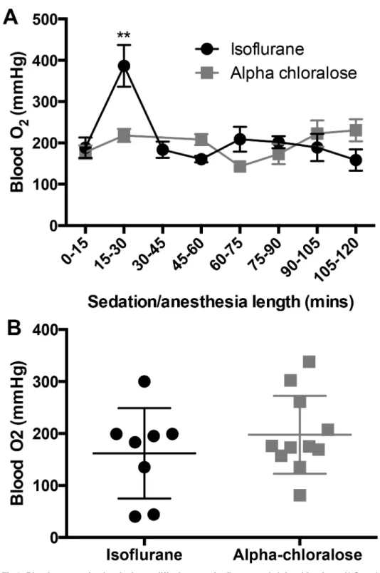
3.3 Blood oxygenation levels do not differ between isoflurane anesthesia
and alpha chloralose sedation
confirmed expected partial pressures of oxygen in the range of PaO2<40mmHg. All partial
pressures of oxygen analyzed as‘arterial’were>100 mmHg, confirming arterial blood
collection.
Arterial blood O2levels were not different between treatment groups over the course of 120
minutes (F(1,81)= 0.6, p = 0.44,Fig 3), but O2levels spiked in the isoflurane-treated animals in
the 15–30 minute time bin (p = 0.002) before returning to baseline levels (Fig 3a). After 120 minutes, O2levels between groups were not different (p = 0.67,Fig 3b).
3.4 Carbon dioxide levels in arterial blood rise significantly under alpha
chloralose sedation
Over time, there is a significant overall increase in blood CO2levels under both treatments
(F(5,82)= 4.06, p = 0.002,Fig 4), but this increase is significantly greater under alpha chloralose
sedation (F(1,82)= 38.06, p<0.0001,Fig 4a). After 120 minutes of sedation, arterial blood
car-bon dioxide levels were significantly higher in alpha chloralose-treated mice than isoflurane-treated mice (p = 0.002,Fig 4b).
Fig 1. Breathing Rate decreases over time during prolonged anesthesia.There were no group differences between isoflurane and alpha chloralose-treated mice at any time (F(1,386)= 0.001, p = 0.97), but there was a significant overall drop in breathing rates
for both groups (F(11,386)= 5.68, p<0.0001, n = 7–24 per time point).
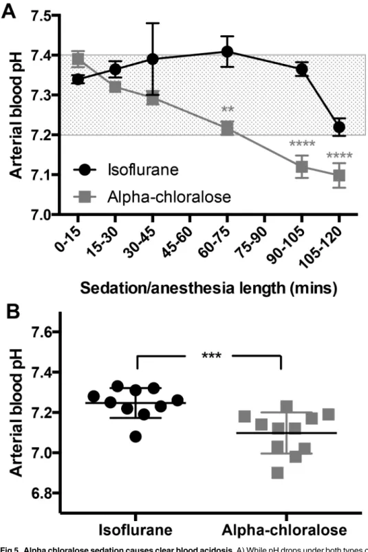
3.5 Alpha chloralose sedation causes blood acidosis over time
During the course of two hours of anesthesia/sedation, blood pH decreases significantly in both groups (F(5,86)= 8.97, p<0.0001,Fig 5a), coinciding with an increase in blood CO2levels.
Despite the decrease, pH remains within a narrow and normal physiological range under iso-flurane (pH 7.2–7.4)[15]. In contrast, alpha chloralose causes a significant drop in arterial blood pH within the first hour, and it continues to fall over time, showing a clear difference between treatment groups (F(1,86)= 35.72, p<0.0001,Fig 5a).
At the 120 minute time point, blood is significantly more acidotic in mice under alpha chlo-ralose sedation (p = 0.0006,Fig 5b). Under isoflurane, 9 of 10 mice still had blood pH levels within a normal range, but only 1 of 11 did not display acidosis (blood pH<7.2) under alpha
chloralose.
4. Discussion
We have shown that isoflurane and alpha chloralose, both commonly utilized for brain imag-ing in mice [39,40], cause differential effects on bodily homeostasis in the mouse. Alpha chlo-ralose causes unstable physiological effects that could confound the outcome measures upon which functional brain imaging relies. Isoflurane anesthesia maintained a stable heart rate, blood pH and carbon dioxide partial pressure (PaCO2) as previously reported [41–43] despite
an overall drop in breathing rate over 120 minutes. In contrast, alpha chloralose sedation
Fig 2. Heart Rate drops under alpha chloralose sedation.Mice retain a steady heart rate under isoflurane anesthesia, ranging from 463±20 beats/min to 489±20 beats/min. However, under alpha chloralose sedation, heart rate rapidly drops to below 350 beats/min, leading to a significant difference in heart rate between the treatment groups (F(1,384)= 516.2,p<0.0001, n = 9–23 per
time point). The shaded region indicates the normal heart rate for an awake mouse at rest (450–500 beats/min).
Fig 3. Blood oxygenation levels do not differ between isoflurane and alpha chloralose.A) Over the course of 120 minutes of sedation/anesthesia, there were no group differences in blood oxygenation levels (F(1,81)= 0.6, p = 0.44, n = 4–19 per time point), but there was a significant effect of time in the isoflurane
group, where blood oxygenation levels spiked between 15 and 30 minutes (F(5,82)= 4.06, p = 0.002), then
returned to baseline levels. B) Blood oxygenation is not different between isoflurane and alpha chloralose after 120 minutes of anesthesia/sedation (p = 0.67).
Fig 4. Blood carbon dioxide levels increase significantly under alpha chloralose sedation over the course of 120 minutes.A) Carbon dioxide levels in arterial blood were significantly higher under alpha chloralose sedation (F(1,82)= 38.06, p<0.0001, n = 4–19 per time point). Although there was an overall effect
of time (F(5,82)= 4.06, p = 0.002), reflecting increasing CO2levels over time, these levels did not differ
significantly from baseline over the course of 120 minutes under isoflurane anesthesia (p>0.05). B) After 120 minutes, carbon dioxide levels are significantly higher in alpha chloralose-sedated animals (p = 0.002).
Fig 5. Alpha chloralose sedation causes clear blood acidosis.A) While pH drops under both types of anesthesia (F(5,86)= 8.97, p<0.0001, n = 3–19 per time point), the drop is much more pronounced under
alpha chloralose sedation (F(1,86)= 35.72, p<0.0001), dropping from a normal reading of pH 7.39±0.05 during
the first 15 minutes, to 7.13±0.07 by 120 minutes. In contrast, blood pH under isoflurane drops from 7.34 ±0.04 to 7.27±0.06 after two hours, which is still within a normal physiological range for the mouse. The shaded region indicates non-acidotic blood pH for the mouse (15). B) Blood is acidotic after 120 minutes of alpha chloralose sedation (p = 0.0006).
caused a significant drop in breathing rates, a much-decreased but stable heart rate, and an increase in blood carbon dioxide levels (PaCO2) with a severe drop in arterial pH over time.
Mice from both groups had no difference in blood oxygenation (PaO2).
Anesthesia is known to affect major organs, especially the cardiopulmonary systems. Anes-thetized animals typically do not breathe as deeply as awake animals. Decreased tidal volumes coupled with decreased respiratory rates can result in hypoventilation and a build up of circu-lating CO2. Thus, respiratory acidosis (low pH and high PaCO2)is not uncommon in
anesthe-tized animals. Rao and Verkman [44] suggest a normal mouse partial CO2pressure (PaCO2) of
34–35 mmHg. As CO2levels increase under normal circumstances, central chemoreceptors
sensitive to arterial CO2tension cause a compensatory increase in respiration and the excess
CO2is exhaled to maintain normal blood gasses and pH [45]. Various drugs (including
anes-thetics) suppress the neurorespiratory center’s response to hypercapnia and are a common cause of respiratory acidosis. We found an increase in PaCO2in both groups over time, but
while the CO2increase in isoflurane mice did not differ significantly from baseline values, the
mice sedated with alpha chloralose showed a steady and significant rise in PaCO2. We suggest
that the hypercapnia is most likely the result of the decreased respiratory rate exacerbating the anesthetic hypoventilation. These effects upon respiration could be prevented by artificially ventilating the animal, although the suitability of this invasive procedure will depend upon the experimental outcome measures under investigation.
While the mouse appears to have a wide range of normal pH in the literature [15,41–44], acidosis in a human is diagnosed when pH falls below 7.36 [41]. Our data show that mice under isoflurane maintained a normal arterial pH for up to 2 hours under anesthesia. In con-trast, the alpha chloralose mice were acidotic after only 30 minutes and pH steadily declined over time. The drop in pH was inversely related to a rise in PaCO2. We diagnose an acute
pri-mary respiratory acidosis by the low pH and high PaCO2. However, we cannot rule out a
mod-est metabolic component to the degree of acidosis seen, as other metabolic changes secondary to anesthesia (lactate, insulin, glucose, free fatty acids, interleukin-6 and TNF-αlevels) were
not measured and others have seen elevated lactate in the anesthetized mouse [41,46,47]. The severe respiratory acidosis of alpha chloralose mice, demonstrated by the marked drop in pH, approached life-threatening levels. When pH drops below 7.2, metabolic enzymes may not function, hemoglobin’s binding affinity for oxygen is decreased, and cardiac arrhythmias may occur due to decreased cardiac contractility [19,48]. These life-threatening conditions could have severe effects on CBF and BOLD outcome measures, thus interfering with func-tional imaging. Although the respiratory rate also decreased with isoflurane over time similar to alpha chloralose, we did not witness the same severity of PaCO2elevation and the end drop
in pH still remained within acceptable mouse limits.
The partial pressure of oxygen was high in both groups. Constantinides et al [43] found that the ideal level of inspired oxygen for maintenance of stable cardiovascular physiology in the mouse was above 50%. When breathing room air (~20% O2) the expected PaO2is 95–100
mmHg with hemoglobin 95–98% saturated. With supplemental oxygen of 60% as in our study, the expected PaO2is 250–300 mmHg [14] as shown in our results.
One major drawback with anesthetized rodents for functional brain scanning is that anes-thesia significantly decreases cortical activation levels [33], which is the outcome that CBF and BOLD indirectly measures. However, lower levels of isoflurane anesthesia (0.75–1.5%) have proven to produce reliable cortical activation in the mouse [20,22,49]. Due to its limited effects on physiological measures, we therefore suggest that isoflurane is a more appropriate tool than alpha chloralose for mouse brain functional scanning.
mice, alpha chloralose cannot be reversed after administration, therefore correct initial dosages are critical, but still not guaranteed to produce similar results in all animals. Alpha chloralose has effects associated with respiratory complications, seizures and prolonged recovery in mam-mals (including dogs, swine and rodents)[32], and so it is not permitted by most animal care and use committees to be used for recovery experiments [50,51].
While isoflurane is a known vasodilator [33], it has been recommended as a valuable alter-native to alpha chloralose [52,53]. We have also shown that it is capable of keeping a mouse physiologically stable over long periods of time. In particular, we show here that heart rate, PaO2, PaCO2and blood pH is well maintained under isoflurane. This is a good indication of a
well-functioning cardiopulmonary system and normal basal physiology.
The purpose of anesthesia is to provide restraint with minimal stress, pain or adverse side effects, and cause amnesia of painful procedures. Adequate agents work because they interfere with the brain’s conscious recognition of pain or stress. Therein lies the challenge of functional imaging of animals, because distress must be prevented while maintaining neural pathways of interest. Additionally, anesthetics cause secondary effects on other organs to various degrees, namely the cardiopulmonary systems. Alternative anesthetic and sedative agents have been successfully used for rodent functional imaging and include halothane [54], ketamine [55] and medetomidine [25,56], although isoflurane and alpha chloralose remain the most utilized in the literature. However, all anesthetics contain caveats for brain imaging; for example, Schro-eter et al [57] compared isoflurane, medetomidine sedation, propofol and urethane on BOLD and cerebral blood volume (CBV) imaging and saw that in the mouse, fMRI responses are influenced by stimulus-induced cardiac changes (an arousal response), potentially masking stimulus-evoked signals. Researchers must therefore carefully consider anesthetic choices based on the research question. We have shown that isoflurane does not interfere with some physiological measures that influence BOLD and CBF outcome measures. As it maintains physiological stability up to two hours, we suggest it is more suitable than alpha chloralose for functional imaging studies.
Supporting Information
S1 Data. All raw data used in this manuscript is contained within the Supporting Informa-tion file.
(XLSX)
Acknowledgments
We are grateful to Dr. M. Catherine Bushnell for support in all aspects of this research. We also thank the staff of the Mouse Imaging Facility at the National Institutes of Health for their tech-nical expertise and help, in particular Dr. Jeeva Munasinghe, Daryl Despres, and Vivian Diaz. We also thank Matthew Grossman for literature searches.
Author Contributions
Conceived and designed the experiments: LL BK. Performed the experiments: LL LB BK. Ana-lyzed the data: LL LB BK. Contributed reagents/materials/analysis tools: LL BK. Wrote the paper: LL LB BK.
References
Reports. 2015; 5:12621. doi:10.1038/srep12621Available: http://www.nature.com/articles/srep12621-supplementary-information. PMID:26218081
2. Iannetti GD, Wise RG. BOLD functional MRI in disease and pharmacological studies: room for improve-ment? Magnetic resonance imaging. 2007; 25(6):978–88. doi:10.1016/j.mri.2007.03.018PMID:
17499469
3. Koretsky AP. Early development of arterial spin labeling to measure regional brain blood flow by MRI. NeuroImage. 2012; 62(2):602–7. doi:10.1016/j.neuroimage.2012.01.005PMID:22245338 4. Nasrallah FA, Tay HC, Chuang KH. Detection of functional connectivity in the resting mouse brain.
NeuroImage. 2014; 86:417–24. doi:10.1016/j.neuroimage.2013.10.025PMID:24157920.
5. Bruijnzeel AW, Alexander JC, Perez PD, Bauzo-Rodriguez R, Hall G, Klausner R, et al. Acute Nicotine Administration Increases BOLD fMRI Signal in Brain Regions Involved in Reward Signaling and Com-pulsive Drug Intake in Rats. International Journal of Neuropsychopharmacology. 2015; 18(2). doi:10. 1093/ijnp/pyu011
6. Liang Z, Liu X, Zhang N. Dynamic resting state functional connectivity in awake and anesthetized rodents. NeuroImage. 2015; 104:89–99. doi:10.1016/j.neuroimage.2014.10.013PMID:25315787 7. Baek K, Shim W, Jeong J, Radhakrishnan H, Rosen B, Boas D, et al. Layer-specific interhemispheric
functional connectivity in the somatosensory cortex of rats: resting state electrophysiology and fMRI studies. Brain Structure and Function. 2015:1–15. doi:10.1007/s00429-015-1073-0
8. Zhang N, Rane P, Huang W, Liang Z, Kennedy D, Frazier JA, et al. Mapping resting-state brain net-works in conscious animals. Journal of neuroscience methods. 2010; 189(2):186–96. doi:10.1016/j.
jneumeth.2010.04.001PMID:20382183
9. Chin CL, Tovcimak AE, Hradil VP, Seifert TR, Hollingsworth PR, Chandran P, et al. Differential effects of cannabinoid receptor agonists on regional brain activity using pharmacological MRI. British journal of pharmacology. 2008; 153(2):367–79. doi:10.1038/sj.bjp.0707506PMC2219521. PMID:17965748 10. Liang Z, King J, Zhang N. Neuroplasticity to a single-episode traumatic stress revealed by resting-state
fMRI in awake rats. NeuroImage. 2014; 103:485–91. doi:10.1016/j.neuroimage.2014.08.050PMID:
25193500
11. Madularu D, Yee JR, Kenkel WM, Moore KA, Kulkarni P, Shams WM, et al. Integration of neural net-works activated by amphetamine in females with different estrogen levels: A functional imaging study in awake rats. Psychoneuroendocrinology. 2015; 56:200–12. doi:10.1016/j.psyneuen.2015.02.022
PMID:25827963
12. Lewis NCS, Messinger L, Monteleone B, Ainslie PN. Effect of acute hypoxia on regional cerebral blood flow: effect of sympathetic nerve activity. Journal of Applied Physiology. 2014; 116(9):1189–96. doi:10.
1152/japplphysiol.00114.2014PMID:24610534
13. Dalkara T, Irikura K, Huang Z, Panahian N, Moskowitz MA. Cerebrovascular Responses under Con-trolled and Monitored Physiological Conditions in the Anesthetized Mouse. Journal of Cerebral Blood Flow & Metabolism. 1995; 15(4):631–8. doi:10.1038/jcbfm.1995.78.
14. Muir WW, Hubbell JA. Handbook of Veterinary Anesthesia. 4th ed. St. Louis, MO: Mosby-Elsevier; 2007. 656 p.
15. Hoyt RF, Hawkins JV, St Clair MB, Kennett MJ. Chapter 2—Mouse Physiology. In: Fox JG, Davisson
MT, Quimby FW, Barthold SW, Newcomer CE, Smith AL, editors. The Mouse in Biomedical Research III. 2nd ed. Burlington: Academic Press; 2007. p. 23–XVI.
16. Scipioni RL, Diters RW, Myers WR, Hart SM. Clinical and clinicopathological assessment of serial phle-botomy in the Sprague Dawley rat. Laboratory animal science. 1997; 47(3):293–9. PubMed Central
PMCID: PMC 9241633. PMID:9241633
17. Diehl K-H, Hull R, Morton D, Pfister R, Rabemampianina Y, Smith D, et al. A good practice guide to the administration of substances and removal of blood, including routes and volumes. Journal of Applied Toxicology. 2001; 21(1):15–23. doi:10.1002/jat.727PMID:11180276
18. Argmann CA, Auwerx J. Collection of Blood and Plasma from the Mouse. Current Protocols in Molecu-lar Biology: John Wiley & Sons, Inc.; 2001.
19. McGuill MW, Rowan AN. Biological Effects of Blood Loss: Implications for Sampling Volumes and Techniques. ILAR Journal. 1989; 31(4):5–20. doi:10.1093/ilar.31.4.5
20. Nair G, Duong TQ. Echo-planar BOLD fMRI of mice on a narrow-bore 9.4 T magnet. Magnetic reso-nance in medicine: official journal of the Society of Magnetic Resoreso-nance in Medicine / Society of Mag-netic Resonance in Medicine. 2004; 52(2):430–4. doi:10.1002/mrm.20158PMID:15282829; PubMed
Central PMCID: PMC2949950.
21. Nasrallah FA, Lee ELQ, Chuang K-H. Optimization of flow-sensitive alternating inversion recovery (FAIR) for perfusion functional MRI of rodent brain. NMR in biomedicine. 2012; 25(11):1209–16. doi:
22. Guilfoyle DN, Gerum SV, Sanchez JL, Balla A, Sershen H, Javitt DC, et al. Functional Connectivity fMRI in Mouse Brain at 7T Using Isoflurane. Journal of neuroscience methods. 2013; 214(2):144–8.
doi:10.1016/j.jneumeth.2013.01.019PMC3644382. PMID:23376497
23. Weber R, Ramos-Cabrer P, Wiedermann D, van Camp N, Hoehn M. A fully noninvasive and robust experimental protocol for longitudinal fMRI studies in the rat. NeuroImage. 2006; 29(4):1303–10. doi:
10.1016/j.neuroimage.2005.08.028PMID:16223588
24. Chao T-HH, Chen J-H, Yen C-T. Repeated BOLD-fMRI Imaging of Deep Brain Stimulation Responses in Rats. PloS one. 2014; 9(5):e97305. doi:10.1371/journal.pone.0097305PMID:24825464
25. Nasrallah FA, Lew SK, Low AS, Chuang KH. Neural correlate of resting-state functional connectivity under alpha2 adrenergic receptor agonist, medetomidine. NeuroImage. 2014; 84:27–34. doi:10.1016/
j.neuroimage.2013.08.004PMID:23948809.
26. Thompson GJ, Pan W-J, Keilholz SD. Different dynamic resting state fMRI patterns are linked to differ-ent frequencies of neural activity. Journal of neurophysiology. 2015; 114(1):114–24. doi:10.1152/jn.
00235.2015PMID:26041826
27. Peeters RR, Tindemans I, De Schutter E, Van der Linden A. Comparing BOLD fMRI signal changes in the awake and anesthetized rat during electrical forepaw stimulation. Magnetic resonance imaging. 2001; 19(6):821–6. doi:10.1016/S0730-725X(01)00391-5PMID:11551722
28. Silva AC, Koretsky AP. Laminar specificity of functional MRI onset times during somatosensory stimula-tion in rat. Proceedings of the Nastimula-tional Academy of Sciences. 2002; 99(23):15182–7. doi:10.1073/
pnas.222561899
29. Shih Y-YI, Chang C, Chen J-C, Jaw F-S. BOLD fMRI mapping of brain responses to nociceptive stimuli in rats under ketamine anesthesia. Medical Engineering & Physics. 2008; 30(8):953–8. doi:10.1016/j.
medengphy.2007.12.004
30. Hutchison RM, Mirsattari SM, Jones CK, Gati JS, Leung LS. Functional Networks in the Anesthetized Rat Brain Revealed by Independent Component Analysis of Resting-State fMRI. Journal of neurophysi-ology. 2010; 103(6):3398–406. doi:10.1152/jn.00141.2010PMID:20410359
31. Ueki M, Mies G, Hossmann KA. Effect of alpha-chloralose, halothane, pentobarbital and nitrous oxide anesthesia on metabolic coupling in somatosensory cortex of rat. Acta anaesthesiologica Scandina-vica. 1992; 36(4):318–22. doi:10.1111/j.1399-6576.1992.tb03474.xPMID:1595336
32. Silverman J, Muir WW. A review of laboratory animal anesthesia with chloral hydrate and chloralose. Laboratory animal science. 1993; 43(3):210–6. PMID:8355479.
33. Masamoto K, Kim T, Fukuda M, Wang P, Kim S-G. Relationship between Neural, Vascular, and BOLD Signals in Isoflurane-Anesthetized Rat Somatosensory Cortex. Cerebral Cortex. 2007; 17(4):942–50.
doi:10.1093/cercor/bhl005PMID:16731882
34. Bruns A, Künnecke B, Risterucci C, Moreau J-L, von Kienlin M. Validation of cerebral blood perfusion imaging as a modality for quantitative pharmacological MRI in rats. Magnetic Resonance in Medicine. 2009; 61(6):1451–8. doi:10.1002/mrm.21779PMID:19358231
35. Ciobanu L, Reynaud O, Uhrig L, Jarraya B, Le Bihan D. Effects of Anesthetic Agents on Brain Blood Oxygenation Level Revealed with Ultra-High Field MRI. PloS one. 2012; 7(3):e32645. doi:10.1371/ journal.pone.0032645PMC3299673. PMID:22427858
36. Itoh K, Sakata M, Watanabe M, Aikawa Y, Fujii H. The entry of manganese ions into the brain is acceler-ated by the activation of N-methyl-d-aspartate receptors. Neuroscience. 2008; 154(2):732–40. doi:10.
1016/j.neuroscience.2008.03.080PMID:18495352
37. Fox JG, Barthold SW, Davisson MT, Newcomer CE, Quimby FW, Smith AL. The Mouse in Biomedical Research: Immunology. 2nd ed: Academic Press; 2006.
38. White WJ, Field KJ. Anaesthesia and surgery of laboratory animals. Veterinary Clinics of North Amer-ica: Small Animal Practice. 1987; 17:989–1017. PMID:3310373
39. Thompson SJ, Bushnell MC. Rodent functional and anatomical imaging of pain. Neuroscience Letters. 2012; 520(2):131–9. doi:10.1016/j.neulet.2012.03.015PMID:22445887
40. Jonckers E, Delgado YPR, Shah D, Guglielmetti C, Verhoye M, Van der Linden A. Different anesthesia regimes modulate the functional connectivity outcome in mice. Magnetic resonance in medicine: official journal of the Society of Magnetic Resonance in Medicine / Society of Magnetic Resonance in Medi-cine. 2013; 72(4):1103–12. doi:10.1002/mrm.24990PMID:24285608.
41. Schwarzkopf TM, Horn T, Lang D, Klein J. Blood gases and energy metabolites in mouse blood before and after cerebral ischemia: the effects of anesthetics. Experimental Biology and Medicine. 2013; 238 (1):84–9. doi:10.1258/ebm.2012.012261PMID:23479767
42. Szczęsny G, Veihelmann A, Massberg S, Nolte D, Messmer K. Long-term anaesthesia using inhalatory
isoflurane in different strains of mice—the haemodynamic effects. Laboratory Animals. 2004; 38(1):64–
43. Constantinides C, Mean R, Janssen BJ. Effects of Isoflurane Anesthesia on the Cardiovascular Func-tion of the C57BL/6 Mouse. ILAR journal / NaFunc-tional Research Council, Institute of Laboratory Animal Resources. 2011; 52:e21–e31. PMC3508701. PMID:21677360
44. Rao S, Verkman AS. Analysis of organ physiology in transgenic mice. American Journal of Physiology
—Cell Physiology. 2000; 279(1):C1–C18. PMID:10898711
45. Dahan A, Teppema LJ. Influence of anaesthesia and analgesia on the control of breathing. British jour-nal of anaesthesia. 2003; 91(1):40–9. doi:10.1093/bja/aeg150PMID:12821564
46. Tanaka T, Nabatame H, Tanifuji Y. Insulin secretion and glucose utilization are impaired under general anesthesia with sevoflurane as well as isoflurane in a concentration-independent manner. Journal of anesthesia. 2005; 19(4):277–81. doi:10.1007/s00540-005-0341-1PMID:16261463
47. Zuurbier CJ, Koeman A, Houten SM, Hollmann MW, Florijn WJ. Optimizing anesthetic regimen for sur-gery in mice through minimization of hemodynamic, metabolic, and inflammatory perturbations. Experi-mental Biology and Medicine. 2014; 239(6):737–46. doi:10.1177/1535370214524877PMID:
24668552
48. Mitchell JH, Wildenthal K, Johnson RL. The effects of acid-base disturbances on cardiovascular and pulmonary function. Kidney International. 1972; 1(5):375–89. doi:10.1038/ki.1972.48PMID:4599247 49. Baltes C, Bosshard S, Mueggler T, Ratering D, Rudin M. Increased blood oxygen level-dependent
(BOLD) sensitivity in the mouse somatosensory cortex during electrical forepaw stimulation using a cryogenic radiofrequency probe. NMR in biomedicine. 2011; 24(4):439–46. doi:10.1002/nbm.1613
PMID:22945293
50. Holzgrefe HH, Everitt JM, Wright EM. Alpha-chloralose as a canine anesthetic. Laboratory animal sci-ence. 1987; 37(5):587–95. PMID:3320515.
51. Balis GU, Monroe RR. The pharmacology of chloralose. A review. Psychopharmacologia. 1964; 6 (1):1–30. PMID:5318644.
52. Sommers MG, van Egmond J, Booij LHDJ, Heerschap A. Isoflurane anesthesia is a valuable alternative forα-chloralose anesthesia in the forepaw stimulation model in rats. NMR in biomedicine. 2009; 22
(4):414–8. doi:10.1002/nbm.1351PMID:19003937
53. Grandjean J, Schroeter A, Batata I, Rudin M. Optimization of anesthesia protocol for resting-state fMRI in mice based on differential effects of anesthetics on functional connectivity patterns. NeuroImage. 2014; 102, Part 2:838–47. doi:10.1016/j.neuroimage.2014.08.043
54. Shah YB, Haynes L, Prior MJW, Marsden CA, Morris PG, Chapman V. Functional magnetic resonance imaging studies of opioid receptor-mediated modulation of noxious-evoked BOLD contrast in rats. Psychopharmacology. 2005; 180(4):761–73. doi:10.1007/s00213-005-2214-6PMID:15778889 55. Chen Y-Y, Shih Y-YI, Lo Y-C, Lu P-L, Tsang S, Jaw F-S, et al. MicroPET imaging of noxious thermal
sti-muli in the conscious rat brain. Somatosensory & Motor Research. 2010; 27(3):69–81. doi:10.3109/
08990220.2010.508222
56. Adamczak JM, Farr TD, Seehafer JU, Kalthoff D, Hoehn M. High field BOLD response to forepaw stim-ulation in the mouse. NeuroImage. 2010; 51(2):704–12. doi:10.1016/j.neuroimage.2010.02.083PMID:
20211267
57. Schroeter A, Schlegel F, Seuwen A, Grandjean J, Rudin M. Specificity of stimulus-evoked fMRI responses in the mouse: The influence of systemic physiological changes associated with innocuous stimulation under four different anesthetics. NeuroImage. 2014; 94:372–84. doi:10.1016/j.neuroimage.