Arabidopsis Small RNA Profile
Quanan Hu1, Jens Hollunder2,3, Annette Niehl4, Camilla Julie Kørner1, Dalya Gereige4, David Windels1, Andreas Arnold1¤, Martin Kuiper5, Franck Vazquez1., Mikhail Pooggin1., Manfred Heinlein1,4
*
1Botanical Institute, Department of Plant Physiology, Zu¨rich-Basel Plant Science Center, University of Basel, Basel, Switzerland,2Department of Plant Systems Biology, Vlaams Interuniversitair Instituut voor Biotechnologie (VIB) - Ghent University, Ghent, Belgium,3Department of Plant Biotechnology and Genetics, Ghent University, Ghent, Belgium,4Institut de Biologie Mole´culaire des Plantes du CNRS (UPR 2357), Universite´ de Strasbourg, Strasbourg, France,5Department of Biology, Norwegian University of Science and Technology, Trondheim, Norway
Abstract
Tobamoviruses encode a silencing suppressor that binds small RNA (sRNA) duplexesin vitro and supposedly in vivoto counteract antiviral silencing. Here, we used sRNA deep-sequencing combined with transcriptome profiling to determine the global impact of tobamovirus infection on Arabidopsis sRNAs and their mRNA targets. We found that infection of Arabidopsisplants withOilseed rape mosaic tobamoviruscauses a global size-specific enrichment of miRNAs, ta-siRNAs, and other phased siRNAs. The observed patterns of sRNA enrichment suggest that in addition to a role of the viral silencing suppressor, the stabilization of sRNAs might also occur through association with unknown host effector complexes induced upon infection. Indeed, sRNA enrichment concerns primarily 21-nucleotide RNAs with a 59-terminal guanine. Interestingly, ORMV infection also leads to accumulation of novel miRNA-like sRNAs from miRNA precursors. Thus, in addition to canonical miRNAs and miRNA*s, miRNA precursors can encode additional sRNAs that may be functional under specific conditions like pathogen infection. Virus-induced sRNA enrichment does not correlate with defects in miRNA-dependent ta-siRNA biogenesis nor with global changes in the levels of mRNA and ta-siRNA targets suggesting that the enriched sRNAs may not be able to significantly contribute to the normal activity of pre-loaded RISC complexes. We conclude that tobamovirus infection induces the stabilization of a specific sRNA pool by yet unknown effector complexes. These complexes may sequester viral and host sRNAs to engage them in yet unknown mechanisms involved in plant:virus interactions.
Citation:Hu Q, Hollunder J, Niehl A, Kørner CJ, Gereige D, et al. (2011) Specific Impact of Tobamovirus Infection on the Arabidopsis Small RNA Profile. PLoS ONE 6(5): e19549. doi:10.1371/journal.pone.0019549
Editor:Eliana Saul Furquim Werneck Abdelhay, Instituto Nacional de Caˆncer, Brazil
ReceivedMarch 8, 2011;AcceptedApril 1, 2011;PublishedMay 10, 2011
Copyright:ß2011 Hu et al. This is an open-access article distributed under the terms of the Creative Commons Attribution License, which permits unrestricted use, distribution, and reproduction in any medium, provided the original author and source are credited.
Funding:We thank the Functional Genomics Center Zu¨rich for providing the resources for RNA profiling using Affimetrix oligonucleotide arrays. This work was supported with financial support by the Human Frontier Science Program Organization (RGP0022/2006) and the Swiss National Science Foundation grants 3100A0-111916 and 31003A-124940 to M.H., 31003A-122469 and 31003A-127514 to M.M.P., and PZ00P3-126329) to F.V. The funders had no role in study design, data collection and analysis, decision to publish, or preparation of the manuscript.
Competing Interests:The authors have declared that no competing interests exist. * E-mail: [email protected]
.These authors contributed equally to this work.
¤ Current address: Friedrich Miescher Institute for Biomedical Research, Basel, Switzerland
Introduction
RNA silencing is a sequence-specific mechanism that coordinates the expression, protection, stability, and inheritance of eukaryotic genomes. It is involved in tuning critical developmental, stress-responses, and bodyguard functions by regulating the expression of genes at the transcriptional and post-transcriptional levels, or by triggering the formation of heterochromatic DNA regions [1,2,3]. RNA silencing is mediated by 21–24 nt small RNAs (sRNAs) that are processed from long dsRNA by RNase III enzymes of the DICER family (DICER-Like - DCL in plants). These sRNAs are classified into small interfering RNAs (siRNAs) and microRNAs (miRNAs) depending on their origin [2,4,5]. siRNAs and miRNAs associate with proteins of the ARGONAUTE (AGO) family to form RNA-Induced Silencing Complexes (RISC) in which they serve as guides to complementary RNA or DNA targets [4,6]. AGO-containing RISCs can then mediate degradation of complementary endogenous or viral RNAs, translational repression of mRNAs, or transcriptional silencing of transposons and DNA repeats.
RNA silencing is known to play a critical role in defense against viruses in plants and insects [20,21,22]. Thus, viral RNAs are used by the RNA silencing machinery to generate viral sRNAs (vsRNAs) that can potentially be loaded into specific AGOs to further target viral RNAs for cleavage and degradation or for translational repression. As part of the ongoing host-virus arms race, viruses have evolved potent RNA silencing suppressors (VSRs). As they evolved independently, the VSRs of different viruses inhibit different RNA silencing pathway components [23,24]. We and others have shown that Tobacco mosaic virus
(TMV) and related tobamoviruses encode a VSR that resides in the small subunit of their replicase [25,26,27]. This subunit binds siRNA and miRNA duplexes in vitro and interferes with their methylation [25,26,28,29], a modus operandi that was also reported for several other VSRs like the Hc-Pro of Tobacco etch virus(TEV) or p19 of tombusviruses [30]. Consistently, miRNAs levels are generally increased in plants infected with TMV, TMV-Cg, cr-TMV, orOilseed rape mosaic virus(ORMV)[25,26,29,31,32]. Studies of few cases have demonstrated that the increased miRNA levels triggered by tobamovirus infection results in increased levels of cognate mRNA targets [25,31] and suggested that sRNA binding by tobamovirus replicase interferes with RISC loading or activity. This model is supported by the ability of cr-TMV suppressor protein to bind sRNA duplexes and to inhibit RISC assembly in vitro [25]. However, a recent report indicates that a positive correlation between miRNA enrichment and increased mRNA target levels may not be necessarily mediated by miRNA sequestration. Thus, it was shown that the enrichment of miR168 in plants infected withCymbidium ringspot virusoccurs through an activity independent of binding by the VSR p19 and that miR168 functions in translational repression rather than in mRNA cleavage despite of the increased AGO1 mRNA levels [33]. Moreover, the movement protein (MP) of TMV enhances the spread of silencing [34] suggesting that the impact of tobamo-viruses on the sRNA profile and gene expression may not be limited to infected cells but may be able to spread ahead of the leading front of infection. These examples clearly indicate that further studies are needed to understand the impact of virus infection on the host sRNA profile and gene expression and its role in virus susceptibility and/or defense.
So far our knowledge on changes of sRNA and transcriptome profiles induced by a virus in its host is limited. Our current knowledge is based on the analysis of a limited number of sRNAs and of their targets by Northern blots or on small-scale sRNA cloning [25,31]. Analysis of the full sRNA complement of cells can be achieved by deep sequencing using a variety of available platforms [35] and first reports describing changes in the plant sRNA profile in response to pathogens are appearing [36]. While deep sequencing analyses of virus-infected plants allowed to describe the profile of virus-derived siRNAs [37,38,39,40,41,42], a comprehensive view on the global impact of virus infection on the profile of host plant sRNAs and their mRNA targets is still lacking. We have used a combination of mRNA profiling and sRNA deep sequencing to understand the impact of ORMV infection on the Arabidopsis sRNA and transcriptome profiles. We describe here a global analysis of viral sRNAs and the changes in cellular sRNA profiles during systemic infection. Our analysis shows that virus infection changes the pattern of sRNAs that are processed from miRNA and ta-siRNA precursors. The virus-induced sRNA profile is inconsistent with the proposed stabilization of sRNA duplexes by the replicase as a sole mechanism and suggests that the observed changes in the sRNA profile involve additional mechanisms. In particular, we found that ORMV infection leads to the specific enrichment of 21 nt sRNAs with a 59 terminal
guanine. This suggests that these sRNAs associate with size-specific and 59 nucleotide-specific effector complexes of yet-unknown nature. Our mRNA profiling data demonstrate that increased levels of miRNAs and siRNAs do not correlate with significant changes in target transcript levels indicating that the virus-induced sRNA fraction is sequestered or active at different levels. Virus infection also leads to the accumulation of novel miRNA-like siRNAs (ml-siRNAs) encoded by miRNA precursors that might be part of a specific plant response to pathogens.
Results
To gain insights into the effects of ORMV infection on the Arabidopsis sRNA profile we conducted Illumina sequencing of sRNA populations extracted from ORMV-infected and mock control-inoculated Arabidopsis Col-0 plants at 7 days post-inoculation (dpi). Following removal of adapter sequences the reads were mapped to the virus andArabidopsis thalianagenomes (Figure S1, A) and only the reads with perfect match were further analyzed. Of 1,787,490 mapped sRNA reads found in the virus-treated sample, 80.1% (1,431,362 reads) originated from Arabi-dopsisand the remaining 19.9% (356,128) of the reads originated from the virus (Figure S1, A and B). Consistent with their biogenesis by DCL4 [29] ORMV vsRNAs are predominantly 21 nt in length (Figure S1, C). The vsRNAs map all along the viral genome, with 88.1% being homologous to the positive strand and 11.9% to the negative strand (Figure S1, E). A similarly strong positive strand bias in vsRNA accumulation has also been reported in the case of TMV-Cg infected samples [40] and may reflect the strong (+)strand-specific accumulation of viral RNA in infected cells [43,44]. The origin of the conspicuous vsRNA hotspots along the viral genome is yet unknown but may be caused by predominant DCL cleavage of structured, double-stranded RNA regions.
ORMV infection causes size-specific and 59 nucleotide-specific enrichment of plant sRNAs
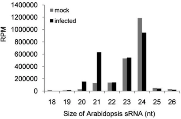
Comparison of the profiles of the mapped Arabidopsis-encoded sRNA obtained for mock and virus-treated samples reveals a significant impact of virus infection. The normalized, size-specific distribution of sRNAs shows a significant decrease in the proportion of 24 nt sRNAs, whereas the proportion of 21 nt sRNAs is increased (Figure 1). Consistently, there is a reduction in the proportion of sRNA reads derived from transposons and
Figure 1. Size-distribution of Arabidopsis sRNAs in ORMV-infected and non-ORMV-infected plants.
centromeric regions, whereas the proportion of reads for miRNAs and ta-siRNAs is increased 4.5-fold (Table 1).
Arabidopsis ta-siRNAs are derived from four families ofTAS
genes. The biogenesis of ta-siRNAs is initiated by AGO-mediated cleavage of TAS transcripts guided by miR173 (TAS1a/b/c and
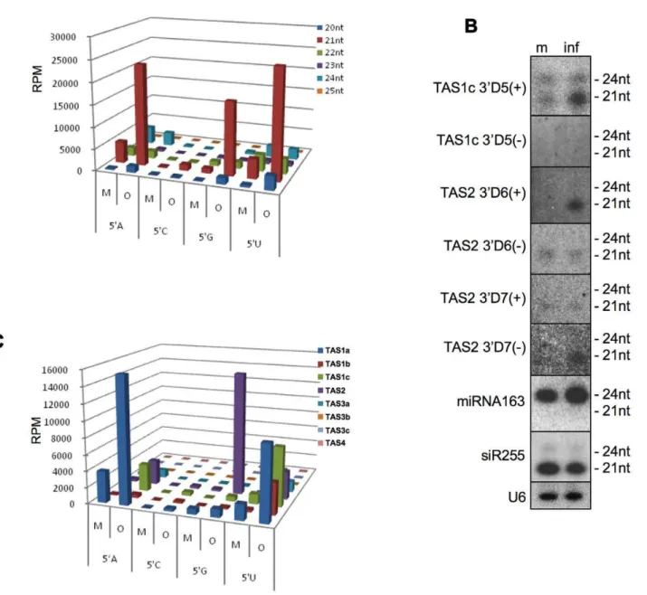
TAS2), miR390 (TAS3a,b,c), or miR828 (TAS4) [14,45,46,47,48]. Cleaved transcripts are then converted by RDR6 to long dsRNAs that are subsequently processed by DCL4 into phased ta-siRNAs that are in register with the cleavage site [45,47,49,50,51]. The majority of ta-siRNAs sequenced in both of our datasets were derived from TAS1a,b,c and TAS2 loci (Table S1). Interestingly, although TAS family loci in Arabidopsis produce ta-siRNAs of different sizes, as observed previously [52], only the 20 and 21 nt ta-siRNAs are significantly enriched (6.5. and 7.5 fold, respective-ly) in ORMV-infected plants (Figure 2A, Table S1). To confirm this size-specific enrichment, we analyzed the sRNAs from mock-and virus-treated samples by RNA blot hybridization using several probes detecting ta-siRNAs of different sizes and from different phases [47,53]. Figure 2B shows for TAS1c 39D5(+) that infection leads to the enrichment of the 21-nt siRNA species, whereas the level of the 24 nt siRNA species remains constant. The blots confirm that the enriched ta-siRNA species can be derived from either strand of the TAS RNA duplex. For example, whereas TAS2 39D6(+) siRNA strand is enriched in infected tissues, the complementary strand (TAS2 39D6(-)) of the ta-siRNA duplex is not. Conversely, specific enrichment is observed for the TAS2 D7 (-) siRNA strand, whereas the level of the complementary TAS2 39D7(+) strand remains unchanged. Consistent with previous observations [25,29], the level of siR255, which derives from
TAS1a,b,c and other loci, does not show any increase upon virus infection.
Since the loading of AGO proteins depends on the identity of the first 59 nucleotide of sRNAs, we analyzed the distribution of the 59nucleotide of the enriched sRNA pool. Figure 2A shows that the degree of over-representation of 21 nt ta-siRNAs caused by ORMV-infection differs according to the 59terminal nucleotide in the order G.A = U.C (Figure 2A). Interestingly, we found a correlation between the 59 nucleotide-specificity of the 21 nt ta-siRNAs and the specific precursorTASRNAs from which they are derived. Specifically, enriched ta-siRNAs with a 59G are predominantly derived from TAS2 including the validated 39D6(+) and 39D7(-) species (Figure 2B and C), although as for the otherTASloci, the unique TAS2 ta-siRNAs with a 59G (and also those with a 59C) are underrepresented compared to ta-siRNAs starting with A or U (Table S2). It should be noted,
however, that size- and 59-terminal guanine are unlikely to represent sufficient criteria to dictate sRNA enrichment since some 21-nt siRNAs initiating with a 59G are not enriched upon ORMV infection (data not shown). Size-specific selection may occur at the level of the sRNA duplex, since infection enriches 24 nt long miRNA163. Although this sRNA is 24 nt in length, it assumes a shorter physical length upon forming a bulge when paired with its 21 nt long passenger strand. Thus, the presence of miRNA163 as a duplex may represent an important prerequisite for its selection and subsequent stabilization in ORMV-infected plants.
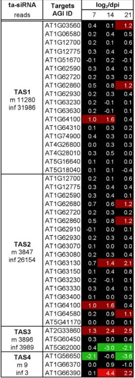
To test whether infection has an effect on the phased processing of ta-siRNAs, we mapped the frequency of unique ta-siRNA species with more than 5 reads toTAS1a,TAS1b, TAS1c,TAS2, andTAS3agenes (Figure 3). A change in ta-siRNA phasing was not observed indicating that the initial miRNA-guided cleavage mediated by AGO1 (TAS1/2) and AGO7 (TAS3a) and subsequent processing of theTASRNA duplexes by DCL4 are not affected in infected cells. The distribution of the mapped ta-siRNA reads mapped to the TAS genes confirms that the enrichment is restricted to specific ta-siRNAs and that in most cases only one of the two strands of the processed ta-siRNA duplexes is enriched. Collectively, these observations indicate that the virus-induced enrichment of ta-siRNAs occurs at a post-processing step, presumably through stabilizing associations with specific effector complexes, as is suggested by the strand, size and nucleotide specificity of the ta-siRNAs.
We note that the specific enrichment of 20–21 nt siRNAs is not restricted toTASloci. A similar effect is indeed also observed for other RDR6/DCL4-dependent, secondary siRNA-generating loci [52] (Table S3) or for IR71, which is one of the long inverted repeats of Arabidopsis that generates all size classes of siRNAs in an RDR-independent fashion [53,54,55,56,57] (Table S4).
Next, we investigated the impact of virus infection on the profile of miRNAs. Previous studies using specific Northern blot probing or small-scale sequencing indicated an enrichment of miRNAs in tobamovirus infected tissues [25,26,31]. The results of our global profiling analysis confirm this trend. We found that all but four of the 35 miRNA families with sequencing reads in both samples showed an increased number of reads upon infection (Table S5). Interestingly, virus infection caused a much higher increased accumulation of miRNA* sequences compared to that of the corresponding miRNAs. Thus, whereas the sRNAs derived from miRNA precursor RNAs in mock-treated plants comprise 97% miRNAs and only 2% miRNA passenger strands, the relative amount of miRNAs in ORMV-infected plants is reduced to 84%
Table 1.sRNA reads* mapped to theArabidopsisgenome.
Mock Infected
Type Unique Reads Total Reads Unique Reads Total Reads
Known miRNA precursors 380 48514 1032 276483
Known TAS precursors 1220 10195 2425 48303
Gene 42003 133451 59901 236165
Tandem repeats 22559 76359 26261 81339
Inverted repeats 14560 66799 19287 78722
Transposons 70568 126655 67768 113376
Centromeric region 2160 11248 2627 10113
rRNA, tRNA, snoRNA and snRNA 8486 98765 10386 161872
*Reads are RPM
whereas that of miRNA* sequences is increased to 14% (Figure 4A). Generally, the number of reads and the degree of accumulation in virus-infected plants differs between miRNAs and their passenger strands (Fig. S5). These observations were confirmed by sRNA blot hybridizations (Figure 4B). Further analysis showed that this strong enrichment of miRNA* sequences primarily concerns those initiating with a 59G resulting in miRNA* sequences with a 59G as the predominant miRNA* species in infected cells (Figure 4A). A summary of the virus-induced enrichment of miRNA gene-derived sRNAs is shown in Figure 4C. The increase in accumulation seen for sRNAs initiating with a 59U is to the majority caused by virus-enriched miRNAs, whereas the increase seen for sRNAs starting with a 59G reflects
the accumulation of miRNA* sequences (see also Figure 4A). The passenger strands of miRNA160, miRNA396, and miRNA398 exhibit the strongest contribution to the increased pool of miRNA* sequences starting with a 59G in virus-infected plants (highlighted in Table S5). However, a 59G is not the only determinant for enrichment of passenger strands. For example, strong increases in accumulation are also shown by the passenger strands of miRNA408 and miRNA472, although these miRNA* sequences initiate with a 59C and 59U, respectively (Table S5). Nevertheless, the overaccumulation of miRNA* strands compared to miRNA guide strands and the preference for 59G-miRNA* is again indicative of specific sRNA-associated effector complexes formed upon virus infection.
Effect of ORMV infection on the host plant transcriptome
Although ORMV starts to spread systemically within 2–3 days after inoculation of individual leaves (Niehl and Heinlein, unpublished), viral symptoms are only observed after 10 days. From this time onward the newly emerging leaves and progressively also the older leaves show curling and retarded growth, and the oldest leaves show signs of necrosis. Three weeks after inoculation, the plants show a clear growth retardation phenotype (Figure 5A).
To test whether the significant virus-induced changes in the sRNA profile seen at 7 dpi correlate with significant effects on transcript levels, we profiled mRNA transcripts at 7, 14, and 21 dpi using Affimetrix ATH1 arrays. In this time-course experiment we observed a gradual increase in accumulation of ORMV genomic RNA and vsRNAs (data not shown) and a gradual increase in symptom severity (Figure 5A). The total RNA extracts for profiling transcripts at 7 dpi were the same as those
data was also validated by principal component analysis (PCA, Figure 5C) and hierarchical clustering (Figure 5D), indicating that the replicates show very similar responses and that the datasets for mock-inoculated and virus-infected samples are clearly separated. Importantly, for the virus-infected samples, the 7 dpi time point is clearly distinct from the later time points. This suggests that expression changes at 14 and 21 dpi may be related to secondary effects that are related to tissue crinkling and chlorosis, whereas the
earlier time point reflects more specific responses to the virus infection and its spread.
Functional GO term enrichment analysis (log2.2; p,0.001) reveals that at 7 dpi there are responses of gene classes related to responses to biotic stimuli, other organisms, stress, defence, and immune system processes, whereas responses at the later time points are focussed to gene classes rather responding to metabolic processes and abiotic stimuli (Table S6). This finding may indicate Figure 4. Effect of ORMV infection on the miRNA profile.(A) ORMV infection produces a significantly higher fold-change in the levels of miRNA* sequences (miRNA passenger strands) than in the level of miRNAs. The virus-induced fold-change is strongest for miRNA* sequences carrying a 59G nucleotide. (B) Northern blots confirming the virus-induced enrichment of miRNAs and the much stronger enrichment for their corresponding miRNA* sequences. m, mock; inf, infected; dpi, days post inoculation. (C) Normalized miRNA reads (RPM) for ORMV-infected (O) and non-infected (mock-treated, M) plants sorted according to size (nt) and 59terminal nucleotide. The majority of miRNAs starts with a 59U nucleotide and these miRNAs are strongly enriched in infected plants. The virus-induced peak of sRNAs starting with a 59G is mostly due to enriched miRNA* sequences (as seen inA).
that at late time points, when the virus has accumulated to high levels, secondary effects on the plant metabolism and nutritional status play a role in addition to the responses to virus.
Effect of ORMV infection on mRNA targets of miRNA and ta-siRNA
We next addressed whether the strong increases in the levels of ta-siRNAs and miRNAs are correlated with similarly strong changes in the level of their mRNA targets. Although our transcriptome data revealed significant changes in the transcript levels of many genes (Figure 5), the levels of the majority of the miRNA and ta-siRNA target transcripts appeared rather stable (Figure 6 and Figure 7). The general down-regulation of targets expected if all over-accumulated miRNAs would engage in target cleavage was not observed. Rather, some of the targets show increases in their abundance. Examples are members of the SPL transcription factor family (targets of miR156/157), a member of the pentatricopeptide family (AT1G62670; target of miR161 and miR400), a GRF gene family transcription factor (AT2G36400,
target of miR396), Auxin response factors (ARF) 16 and 17 (AT1G77850 and AT4G30080; targets of miR160), and genes encoding LRR disease resistance gene motifs (AT1G122280 and AT1G15890; targets of miR472). Consistent with previously published observations by others [25,33], a strong increase was also found for AGO1 (AT1G48410), the target of miR168. Overall, stronger changes in miRNA and ta-siRNA target transcript levels are seen at later infection stages (21 dpi), whereas there are rather mild changes, if any, at the time point of sRNA analysis (7 dpi). Only two of the 248 tested miRNA and ta-siRNA target transcripts show reduced levels at all three time points, while 16 targets exhibit increased levels at all three time points. Twenty-one targets display increased expression levels at later time points (starting at 14 or at 21 dpi), and 20 targets show reduced expression levels at later time points (starting at 14 or at 21 dpi). Notably, changes in the levels of miRNAs targeting multiple genes (e.g. miR163, 164, 169, 171, 393, 156/157) did not trigger similar changes in all their known target transcripts. Rather, the targets belonging to groups controlled by the same miRNAs exhibit Figure 5. Effect of ORMV infection on the Arabidopsis transcriptome.(A) Disease symptoms of ORMV-infected plants as compared to mock-inoculated, non-infected plants at 7, 14, and 21 dpi. (B) Venn-diagram depicting the genes differentially expressed upon ORMV infection. A log2
fold-change cut-off value of 2 and a significance threshold of 0.001 were used for data analysis. (C) Principal component analysis on RMA-normalised expression values illustrating reproducibility between specific profiles and a clear data separation between the specific treatments. (D) Hierarchical clustering of expression values.
diverse changes. These observations indicate that the changes in miRNA and ta-siRNA levels in ORMV-infected cells do not lead to corresponding changes in the transcriptome.
ORMV infection promotes expression of novel miRNA-like sRNAs from miRNA precursors
Our sequencing data revealed the presence of a substantial number of unique sRNAs derived from miRNA primary transcripts that do not represent the known miRNA or miRNA* sequences (Figure 8). Their unique accumulation upon virus infection was verified by sRNA blot hybridization (Figure 9). This finding indicates that miRNA precursors can generate multiple sRNAs including novel pathogen-inducible species. Apparently, these miRNA-like sRNAs (ml-sRNAs) are produced at very low levels under normal conditions whereas they are better processed or stabilized, and thus enriched, in plants challenged with ORMV. These ml-sRNAs are in phase with the canonical miRNAs, suggesting that they are produced during phased processing of the miRNA precursor RNAs by DCL1 [58]. Among the 19 ml-sRNAs identified in our experiment, eight were recently shown to be also induced by the bacterial pathogen Pseudomonas [36] (Table 2). Thus, accumulation of ml-sRNAs appears to be a common feature of bacterial and viral infection.
To determine whether enriched ml-sRNAs could play a role in RISC-mediated degradation of target transcripts, we used them as queries for a DegradomeSearch with StarBase (http://starbase. sysu.edu.cn/index.php), a public platform for exploring micro-RNA-target interaction maps from Argonaute CLIP-Seq (HITS-CLIP) and degradome sequencing (Degradome-Seq, PARE) data [59]. Using a penalty score of $4.5 and searching for targets indicated by$1 cleavage tags, we identified potential targets of 4 of the eleven newly identified ml-siRNAs in our database (miR163-IP1, miR163-IP2, miR841-IP1, and miR841-IP2). To further investigate the potential role of ml-siRNAs in target mRNA degradation we searched sequenced libraries of AGO-associated sRNAs [14,19]. Indeed, here we found several indications of associations of ml-sRNAs with AGO proteins. Thus, miR163-IP1 was associated with AGO1, 2, 4, 5, and 7, miR163-IP2 was associated with AGO1, 4, 5, and 7, and miR846-IP1occurred in association with AGO2. These findings suggest that at least some of the virus-induced ml-sRNAs may function in the regulation of mRNA targets in association with specific AGO complexes. It will be interesting to determine the functions of these ml-siRNAs during infection and during normal plant develop-ment.
Discussion
Plant-virus interaction triggers multiple plant defense pathways [60], including RNA silencing [21,22]. To counteract RNA silencing, plant viruses encode diverse types of VSR that act at different steps in the silencing pathway [24]. Up to now, most of the investigated plant VSRs are pathogenic proteins [61,62,63]. Their activities contribute to the development of virus-induced disease symptoms by interfering with endogenous gene expression and endogenous sRNA pathways [25,31,64]. The small replicase subunit of tobamoviruses was identified as a pathogenicity determinant [65] and was shown to function as VSR [25,26,27,66].In vitroexperiments indicate that this protein binds double-stranded sRNAs [25,28,30] and this binding has been proposed to account for the enrichment of both miRNAs and miRNA* sequences seen in infected plants [25,26,31]. A fraction of the accumulated sRNAs lacks methylation at the 39 end, suggesting that replicase binding to sRNAs may interfere with
their methylation by HEN1 [25,26]. Point mutation in the methyltransferase domain of the replicase interferes with the silencing suppressing capacity of the protein, coupled with weakened pathogenic symptoms in plants and decreased accumu-lation of non-methylated miRNAs/siRNAs [26]. Although the ability of the replicase to bind sRNA duplexesin vitromay explain the enrichment for both miRNAs and miRNA* sequences seen in infected plants [25,26,31], it remained unclear whether thein vivo -enriched miRNAs and miRNA* sequences are indeed derived from replicase-stabilized duplexes or whether other virus-induced mechanisms play a role in sRNA enrichment. Moreover, it remained unclear whether the sRNA enrichments would impose changes in the host transcriptome. We have now gained critical insights into the tobamovirus interaction with RNA silencing, and sRNAs in particular, by characterizing the sRNA and transcrip-tome profiles of ORMV-infectedArabidopsisplants.
ORMV infection causes global size-specific sRNA enrichment in systemically infected plants
Our deep sequencing data indicate that ORMV infection leads to a global enrichment of sRNA species that are predominantly 21 nt in length, including miRNAs and ta-siRNAs, other RDR6-dependent siRNAs, as well as siRNAs processed from inverted repeat loci. The observation that ORMV infection enriches 20– 21 nt sRNAs but not 24 nt sRNAs is in agreement with an earlier study [29] and consistent with the observation madein vitrothat the tobamoviral VSR protein binds sRNA duplexes in a size-specific manner [25]. Tagami et al (2007) found that infection by TMV-Cg leads to specific enrichment of miRNAs [31]. Our work now extends this conclusion by the demonstration that ORMV infection causes a general enrichment of 20–21 nt RNAs irrespective of their origin.
Viral suppressor binding may not be the sole determinant of the enriched sRNA profile
The ability of the tobamoviral silencing suppressor to bind sRNA duplexes in a size-specific mannerin vitro [25,28,30] has been proposed to explain the enrichment of miRNA* sequences along with their corresponding miRNAs observed in vivo
AGO-associated effector complexes in the virus-induced sRNA enrich-ment. Interestingly, we found that the 59nucleotide specific pattern of enrichment of ta-siRNAs (G.A = U.C) mimics that of
vsRNAs (Figure S1, D). This may suggest that vsRNAs and ta-siRNAs are stabilized by similar complexes.
A role for host effector complexes in addition to the binding by the viral suppressor in the stabilization of sRNAs is also supported by the observation that ORMV infection causes a stronger increase in the levels of miRNA* sequences relative to that of the respective miRNAs. For example, miR160*, miR396*, and miR398*, are particularly highly over-represented as compared to their miRNA. This observation cannot solely be explained by the ability of the tobamoviral replicase to bind sRNA duplexes since this should lead to equal accumulation of both strands of the different duplexes. Another less likely possibility, which is in contradiction with earlier in vitro binding assays indicating that replicase does not bind single-stranded sRNAs [25,28], is that the replicase binds only one of the two strands of sRNA duplexes and exhibits some kind of 59 nucleotide preference similar to that of AGO proteins. However, we noted the enrichment of miRNA163 in infected plants. Given the preference for enrichment of 21 nt sRNAs, this 24 nt long miRNA may only be enriched when present in a duplex with its passenger strand, which forces the 24 nt miRNA to bulge out and thus to assume a length characteristic for 21 nt sRNA. This finding may suggest that the size selection for enrichment occurs at the level of duplexes. Thus, it may be conceivable that the ability of the viral suppressor to bind 21 nt sRNA duplexes plays a role in sRNA size selection whereas the final enrichment of single strands is caused by association with other proteins.
Possible functions of enriched sRNAs during ORMV infection
Our observation that the enriched miRNA and ta-siRNA levels have no strong effects on the levels of their mRNA targets does not necessarily indicate that they are inactive. First, the targets of these sRNAs may be robustly regulated by feedback mechanisms (e.g. at the level of transcription) or may be controlled by established RISCs that are stable and thus resistant against virus-induced changes in sRNA levels. Second, since some of the mRNA targets show changes in expression levels, the virus-induced sRNA enrichment may occur predominantly in specific tissues in which the majority of the corresponding targets is not expressed. The enriched sRNAs may also function in sRNA-guided translational repression rather than in target transcript cleavage. Translational suppression by sRNA-guided AGO complexes is a common phenomenon in plants and animals [12,67,68]. For example, as we could confirm in parallel experiments (not shown), in Arabidopsis, accumulation of miR168 caused by distinct RNA viruses leads to AGO10-mediated translational inhibition rather than to AGO1-mediated cleavage of the AGO1 mRNA [33]. Moreover, recent reports suggest that plant miRNAs can also mediate DNA methylation [11,69], which adds yet another dimension by which the enriched sRNAs may act.
With regard to functional diversification it is also important to note that a large proportion of the sRNAs that accumulate in tobamovirus-infected plants is not methylated [25,26,29]. Al-though non-methylated sRNAs are usually degraded [70,71], they may be stabilized upon association with specific effectors. A precedent for this hypothesis is provided by miRNAs in Drosophila that bind to either AGO1 or AGO2 proteins, Figure 6. Changes in the level of miRNA target transcripts upon ORMV infection at 7, 14, and 21 dpi.Heatmap shows log2-fold change
values for the mRNA targets of specific miRNAs. The miRNA reads in mock-treated (m) and ORMV-infected (inf) plants is shown. Although some miRNA targets show increased (red) and decreased (green) levels of expression upon infection, the majority of the miRNA target mRNAs does not show a change in the level of expression.
doi:10.1371/journal.pone.0019549.g006
Figure 7. Changes in the level of ta-siRNA target transcripts upon ORMV infection at 7, 14, and 21 dpi.Heat map shows log2-fold change
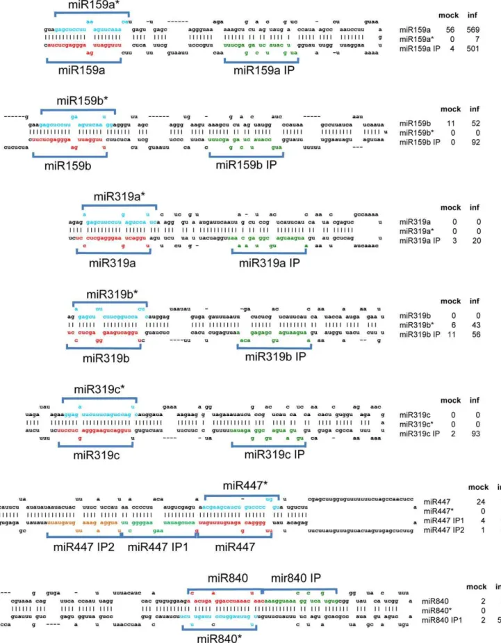
Figure 8. ml-siRNAs produced from miRNA precursor RNAs upon infection.The foldback structures of several miRNA precursor RNAs are shown. The sequences of mature miRNAs and miRNA* sequences are indicated in red and blue color, respectively. ml-siRNAs are depicted in green and yellow color. Since the ml-siRNAs are produced in phase (IP) with the miRNA and miRNA* sequences, they are designated according to the particular miRNA with the extension ‘‘IP’’ (for example, ‘‘miR159 IP’’). The normalized reads (RPM) obtained for miRNAs, miRNA*, and ml-siRNA sequences for mock-treated (mock) and ORMV-infected plants (inf) are shown.
dependent on whether they are methylated at the 39 end by HEN1, or not [72,73,74]. Moreover, unlike the methylated sRNAs associated with AGO2, the AGO1-bound non-methylated sRNAs undergo target-guided tailoring and trimming, which contributes to the efficiency of sRNAs with limited complementarity to the target [75]. Thus, the enrichment of size- and nucleotide-specific Arabidopsis sRNAs that are not methylated and thus potentially changed at their 39end may represent an important mechanism by which ORMV and potentially other viruses could diversify sRNA function during infection. Our analysis revealed the particular enrichment of sRNAs with 59 terminal guanine. Notably, it is not known which of the AGO proteins is predominantly associated with 21 nt 59G-sRNAs. It is also possible that non-AGO proteins may functionally associate with 59 G-sRNAs and other enriched G-sRNAs to form effector complexes with potentially diverse and yet unknown functions.
The enrichment of miRNA*s suggests that they might have a conditional function, as may also be supported by their association with predicted targets and the presence of potential target RNA cleavage products in degradome databases [76].
miRNA precursors produce novel sRNAs in response to pathogen infection
In addition to miRNA* sequences, virus infection also causes the accumulation of ml-sRNAs, which also may play important roles during infection. The detection of ml-sRNAs indicates that miRNA precursors can produce additional sRNAs in Arabidopsis upon virus infection. Processing of multiple miRNAs from one precursor is a common phenomenon in animals where miRNA precursors often fold into complex secondary structures with Figure 9. Northern blot analysis confirms the accumulation of
ml-siRNAs in infected tissues.m, mock; inf, infected. doi:10.1371/journal.pone.0019549.g009
Table 2.MIR-encoded ml-siRNAs.
Reads
MIR ml-siRNA ml-siRNA sequence size m inf AGO1 PARE2
miR159A 159A_IP* ATTGCATATCTCAGGAGCTTT 21 2 350 1,2,7 AT5G24620
miR159B 159B_IP* ATGCCATATCTCAGGAGCTTT 21 0 64 1,2,7
miR319A 319A_IP AATGAATGATGCGGTAGACAA 21 2 14 1,2,4,5
319A_IP1 AATGAATGATGCGGTAGACAAA 22 0 2 1,2,4,5
miR319B 319B_IP* AATGAATGATGCGAGAGACAA 21 6 39 1,2
miR319C 319C_IP TGTGAATGATGCGGGAGATAT 21 1 65 1
miR163 163_IP1 ATTATCCCCCGTGTTTTGTCC 21 0 3 1,2,4,5,7 AT4G11680
163_IP2 CCAAAACCCGGTGGATAAAAT 21 0 5 1,4,5,7 AT3G52150
miR169F 169F_IP* TGAAGGAATAACGAATGGAAT 21 2 0 1
miR169L 169L_IP TGGCGAAAAGAGTCATGTTTAA 22 1 6
miR447 447_IP1* ACTCGATATAAGAAGGGGCTT 21 2 44 1,2,4,5,7
447_IP2* TATGGAAGAAATTGTAGTATT 21 1 61 1,2,4,5,7
miR822 822_IP* AAACAATATACGTTGCATCCC 21 2 15 1,2,4,7
miR840 840_IP AAAGGTAAACGGCTCAGTGTG 21 1 370
miR841 841_IP1 CACATGCAACTCAAGACTAGA 21 10 1 AT1G01790
miR846 846_IP1 AATTGGATATGATAAATGGTA 21 0 8 2 AT4G38740,
AT5G57520
846_IP2* AATTGGATATGATAAATGGTAA 22 1 9
miR863 863_alter ATGCGATTGAGAGCAACAAGACAT 24 158 207
863_alter TGCGATTGAGAGCAACAAGAC 21 17 136 1,4
*Present also in plants infected withPseudomonas syringae(Zhang et al., 2010).1Potential association of ml-siRNA with specific AGO proteins.2Potential targets of ml-siRNA. Reads are RPM.
multiple hairpins, each coding for one or more miRNAs. In plants, miRNA precursors usually form one hairpin coding for one miRNA. However, in rare cases, plant miRNA precursors can code for more than one miRNA. In these cases, miRNAs were shown to be processed sequentially from the longer hairpins by DCL1 from the hairpin base [58]. In rice, in addition to DCL1, other DCLs are also involved in processing multiple miRNAs from one-hairpin precursors [77].
The ml-sRNAs that we have identified here are rare or absent in non-infected tissues and accumulate to high levels upon ORMV infection. We identified 19 ml-siRNAs derived from 14 precursors (Table 2). The enrichment and identification of ml-siRNAs emphasizes the advantage of virus infection for the identification of novel sRNAs [31]. Interestingly, a significant number of different ml-siRNAs is also generated in Arabidopsis plants infected withPseudomonas syringae[36] suggesting that the synthesis or stabilization of ml-siRNAs respond to common factors triggered by infection with bacterial or viral pathogens. Sequences with homology to ml-siRNAs occur in sRNA databases of a wide range of organisms indicating that they play important roles and have been conserved during evolution [36]. Similar to Zhang and colleagues (2010), we found ml-siRNAs arranged in phase with miRNAs and miRNA*s. The ml-siRNAs of miR159 and miR319 precursors are located towards the loop of the precursors, separated by one phase from the miRNA sequence at the lower stems. Since miR159 and miR319 are generated by sequential DCL cleavage of the precursors starting at the loop [78,79], the ml-siRNAs are likely generated during normal miRNA processing. However, whereas the miR159/319 ml-siRNAs may be unstable under normal conditions, they may be stabilized in virus-infected tissues. A loop-based processing mechanism may also apply to the precursors of miR840 and miR846, since also here the ml-siRNAs identified in our data are located towards the loop. We also found other cases, for example pre-miR447, where two ml-siRNAs located near the hairpin base are processed from phases directly adjacent to the miRNA located next to the loop. This situation is consistent with the canonical base-to-loop processing mode, where miRNA processing is initiated by a cut close to the base of the stem [80]. Although the same applies to ml-siRNAs of the miR169 and miR822 precursors according to our data, Zhang et al. (2010) found ml-siRNAs located on either side of miR822. Whether the ml-siRNAs and miRNAs of the stem-based pathway may originate by consecutive, in-phase cleavage as in the case of the miR159/ 319 precursors or whether these sRNAs are produced via independent cleavage and release from different precursor molecules remains to be seen. Although the number of unique siRNAs found by Zhang et al (2010) is higher, we found ml-siRNAs derived from miR840 and miR319c precursors, which were not detected in the previous study. Further studies are needed to determine whether this difference is specific to stimuli (e.g. infection by bacteria versus virus) or whether this only reflects the efficiency by which unique sRNAs are sequenced. It appears likely that the ml-siRNAs accumulating in virus-infected tissues are caused by stabilization by the VSR or by another virus-induced effector complex. Bacteria were recently shown to encode silencing suppressors [81]. Thus, ml-siRNAs may represent potential anti-pathogen agents triggered by both viral and bacterial anti-pathogens.
Materials and Methods
Plant materials and virus infection
Arabidopsis thaliana col-0 plants were grown in humidity-controlled growth chambers at 21uC using a 12 h/12 h light/ dark cycle. Nicotiana benthamiana plants were grown under
greenhouse conditions at 24uC using a 16 h/8 h light/dark cycle. To generate the inoculum for ORMV infection, N. benthamiana
plants were inoculated with ORMV RNAin vitrotranscribed from an infectious clone. Crude, virus-containing extracts (sap) from these infected plants as well as virus-free extracts from non-infected plants were used for mechanical inoculation of young Arabidopsis plants (5 leaves stage). The rosette leaves were harvested at 7 dpi, 14 dpi, and 21 dpi. The harvested leaves of 16–20 mock- or ORMV-inoculated plants were pooled per sample. Three independent samples of each treatment and time point were prepared.
Sample preparation for RNA profiling and deep sequencing
Total RNA extracts were prepared by using classical Trizol (Invitrogen, Switzerland) extraction protocols following the instruc-tions of the producer. The quality of the samples was verified by hybridization with specific probes to detect viral RNA and viral/ endogenous sRNAs. Affimetrix gene chip hybridization for RNA profiling was performed at the Functional Genomics Center Zurich. The resulting Affimetrix microarray dataset was analyzed by using the R environment (http://www.R-project.org) [82] and Bioconductor software (http://www.biocinductor.org). For sRNA analysis, the total RNA samples were separated by gel electro-phoresis. 18–30 nt sRNAs were isolated form the gel, ligated with adapters and sequenced using Solexa technology (ServiceXS B.V., Leiden, Netherlands; http://www.servicexs.com/).
Processing of deep sequencing data
sRNA libraries were sequenced on an Illumina Genome Analyzer using the 36-cycle Solexa Sequencing Kit. The Illumina Gerald pipeline was used to process and extract the first 36 bases of the runs and a total of 6,658,605 raw sequence tags (3,447,032 reads for mock-inoculated samples and 3,211,573 reads for virus-inoculated samples) were generated. Following removal of adapter sequences the reads were grouped and counted according to sequence identity using a customized Python script (available upon request). The reads were mapped against the ORMV and A. thalianagenomes using Bowtie software [83]. All read counts were normalized to adjust for differences in library size and coverage to reads per million (RPM) according to the total read count in each library. Thus, each raw read count is multiplied by 106and then divided by the total read count of the whole library. This normalization step allows for direct comparisons between the data sets.
Verification of sRNA and mRNA levels
sRNAs were detected by Northern blot hybridization using radiolabeled oligonucleotide probes as previously described [84]. The quality of RNA profiling and the infection-induced response at the level of transcripts were verified by quantifying several transcripts by qRT-PCR.
Target prediction and PARE database mining
sRNA targets were identified by searching degradome databases with the StarBase on-line tool [59], using a penalty score$4.5 and $1 cleavage tags.
Supporting Information
Figure S1 The viral and endogenous sRNA profile. (A)
Number of normalized sRNA reads (RPM) mapped to the
Arabidopsis thaliana (A.t.) and viral genomes. (B) Proportion of virus- and plant-derived sRNA reads in the population of sequenced and mapped sRNAs of ORMV-infected plants. (C)
Size distribution of vsRNAs. Virus infection increases the number of 21 nt sRNAs whereas the number of 24 nt sRNAs is reduced.
(D) The normalized frequency (RPM) of vsRNAs according to their specific 59nucleotide.(E)vsRNAs mapped to the plus strand (black) and minus strand (grey) of the ORMV genome.
(TIF)
Table S1 Size-specific profile of ta-siRNAs in mock- and ORMV-treated plants (7 dpi).
(DOC)
Table S2 59nucleotide-specific accumulation of TAS2-derived 21 nt siRNAs in ORMV-infected tissue (7 dpi).
(DOC)
Table S3 Size-specific profile of siRNAs encoded by RDR6-dependent loci in mock- and ORMV-treated plants.
(DOC)
Table S4 Size-specific profile of siRNAs encoded by IR71 in mock- and ORMV-treated plants (7 dpi).
(DOC)
Table S5 miRNA and miRNA* reads in mock and
ORMV-infected plants.
(DOC)
Table S6 Functional annotations for genes with modu-lated expression (log2-fold change, p,0.001) upon
ORMV infection.
(DOC)
Acknowledgments
We thank the Functional Genomics Center Zu¨rich for providing the resources for RNA profiling using Affimetrix oligonucleotide arrays.
Author Contributions
Conceived and designed the experiments: MH MP. Performed the experiments: QH CK AA AN DG DW FV. Analyzed the data: QH MK AN JH MP MH. Contributed reagents/materials/analysis tools: MK JH MP MH. Wrote the paper: QH AN MP MH FV
whole activity MH.
References
1. Ghildiyal M, Zamore PD (2009) Small silencing RNAs: an expanding universe. Nat Rev Genet 10: 94–108.
2. Vazquez F, Legrand S, Windels D (2010) The biosynthetic pathways and biological scopes of plant small RNAs. Trends Plant Sci 15: 337–345. 3. Charon C, Moreno AB, Bardou F, Crespi M (2010) Non-protein-coding RNAs
and their interacting RNA-binding proteins in the plant cell nucleus. Mol Plant 3: 729–739.
4. Vaucheret H (2008) Plant ARGONAUTES. Trends Plant Sci 13: 350–358. 5. Meyers BC, Axtell MJ, Bartel B, Bartel DP, Baulcombe D, et al. (2008) Criteria
for annotation of plant MicroRNAs. Plant Cell 20: 3186–3190.
6. Mallory A, Vaucheret H (2010) Form, function, and regulation of ARGO-NAUTE proteins. Plant Cell 22: 3879–3889.
7. Zheng X, Zhu J, Kapoor A, Zhu JK (2007) Role of Arabidopsis AGO6 in siRNA accumulation, DNA methylation and transcriptional gene silencing. EMBO J 26: 1691–1701.
8. Havecker ER, Wallbridge LM, Hardcastle TJ, Bush MS, Kelly KA, et al. (2010) The Arabidopsis RNA-directed DNA methylation argonautes functionally diverge based on their expression and interaction with target loci. Plant Cell 22: 321–334.
9. Zilberman D, Cao X, Jacobsen SE (2003) ARGONAUTE4 control of locus-specific siRNA accumulation and DNA and histone methylation. Science 299: 716–719.
10. Xie Z, Johansen LK, Gustafson AM, Kasschau KD, Lellis AD, et al. (2004) Genetic and functional diversification of small RNA pathways in plants. PLoS Biol 2: e104.
11. Chellappan P, Xia J, Zhou X, Gao S, Zhang X, et al. (2010) siRNAs from miRNA sites mediate DNA methylation of target genes. Nucl Acids Res 38: 6883–6894.
12. Brodersen P, Sakvarelidze-Achard L, Bruun-Rasmussen M, Dunoyer P, Yamamoto YY, et al. (2008) Widespread translational inhibition by plant miRNAs and siRNAs. Science 320: 1185–1190.
13. Vaucheret H, Vazquez F, Crete P, Bartel DP (2004) The action of ARGONAUTE1 in the miRNA pathway and its regulation by the miRNA pathway are crucial for plant development. Genes Dev 18: 1187–1197. 14. Montgomery TA, Howell MD, Cuperus JT, Li D, Hansen JE, et al. (2008)
Specificity of ARGONAUTE7-miR390 interaction and dual functionality in TAS3 trans-acting siRNA formation. Cell 133: 128–141.
15. Ramachandran V, Chen X (2008) Degradation of microRNAs by a family of exoribonucleases in Arabidopsis. Science 321: 1490–1492.
16. Yu B, Yang Z, Li J, Minakhina S, Yang M, et al. (2005) Methylation as a crucial step in plant microRNA biogenesis. Science 307: 932–935.
17. Boutet S, Vazquez F, Liu J, Beclin C, Fagard M, et al. (2003) Arabidopsis HEN1: a genetic link between endogenous miRNA controlling development and siRNA controlling transgene silencing and virus resistance. Curr Biol 13: 843–848.
18. Takeda A, Iwasaki S, Watanabe T, Utsumi M, Watanabe Y (2008) The mechanism selecting the guide strand from small RNA duplexes is different among Argonaute proteins. Plant Cell Physiol 49: 493–500.
19. Mi S, Cai T, Hu Y, Chen Y, Hodges E, et al. (2008) Sorting of small RNAs into Arabidopsis argonaute complexes is directed by the 59terminal nucleotide. Cell 133: 116–127.
20. Csorba T, Pantaleo V, Burgyan J (2009) RNA silencing: an antiviral mechanism. Adv Virus Res 75: 35–71.
21. Ding SW (2010) RNA-based antiviral immunity. Nat Rev Immunol 10: 632–644.
22. Ding SW, Voinnet O (2007) Antiviral immunity directed by small RNAs. Cell 130: 413–426.
23. Diaz-Pendon JA, Ding S-W (2008) Direct and indirect roles of viral suppressors of RNA silencing in pathogenesis. Annu Rev Phytopathol 46: 303–326. 24. Wu Q, Wang X, Ding SW (2010) Viral suppressors of RNA-based viral
immunity: host targets. Cell Host Microbe 8: 12–15.
25. Csorba T, Bovi A, Dalmay T, Burgyan J (2007) The p122 subunit ofTobacco mosaic virusreplicase is a potent silencing suppressor and compromises both small interfering RNA- and microRNA-mediated pathways. J Virol 81: 11768–11780. 26. Vogler H, Akbergenov R, Shivaprasad PV, Dang V, Fasler M, et al. (2007) Modification of small RNAs associated with suppression of RNA silencing by tobamovirus replicase protein. J Virol 81: 10379–10388.
27. Kubota K, Tsuda S, Tamai A, Meshi T (2003)Tomato mosaic virusreplication protein suppresses virus-targeted posttranscriptional gene silencing. J Virol 77: 11016–11026.
28. Kurihara Y, Inaba N, Kutsuna N, Takeda A, Tagami Y, et al. (2007) Binding of tobamovirus replication protein with small RNA duplexes. J Gen Virol 88: 2347–2352.
29. Blevins T, Rajeswaran R, Shivaprasad PV, Beknazariants D, Si-Ammour A, et al. (2006) Four plant Dicers mediate viral small RNA biogenesis and DNA virus induced silencing. Nucl Acids Res 34: 6233–6246.
30. Lakatos L, Csorba T, Pantaleo V, Chapman EJ, Carrington JC, et al. (2006) Small RNA binding is a common strategy to suppress RNA silencing by several viral suppressors. EMBO J 25: 2768–2780.
31. Tagami Y, Inaba N, Kutsuna N, Kurihara Y, Watanabe Y (2007) Specific enrichment of miRNAs inArabidopsis thalianainfected withTobacco mosaic virus. DNA Res 14: 227–233.
32. Bazzini AA, Hopp HE, Beachy RN, Asurmendi S (2007) Infection and coaccumulation of Tobacco mosaic virus proteins alter microRNA levels, correlating with symptom and plant development. Proc Natl Acad Sci U S A 104: 12157–12162.
33. Varallyay E, Valoczi A, Agyi A, Burgyan J, Havelda Z (2010) Plant virus-mediated induction of miR168 is associated with repression of ARGONAUTE1 accumulation. EMBO J 29: 3507–3519.
34. Vogler H, Kwon MO, Dang V, Sambade A, Fasler M, et al. (2008)Tobacco mosaic virusmovement protein enhances the spread of RNA silencing. PLoS Pathog 4: e1000038.
35. Morozova O, Marra MA (2008) Applications of next-generation sequencing technologies in functional genomics. Genomics 92: 255–264.
36. Zhang W, Gao S, Zhou X, Xia J, Chellappan P, et al. (2010) Multiple distinct small RNAs originate from the same microRNA precursors. Genome Biol 11: R81.
through governmental grants and co-ordinated the
37. Martinez G, Donaire L, Llave C, Pallas V, Gomez G (2010) High-throughput sequencing ofHop stunt viroid-derived small RNAs from cucumber leaves and phloem. Mol Plant Pathol 11: 347–359.
38. Donaire L, Wang Y, Gonzalez-Ibeas D, Mayer KF, Aranda MA, et al. (2009) Deep-sequencing of plant viral small RNAs reveals effective and widespread targeting of viral genomes. Virology 392: 203–214.
39. Wu Q, Luo Y, Lu R, Lau N, Lai EC, et al. (2010) Virus discovery by deep sequencing and assembly of virus-derived small silencing RNAs. Proc Natl Acad Sci U S A 107: 1606–1611.
40. Qi X, Bao FS, Xie Z (2009) Small RNA deep sequencing reveals a role for
Arabidopsis thalianaRNA-dependent RNA polymerases in viral siRNA biogenesis. PLoS One 4: e4971.
41. Pantaleo V, Saldarelli P, Miozzi L, Giampetruzzi A, Gisel A, et al. (2010) Deep sequencing analysis of viral short RNAs from an infected Pinot Noir grapevine. Virology 408: 49–56.
42. Szittya G, Moxon S, Pantaleo V, Toth G, Rusholme Pilcher RL, et al. (2010) Structural and functional analysis of viral siRNAs. PLoS Pathog 6: e1000838. 43. Buck KW (1999) Replication ofTobacco mosaic virusRNA. Phil Trans R Soc
London B 354: 613–627.
44. Kielland-Brandt MC (1974) Studies on biosynthesis ofTobacco mosaic virus. VII. Radioactivity of plus and minus strands in different forms of viral RNA after labelling of infected tobacco leaves. J Mol Biol 87: 489–503.
45. Yoshikawa M, Peragine A, Park MY, Poethig RS (2005) A pathway for the biogenesis of trans-acting siRNAs in Arabidopsis. Genes Dev 19: 2164–2175. 46. Baumberger N, Baulcombe DC (2005) Arabidopsis ARGONAUTE1 is an RNA
slicer that selectively recruits microRNAs and short interfering RNAs. Proc Natl Acad Sci U S A 102: 11928–11933.
47. Allen E, Xie Z, Gustafson AM, Carrington JC (2005) MicroRNA-directed phasing during trans-acting siRNA biogenesis in plants. Cell 121: 207–221. 48. Rajagopalan R, Vaucheret H, Trejo J, Bartel DP (2006) A diverse and
evolutionarily fluid set of microRNAs inArabidopsis thaliana. Genes Dev 20: 3407–3425.
49. Peragine A, Yoshikawa M, Wu G, Albrecht HL, Poethig RS (2004) SGS3 and SGS2/SDE1/RDR6 are required for juvenile development and the production of trans-acting siRNAs in Arabidopsis. Genes Dev 18: 2368–2379.
50. Vazquez F, Vaucheret H, Rajagopalan R, Lepers C, Gasciolli V, et al. (2004) Endogenous trans-acting siRNAs regulate the accumulation of Arabidopsis mRNAs. Mol Cell 16: 69–79.
51. Gasciolli V, Mallory AC, Bartel DP, Vaucheret H (2005) Partially redundant functions of Arabidopsis DICER-like enzymes and a role for DCL4 in producing trans-acting siRNAs. Curr Biol 15: 1494–1500.
52. Howell MD, Fahlgren N, Chapman EJ, Cumbie JS, Sullivan CM, et al. (2007) Genome-wide analysis of the RNA-DEPENDENT RNA POLYMERASE6/ DICER-LIKE4 pathway in Arabidopsis reveals dependency on miRNA- and tasiRNA-directed targeting. Plant Cell 19: 926–942.
53. Henderson IR, Zhang X, Lu C, Johnson L, Meyers BC, et al. (2006) Dissecting
Arabidopsis thalianaDICER function in small RNA processing, gene silencing and DNA methylation patterning. Nat Genet 38: 721–725.
54. Lindow M, Krogh A (2005) Computational evidence for hundreds of non-conserved plant microRNAs. BMC Genomics 6: 119.
55. Fahlgren N, Howell MD, Kasschau KD, Chapman EJ, Sullivan CM, et al. (2007) High-throughput sequencing of Arabidopsis microRNAs: evidence for frequent birth and death of MIRNA genes. PLoS One 2: e219.
56. Vazquez F, Blevins T, Ailhas J, Boller T, Meins F, Jr. (2008) Evolution of Arabidopsis MIR genes generates novel microRNA classes. Nucl Acids Res 36: 6429–6438.
57. Dunoyer P, Schott G, Himber C, Meyer D, Takeda A, et al. (2010) Small RNA duplexes function as mobile silencing signals between plant cells. Science 328: 912–916.
58. Kurihara Y, Watanabe Y (2004) Arabidopsis micro-RNA biogenesis through Dicer-like 1 protein functions. Proc Natl Acad Sci U S A 101: 12753–12758. 59. Yang JH, Li JH, Shao P, Zhou H, Chen YQ, et al. (2010) starBase: a database
for exploring microRNA-mRNA interaction maps from Argonaute CLIP-Seq and Degradome-Seq data. Nucl Acids Res.
60. Soosaar JL, Burch-Smith TM, Dinesh-Kumar SP (2005) Mechanisms of plant resistance to viruses. Nat Rev Microbiol 3: 789–798.
61. Brigneti G, Voinnet O, Li WX, Ji LH, Ding SW, et al. (1998) Viral pathogenicity determinants are suppressors of transgene silencing inNicotiana benthamiana. EMBO J 17: 6739–6746.
62. Voinnet O, Pinto YM, Baulcombe DC (1999) Suppression of gene silencing: a general strategy used by diverse DNA and RNA viruses of plants. Proc Natl Acad Sci U S A 96: 14147–14152.
63. Voinnet O (2005) Induction and suppression of RNA silencing: insights from viral infections. Nat Rev Genet 6: 206–220.
64. Endres MW, Gregory BD, Gao Z, Foreman AW, Mlotshwa S, et al. (2010) Two plant viral suppressors of silencing require the ethylene-inducible host transcription factor RAV2 to block RNA silencing. PLoS Pathog 6: e1000729. 65. Lewandowski DJ, Dawson WO (1993) A single amino acid change inTobacco mosaic virusreplicase prevents symptom production. Mol Plant Microbe Interact 6: 157–160.
66. Ding XS, Liu J, Cheng NH, Folimonov A, Hou YM, et al. (2004) TheTobacco mosaic virus126-kDa protein associated with virus replication and movement suppresses RNA silencing. Mol Plant Microbe Interact 17: 583–592. 67. Brodersen P, Voinnet O (2009) Revisiting the principles of microRNA target
recognition and mode of action. Nat Rev Mol Cell Biol 10: 141–148. 68. Chen XM (2009) Small RNAs and their roles in plant development. Ann Rev
Cell Dev Biol 25: 21–44.
69. Wu L, Zhou HY, Zhang QQ, Zhang JG, Ni FR, et al. (2010) DNA methylation mediated by a microRNA pathway. Mol Cell 38: 465–475.
70. Park W, Li J, Song R, Messing J, Chen X (2002) CARPEL FACTORY, a Dicer homolog, and HEN1, a novel protein, act in microRNA metabolism in
Arabidopsis thaliana.Curr Biol 12: 1484–1495.
71. Li J, Yang Z, Yu B, Liu J, Chen X (2005) Methylation protects miRNAs and siRNAs from a 39-end uridylation activity in Arabidopsis. Curr Biol 15: 1501–1507.
72. Saito K, Sakaguchi Y, Suzuki T, Suzuki T, Siomi H, et al. (2007) Pimet, the Drosophila homolog of HEN1, mediates 29-O-methylation of PIWI-interacting RNAs at their 39ends. Genes Dev 21: 1603–1608.
73. Horwich MD, Li C, Matranga C, Vagin V, Farley G, et al. (2007) The Drosophila RNA methyltransferase, DmHen1, modifies germline piRNAs and single-stranded siRNAs in RISC. Curr Biol 17: 1265–1272.
74. Pelisson A, Sarot E, Payen-Groschene G, Bucheton A (2007) A novel repeat-associated small interfering RNA-mediated silencing pathway downregulates complementary sense gypsy transcripts in somatic cells of the Drosophila ovary. J Virol 81: 1951–1960.
75. Ameres SL, Horwich MD, Hung JH, Xu J, Ghildiyal M, et al. (2010) Target RNA-directed trimming and tailing of small silencing RNAs. Science 328: 1534–1539.
76. German MA, Pillay M, Jeong DH, Hetawal A, Luo SJ, et al. (2008) Global identification of microRNA-target RNA pairs by parallel analysis of RNA ends. Nat Biotech 26: 941–946.
77. Wu L, Zhou H, Zhang Q, Zhang J, Ni F, et al. (2010) DNA methylation mediated by a microRNA pathway. Mol Cell 38: 465–475.
78. Bologna NG, Mateos JL, Bresso EG, Palatnik JF (2009) A loop-to-base processing mechanism underlies the biogenesis of plant microRNAs miR319 and miR159. EMBO J 28: 3646–3656.
79. Addo-Quaye C, Snyder JA, Park YB, Li YF, Sunkar R, et al. (2009) Sliced microRNA targets and precise loop-first processing of MIR319 hairpins revealed by analysis of thePhyscomitrella patensdegradome. RNA 15: 2112–2121. 80. Schwab R, Voinnet O (2009) miRNA processing turned upside down. EMBO J
28: 3633–3634.
81. Navarro L, Jay F, Nomura K, He SY, Voinnet O (2008) Suppression of the microRNA pathway by bacterial effector proteins. Science 321: 964–967. 82. Ihaka R, Gentleman R (1996) R: A language for data analysis and graphics.
J Comp Graph Stat 3: 299–314.
83. Langmead B, Trapnell C, Pop M, Salzberg SL (2009) Ultrafast and memory-efficient alignment of short DNA sequences to the human genome. Genome Biol 10: R25.