INTRODUCTION
Address to: Dra. Luzia Helena Queiroz. FMVA/UNESP. Rua Clóvis Pestana 793,
16060-680 Araçatuba, SP, Brasil.
Phone: 55 18 3636-1360; Fax: 55 18 3636-1352
e-mail: [email protected]; [email protected] Received 15 August 2014
Accepted 30 October 2014
Rabies surveillance in bats
in Northwestern State of São Paulo
Daiene Karina Azevedo Casagrande
[1],
Ana Beatriz Botto de Barros da Cruz Favaro
[1],
Cristiano de Carvalho
[1],
Mileia Ricci Picolo
[2],
Janaína Camila Borges Hernandez
[3],
Monique Serra Lot
[3],
Avelino Albas
[2],
Danielle Bastos Araújo
[4],
Wagner André Pedro
[1]and
Luzia Helena Queiroz
[1][1]. Faculdade de Medicina Veterinária de Araçatuba, Universidade Estadual Paulista, Araçatuba, SP. [2]. Polo Regional de Desenvolvimento Tecnológico dos Agronegócios da Alta Sorocabana, Agência Paulista de Tecnologia Agropecuária, Presidente Prudente, SP. [3]. Curso de Farmácia e Bioquímica, Universidade Paulista, Araçatuba, SP. [4]. Núcleo de Pesquisas em Raiva, Instituto de Ciências Biomédicas, Universidade de São Paulo, São Paulo, SP.
ABSTRACT
Introduction: Rabies is an important zoonosis that occurs in mammals, with bats acting as Lyssavirus reservoirs in urban, rural and natural areas. Rabies cases in bats have been recorded primarily in urban areas in Northwestern State of São Paulo since 1998. This study investigated the circulation of rabies virus by seeking to identify the virus in the brain in several species of bats in this region and by measuring rabies-virus neutralizing antibody levels in the hematophagous bat Desmodus rotundus. Methods: From 2008 to 2012, 1,490 bat brain samples were sent to the Universidade Estadual Paulista (UNESP) Rabies Laboratory in Araçatuba, and 125 serum samples from vampire bats that were captured in this geographical region were analyzed. Results: Rabies virus was detected in the brains of 26 (2%) of 1,314 non-hematophagous bats using the fl uorescent antibody test (FAT) and the mouse inoculation test (MIT). None of the 176 hematophagous bat samples were positive for rabies virus when a virus detection test was utilized. Out of 125 vampire bat serum samples, 9 (7%) had levels of rabies virus neutralization antibodies (RVNAs) that were higher than 0.5IU/mL; 65% (81/125) had titers between 0.10IU/mL and 0.5IU/mL; and 28% (35/125) were negative for RVNAs using the simplifi ed fl uorescent inhibition microtest (SFIMT) in BHK21 cells. The observed positivity rate (1.7%) was higher than the average positivity rate of 1.3% that was previously found in this region. Conclusions: The high percentage of vampire bats with neutralizing antibodies suggests that recent rabies virus exposure has occurred, indicating the necessity of surveillance measures in nearby regions that are at risk to avoid diffusion of the rabies virus and possible rabies occurrences.
Keywords: Rabies virus. Antibodies. Viral detection. Desmodus rotundus. Non-hematophagous bats.
Rabies encephalitis is almost always fatal; it is caused by a virus of the Lyssavirus genus, and all mammalian species are susceptible1,2. Although it is described as one of the most ancient
diseases3, it was only in 1911 that the role of bats as reservoirs and transmitters of rabies in Brazil was fi rst considered4. In
a study conducted in Brazil in 1931, rabies was diagnosed in hematophagous bats, and the epidemiological importance of these species in disease transmission, especially to herbivores, was recognized5.
Bats are the second largest group of mammals, with more than 1,300 species. Among the 178 species that occur in Brazil6,
41 have already been found to be infected with the rabies virus (RABV) species of Lyssavirus7.
Recently, rabies virus infections in bats have become more evident because of the success of measures that have been taken to control rabies in dogs in Brazil. Bats have been responsible for 70% of the human rabies cases over the past decade (2004-2013), followed by dogs, which caused 22% of the human cases8,9.
In Latin America, bats were responsible for 727 known human rabies cases between 1990 and 2013; 243 of these cases occurred in the past ten years (2004-2013). Of these 243 cases, 91.4% were caused by hematophagous bats and 8.6% were caused by either non-hematophagous or non-identifi ed species8.
The high prevalence of bats as transmitters of rabies to humans is primarily a consequence of outbreak reporting in the States of Pará and Maranhão in Brazil in 2004 and 20058.
RESULTS METHODS
in the State of Sao Paulo7, most of them in the Northwest
region; among these cases, 50 cases were detected in non-hematophagous species between 1998 and 2007, corresponding to a 1.2% infection positivity rate11.
Bats become infected with RABV and develop rabies primarily through bites resulting from interactions with individuals of the same or different species12, and rabies infections
usually lead to the deaths of infected animals. In hematophagous bats, the disease is identifi ed only when a high percentage of the population is already infected, suggesting that a portion of the animals can be infected without clinical signs13 while
developing an antibody response14. Thus, seroprevalence studies
can provide valuable information about rabies virus circulation, with the detection of anti-RABV antibodies indicating that seropositive animals were exposed to the virus and that there may have been a recent disease outbreak in the colony12,15.
The aim of this work was to investigate rabies virus circulation in bats in Northwestern State of São Paulo by seeking to identify the virus in the brain in several bat species and by measuring rabies virus neutralization antibodies (RVNAs) in the hematophagous bat Desmodus rotundus.
Bat samples for serology and virus detection
Sera and brain tissue samples from the common vampire bat,
Desmodus rotundus, wereobtained from January 2009 to July 2012 from shelters in the Araçatuba region of Northwestern São Paulo. The shelters were identifi ed using data from the Center for Hematophagous Bats Control of Penápolis (SP) of the Offi ce of Agriculture Defense [Escritório de Defesa Agropecuária de
Araçatuba (EDA)].
Ranches with cases of rabies in herbivores in the Cities of Gabriel Monteiro and Guararapes were visited during the study period. Bat colonies previously identifi ed by the Offi ce of Agricultural Defense and farmers with a history of spoliation in their herds were also visited.
All of the activities in this study were offi cially authorized by the responsible offices (the Ministry of Environment [Ministério do Meio Ambiente (MMA)], the Brazilian Institute for the Environment and Renewable Natural Resources [Instituto Brasileiro do Meio Ambiente e dos Recursos Naturais
Renováveis (IBAMA)] and the Chico Mendes Institute for
the Conservation of Biodiversity [Instituto Chico Mendes de
Conservação da Biodiversidade (ICMBio)]; protocol numbers
12.751-3/2009 and 27.346-1/2011).
Bats were captured using mist nets of different sizes (7m x 2m, 10m x 2m and 12m x 2m); the nets were placed inside or at the entrances of bat shelters. Dip nets were also used inside the shelters. In colonies with ten or more vampire bats, 20% of the bats were captured; blood was sampled from these bats under anesthesia with ketamine hydrochloride (Ketamina®), after which they were euthanized by cervical dislocation, and their brains were collected. All samples were shipped to the Rabies Laboratory at the Universidade Estadual Paulista (UNESP) in Araçatuba for virus detection.
The non-hematophagous bat samples were primarily from urban areas and were sent to the UNESP Rabies Laboratory in Araçatuba by the Zoonosis Control Centers and occasionally sent directly by the population from January 2008 through December 2012. Most of the bats, alive or dead, were found inside or outside houses or on sidewalks, and no information about clinical signs in the bats was available.
Rabies virus detection
Assays to detect the rabies virus were performed in the UNESP Rabies Laboratory in Araçatuba using the techniques recommended by the World Health Organization: the fl uorescent antibody test (FAT)16 and the mouse inoculation test (MIT)17.
Rabies virus neutralizing antibody research
Blood samples with volumes of at least 500µL were obtained from Desmodus rotundus under anesthesia, by intracardiac puncture, and centrifuged. Serum samples were stored at -20ºC and inactivated at 56ºC for 1h before serial dilution in 96-well plates in minimal essential medium (MEM).
The simplifi ed fl uorescent inhibition microtest (SFIMT)18
was performed in the São Paulo Agency of Agribusiness Technology [Agência Paulista de Tecnologia dos Agronegócios
(APTA)] Rabies Laboratory in Presidente Prudente. A 200-IU/ mL national reference equine serum obtained from the Butantan Institute was used. The results of these tests were expressed in terms of the dilution of a test serum required to achieve a 50% decrease in the number of cells infected with the virus, i.e., the 50% virus neutralization titer.
The resulting titers were then expressed in International Units per mL (IU/mL) by comparing the test results with the results obtained using the reference serum (200IU/mL). The initial dilution of the test samples (1:5) corresponded to a 0.06IU/mL titer, and all samples that resulted in 50% virus neutralization at all dilutions were considered positive19.
Serum samples were categorized as non-reactive (%), negative (< 0.06IU/mL) and reactive with low (0.06IU/mL to 0.19IU/mL), medium (0.20IU/mL to 0.49IU/mL) and protective (≥ 0.50IU/mL) titers. RABV seroprevalence rate was calculated considering titers ≥ 0.50IU/mL to be seroprotective, as previously described12,14.
Ethical considerations
All sampling and laboratory procedures were performed in accordance with the ethical principles of the Brazilian Committee on Animal Experimentation [Colégio Brasileiro de Experimentação Animal (COBEA)], (protocol number 00858-2012).
DISCUSSION
TABLE 1 -Detection of rabies virus in bats in Northwestern State of São Paulo, Brazil, from 2008 to 2012.
Feeding habits
non-hematophagous hematophagous(Desmodus rotundus)
Positivity Year positive negative total positive negative total (% of total)
2008 1 237 238 0 0 0 0.4
2009 9 363 3 72 0 16 16 2.3
2010 9 349 358 0 37 37 2.3
2011 1 113 114 0 71 71 0.5
2012 6 226 232 0 52 52 2.1
Total 26 1,288 1,314 0 176 176 1.7
TABLE 2 -Distribution of rabies virus-positive bats according to species and feeding habits in Northwestern State of São Paulo, Brazil, from 2008 to 2012.
Family/species Feeding habits Positive Phyllostomidae Artibeus lituratus frugivorous 3 Molossidae Molossus molossus insetivorous 3
Molossus rufus insetivorous 7
Vespertilionidae Eptesicus diminutus insetivorous 2
Eptesicus furinalis insetivorous 7
Myotis nigricans insetivorous 4
Total 26
non-hematophagous species, 26 (2%) brains were positive, the majority of which were from 2009 and 2010 (nine cases per year in both of these years; Table 1). Overall, 1.7% of all the samples analyzed were positive for the rabies virus.
Insectivorous bats from the Molossidae (38.5%) and Vespertilionidae (50%) families represented 88.5% (23/26) of the total positive cases among non-hematophagous bats. Among the rabies virus-positive bats, 43.5% (10/23) were of the genus
Molossus, with a predominance of Molossus rufus; 39.1% (9/23) were of the genus Eptesicus, with a predominance of Eptesicus furinalis, and 17.4% (4/23) were of the species Myotis nigricans. Only 11.5% (3/26) of the RABV-positive bats were of the frugivorous species Artibeus lituratus (Phyllostomidae; Table 2).
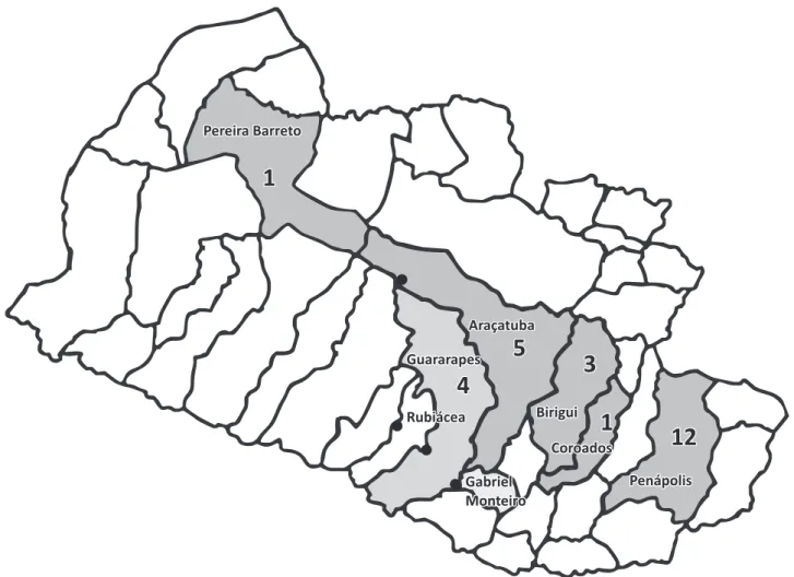
Evidence of rabies virus infection was found in bats from six cities in the Northwestern region (Figure 1). The most cases were recorded in Penápolis (12), followed by Araçatuba (5), Guararapes (4), Birigui (3), Pereira Barreto (1) and Coroados (1). In Guararapes, rabies was found in bats (September and October 2009) and in one head of cattle (April/2009), whereas in Gabriel Monteiro, rabies was only found in two heads of cattle (May and June/2011).
Serum samples from 125 (71%) of the 176 hematophagous bats captured in the region were submitted to simplified
fl uorescent inhibition microtest (SFIMT). From this total, 86 were from a roost in a tree hollow in Araçatuba, 33 were from a tree in Gabriel Monteiro, four were from a tunnel on Marechal Rondon Road in Rubiacea, and two were from an abandoned shed on a farm in Guararapes.
According to the criteria defi ned above, 72% (90/125) of the serum samples were positive, and of these, 39% (35/90) were from males and 61% (55/90) were from females. Low and medium titers (0.10IU/mL to 0.50IU/mL) were observed in 90% (81/90) of the bats, and titers ≥ 0.50IU/mL were observed in 10% (9/90), which translates to a seroprevalence of 7.2% (9/125; Figure 2). Only three samples with titers ≥ 0.50IU/mL were from bats captured in shelters close to properties with confi rmed bovine rabies cases; these bats were captured in Guararapes (1), Rubiácea (1) and Gabriel Monteiro (1). The six remaining samples were from animals captured in a tree in Araçatuba, a city without any recorded bovine rabies cases (Figure 1).
88% of the bats sent for diagnosis from 2008 to 2012. However, bovine cases were also recorded in the region. This pattern of RABV detection has been observed since 1998 in the Northwestern region11,20,21, where rabies in bats, cattle, dogs and cats was diagnosed
in relation to vampire bat variants. However, in these previous studies, the virus was not isolated from the brains of this bat species.
The diffi culty of fi nding hematophagous bats with clinical signs of rabies and/or RABV brain infections has been reported in other studies, even in regions considered epidemic for rabies in cattle. Sugay and Nilsson22 investigated the prevalence of rabies
virus in the Vale do Paraíba region, SP, and reported that only 3.8% of bats were positive for RABV in an epidemic region; this prevalence was considered to be low, given the number of bovine cases in the area. Souza et al.19 observed a similarly low
prevalence of RABV in the Vale do Paraiba region; only 5.1% (7/138) of samples from hematophagous bats captured in caves, all from the same capture site, were positive for RABV.
Rabies virus circulation or the risk of rabies occurrence in vampire bats is usually monitored by active surveillance, i.e., by capturing bats in shelters. The prevalence rate in those cases is usually lower than 1%, considering that apparently healthy 0
5 10 15 20 25 30 35 40 45
<0.06 0.10- 0.19 0.20- 0.49 0.50 Antibody titer (UI/ml)
N
u
m
b
e
r
o
f
a
n
im
a
ls
>
FIGURE 1 - A map of the Araçatuba region showing the counties in which only bat cases of rabies were diagnosed (dark grey), both bat and bovine cases (medium grey) and only bovine cases (light grey). The numbers indicate the numbers of bat cases, and the dots indicate the locations of shelters in which bats with rabies virus neutralizing antibodies were found.
bats are monitored23. Information about the confi rmed Desmodus
rotundus rabies cases describes the bats feeding during the day or being found dead in places with cattle or in caves with a large concentration of individuals, in areas with high numbers of bovine cases19,22. Only one case of a Desmodus rotundus bat
positive for rabies in an urban area has been described in the literature; this bat was found in Ubatuba, State of São Paulo24.
The circulation of rabies virus in non-hematophagous bats was investigated in the present study by passive surveillance in urban areas, and 1.7% (26/1,490) of the samples that were collected were found to be RABV-positive. This detection rate was greater than the 1.3% (98/7393) reported by Cunha et al.20 from 1997 to
2002 for Northwestern State of São Paulo. Considering only the Araçatuba region, the positivity rate found in the present study exceeded the 1.2% (50/4035) found for samples collected from 1993 to 200711 and the 1.1% found in Campo Grande, State of
Mato Grosso do Sul from February to December 200125. In the
present study, the highest positivity rate, 2.3% (9/388) in 2009, did not reach the maximum positivity rate of 3.3% (10/304) recorded in this geographical region, in 200111.
Variations between years are observed when these percentages are compared with those observed for other regions in State of São Paulo. The positivity rate increased in the metropolitan São Paulo region from 0.7% (1988-1992)26 to
0.8% (1993-2003)11, reaching 2.2% and 1.4% in 2002 and 2003,
respectively. However, in other studies, the rabies virus was not detected in bats from urban areas, including Japurá, Paraná27 and
municipalities in Roraima28. Positivity rates ranging from 0 to
0.9% in the same geographical area have also been observed, for example in the Botucatu region, SP14,29,30.
In the present study, 11.5% of rabies virus cases detected during the study period (2008-2012) were in the frugivorous species Artibeus lituratus. This percentage represented a decrease from 30% of rabies virus cases being detected in these bats in the period from 1998 to 200711. The positivity
rate in insectivorous bats from the Vespertilionidae family remained unchanged at 50% (13/26). However, E. furinalis
was the predominant species affected in this family,rather than
M. nigricans,as was observed from 1998 to 2007. The positivity rate for bats of the Molossidae family increased from 20% (1998 to 2007) to 38.5% (2008 to 2012), whereas M. rufus was the predominant RABV-positive species in both periods. Five of the seven positive E. furinalis cases came from the same shelter in Penapolis, with three- to four-month intervals between cases.
All bats with RVNA titers ≥ 0.10IU/mL were considered to have reactive titers (the lowest titer for which fl uorescence inhibition was observed), in contrast with titers ≥ 0.50IU/mL, which are required for a sample to be considered seropositive. This criterion for seropositivity of a titer ≥ 0.50IU/mL was also adopted in a serological surveillance of terrestrial wild mammals31. The positivity rate in our study was 72%;
seropositivity indicates a previous contact with RABV in the colony or a recent disease outbreak15,31, even though no ill
animals were observed. Similar situations have been described in the literature by Souza et al.19 for Vale do Ribeira, which had
a 6.7% positivity rate, and by Langoni et al.14 in Botucatu, in
which 65.2% of all bats examined had reactive antibody titers.
RABV seroprevalence was 7.2% in this study, if only those bats with titers ≥ 0.50IU/mL were considered to be seropositive, as has been reported by some authors. This result is similar to the 7.4% positivity rate found among hematophagous bats14
in Botucatu, State of São Paulo, and higher than the 5.9% seroprevalence rate observed in the metropolitan São Paulo region for bats of a variety of species, including hematophagous bats12.
Serological studies have been conducted in bats and terrestrial wild species in Brazil and other countries as a means of determining the risk of rabies occurrence and the need for RABV surveillance14,31-35. All of these studies detected
the presence of RVNAs; although the OIE recommends the standardization of serological techniques for the detection of RVNAs36, seroprevalence studies conducted using a variety of
methods are still important tools for investigating viral activity levels in wild populations.
No virus was detected in the Desmodus rotundus samples, and the rabies risk in herbivoresin Northwestern São Paulo varied from medium to negligible, according to the municipality37.
However, the high prevalence of RVNA-reactive bats (72%) and the substantial seroprevalence (7.2%) of RVNAs in vampire bats from shelters close to sites with bovine rabies cases indicate that rabies virus infections occur in Desmodus rotundus in the Araçatuba region of Northwestern State of São Paulo.
The detection of RABV in non-hematophagous bats in urban areas demonstrates the roles and importance of these species as transmitters of RABV. The presence of vampire bats with RVNAs indicates recent exposure to RABV antigens or to the virus itself, confi rming that even in the absence of virus detection, seroprevalence studies can provide important epidemiological data on rabies infections in bats and can generate valuable RABV surveillance data for the affected species.
The authors declare that there is no confl ict of interest. CONFLICT OF INTEREST
FINANCIAL SUPPORT ACKNOWLEDGMENTS
To Dr. Vladimir de Souza Nogueira Filho, Coordinator of the State Program of herbivores rabies control (EDA Bauru - CDA-SAA / SP) and vampire bat control teams of CDA for providing the data on shelters of bats registered in Northwestern São Paulo.
1. Acha PN, Szyfres B. Rabia. Zoonosis y enfermedades transmisibles comunes al hombre y a los animales. 3rd ed. Washington:
Organización Panamericana de la Salud. Ofi cina Sanitaria Panamericana, Ofi cina Regional de la Organización Mundial de la Salud; 2003. p. 351-383.
2. Rupprecht CE, Stöhr K, Meredith C. Rabies. In: Williams ES, Barker IK, editors. Infectious disease of wild mammals. Iowa: Iowa State University Press; 2001. p. 3-36.
3. Jackson AC. History of Rabies Research. In: Jackson AC, editor. Rabies: scientifi c basis of the disease and its management. 3rd ed.
San Diego, USA: Acad. Press; 2013. p 1-15.
4. Carini A. Sur une grande épizootie de rage. Annales de I’Institut Pasteur 1911; 25:843-846.
5. Carneiro DVM. Transmission of rabies by bats in Latin America. Bull World Health Org 1954; 10:775-780.
6. Nogueira MR, Lima IP, Moratelli R, Tavares VC, Gregorin R, Peracchi AL. Checklist of Brazilian bats, with comments on original records. Check List 2014; 10:808-821.
7. Sodre MM, Gama AR, Almeida MF. Updated list of bat species positive for rabies in Brazil. Rev Inst Med Trop Sao Paulo 2010; 52:75-81.
8. Organização Panamericana da Saúde, Organização Mundial da Saúde. Sistema de informacão epidemiológica, database 2014, históricos anuais [Internet]. [Cited 2014 April 03]. Available at: http://siepi2.panaftosa.org.br/Export.aspx/.
9. Schneider MC, Romijn PC, Uieda W, Tamayo H, Silva DF, Belotto A, et al. Rabies transmitted by vampire bats to humans: An emerging zoonotic disease in Latin America? Rev Panam Salud Publica 2009; 25:260-269. 10. Caldas EP. Raiva Silvestre no Brasil: situação, riscos, desafi os e
perspectivas de controle. In: 14ª. REDIPRA - Reunião de Diretores de Programas Nacionais de Controle da Raiva na América Latina. Lima, Perú, 2013. [Cited 2014 April 03]. Available at: http://www. paho.org/panaftosa/index.php?option=com_content&view=article &id=799&Itemid=336/.
11. Queiroz LH, Carvalho C, Buso DS, Ferrari CIL, Pedro WA. Perfi l epidemiológico da raiva na região Noroeste do Estado de São Paulo no período de 1993 a 2007. Rev Soc Bras Med Trop 2009; 42:9-14. 12. Almeida MF, Martorelli MFA, Sodré MM, Kataoka APAG, Rosa AR,
Oliveira ML, et al. Diagnóstico e sorologia de raiva em morcegos do Estado de São Paulo, Brasil. Rev Soc Bras Med Trop 2011; 44:140-145. 13. Mayen F. Hematophagous bats in Brazil, their role in rabies
transmission, impact on public health, livestock industry and alternatives to an indiscriminate reduction of bat population. J Vet Med B Infect Dis Vet Public Health 2003; 50:469-472.
14. Langoni H, Souza LC, Zetun CB, Silva TCC, Hofmann JL, Silva RC. Serological survey for rabies in serum samples from vampire bats
(Desmodus rotundus) in Botucatu region, SP, Brazil. J Venom Anim
Toxins Incl Trop Dis 2008; 14:651-659.
15. Megid J. Raiva. In: Cubas ZS, Silva JCR, Catão Dias JL, editors. Tratado de animais selvagens – medicina veterinária. São Paulo: Roca; 2007. p. 785-798
16. Dean DJ, Abelseth MK, Atanasiu P. The fl uorescent antibody test.
In: Meslin FX, Kaplan MM, Koprowski H. Laboratory techniques
in rabies. 4th ed. Geneva Switzerland: World Health Organization;
1996. p. 476.
17. Koprowski H. The mouse inoculation test. In: Meslin FX, Kaplan
MM, Koprowski H. Laboratory techniques in rabies. 4th. ed. Geneva
Switzerland: World Health Organization; 1996. p. 476.
18. Favoretto SR, Carrieri ML, Tino MS, Zanetti CR, Pereira OAC. Simplifi ed Fluorescent Inhibition Microtest for the Rabies Neutralizing Antibodies. Rev Inst Med Trop São Paulo 1993; 35:171-175.
19. Souza MCAM, Bernardi F, Ito FH. Epidemiology of rabies: biological and serological aspects of rabies in vampire bats
Desmodus rotundus (E. Geoffroy) captured in Vale do Paraíba,
southeastern of Brazil. Arq Int Biol São Paulo 1997; 64:91-101.
20. Cunha EMS, Queiroz Da Silva LH, Lara MCCSH, Nassar AFC, Albas A, Sodre MM, et al. Bat rabies in the North-northwestern regions of São Paulo State – Brazil, 1997-2002. Rev Saude Publica 2006; 40:1082-1086.
21. Queiroz LH, Favoretto SR, Cunha EMS, Campos ACA, Lopes MC, Carvalho C, et al. Rabies in southeast Brazil: a change in the epidemiological pattern. Arch Virology 2012; 157:93-105.
22. Sugay W, Nilsson MR. Isolamento do vírus da raiva em morcegos hematófagos do Estado de São Paulo, Brasil. Bol Ofi c Sanit Panamericana 1966; 50:310-315.
23. Kotait I. Infecção de morcegos pelo vírus da raiva. Bol Inst Pasteur (São Paulo) 1996; 1:51-58.
24. Ferraz C, Achkar SM, Kotait I. First report of rabies in vampire bats
(Desmodus rotundus) in an urban area, Ubatuba, São Paulo State,
Brazil. Rev Inst Med Trop São Paulo 2007; 49:389-390.
25. Deus GT, Becer M, Navarro IT. Diagnóstico da raiva em morcegos não hematófagos na cidade de Campo Grande, Mato Grosso de Sul, Centro Oeste do Brasil: descrição de casos. Semina: Cienc Agr, Londrina 2003; 24:171-176.
26. Almeida MF. Diagnóstico laboratorial de raiva em quirópteros realizado em área metropolitana na região sudeste do Brasil. Rev Saude Publica 1994; 28:341-344.
27. Marques MA, Bonani GA, Filho HO. Ocorrência do vírus rábico em morcegos do município de Japurá-PR - um estudo preliminar. Arq Ciênc Vet Zoo 2010; 13:33-36.
28. Cardoso MN, Passos MCV, Melo SAN, Souza JR, Acosta PO. Diagnóstico do vírus da raiva por RT-PCR em morcegos de municípios do estado de Roraima. In: Reunião Regional da Sociedade Brasileira para o Progresso da Ciência, 2010, Boa Vista. Anais. Boa Vista: SBPC, 2010. [Cited: 2014 April 17]. Available at: http://www.sbpcnet.org.br/livro/boavista/resumos/1322.htm/.
29. Cortes VA, Souza LC, Uieda W, Figueiredo AC. Abrigos diurnos e infecção rábica em morcegos de Botucatu, São Paulo, Brasil. Vet Zootec 1994; 6:179-186.
30. Souza LC, Langoni H, Silva RC, Luchesi SB. Vigilância epidemiológica da raiva na região de Botucatu-SP: importância dos quirópteros na manutenção do vírus na natureza. Ars Veterinária 2005; 21:62-68.
31. Araujo DB, Martorelli LA, Kataoka APGA, Campos ACA, Rodrigues CS, Sanfi lippo LF, et al. Antibodies against rabies virus in terrestrial wild mammals in native rainforest on the north coast of São Paulo State, Brazil. J Wild Dis 2014; 50:469-477.
32. Almeida MF, Massad E, Aguiar EAC, Martorelli LFA, Joppert AMS. Neutralizing antirabies antibodies in urban terrestrial wildlife in Brazil. J Wildl Dis 2001; 37:394-398.
34. Turmelle AS, Allen LC, Jackson FR, Kunz TH, Rupprecht CE, Mccranken GF. Ecology of rabies virus exposure in colonies of Brazilian free-tailed bats (Tadarida brasiliensis) at natural and man-made roosts in Texas. Vector-borne Zoon Dis 2010; 10:165-175.
35. Bowen RA, O’shea TJ, Shankar V, Neubaum MA, Neubaum DJ, Rupprecht CE. Prevalence of neutralizing antibodies to rabies virus in serum of seven species of insectivorous bats from Colorado and New Mexico, United States. J Wildl Dis 2013; 49:367-374.
36. World Organization for Animal Health (OIE). Rabies. In: Manual of diagnostic tests and vaccines for terrestrial animals. OIE: 2013 [Cited: 2014 May 22]. Available at: http://www.oie.int/fi leadmin/ Home/eng/Health_standards/tahm/2.01.13_RABIES.pdf. 37. Braga GB, Grisi-Filho JHH, Leite BM, Sena EF, Dias RA. Predictive