INTRODUCTION
Address to: Dra. Jacqueline Pontes Monteiro. Depto. Puericultura e Pediatria/
FMRP/USP. Av. Bandeirantes 3900, Campus USP, 14049-900 Ribeirão Preto, SP, Brasil.
Phone: 55 16 3602-2477; Fax: 55 16 3602-2700 e-mail: [email protected]
Received 13 October 2014 Accepted 18 November 2014
Vitamin A, vitamin E, iron and zinc status in a cohort
of HIV-infected mothers and their uninfected infants
Jacqueline Pontes Monteiro
[1],
Maria Letícia Santos Cruz
[2],
Marisa Márcia Mussi-Pinhata
[1],
Roberta Garcia Salomão
[1],
Alceu Jordão Junior
[3],
Laura Freimanis Hance
[4],
Jennifer Suzanne Read
[5],[6],
José Henrique da Silva Pilotto
[7],[8],
Rachel Ann Cohen
[4],
Sonia Karolina Stoszek
[4]and
George Kelly Siberry
[5][1]. Departamento de Puericultura e Pediatria, Faculdade de Medicina de Ribeirão Preto, Universidade de São Paulo, Ribeirão Preto, SP, Brasil. [2]. Departamento de Doenças Infecciosas, Hospital Federal dos Servidores do Estado, Rio de Janeiro, RJ, Brasil. [3]. Departamento de Clínica Médica, Faculdade de Medicina de Ribeirão Preto, Universidade de São Paulo, São Paulo, SP, Brasil. [4]. Westat, Rockville, Maryland, USA. [5]. Maternal and Pediatric Infectious Disease Branch, National Institute of Child Health and Human Development, National Institute of Health, Bethesda, Maryland, USA. [6].Division of Infectious Diseases, Department of Pediatrics, The George Washington University School of Medicine, Washington, District of Columbia, USA. [7]. Departamento de Doenças Infecciosas, Hospital Geral de Nova Iguaçu, Nova Iguaçu, RJ, Brasil. [8]. Laboratório de AIDS e Imunologia Molecular, Instituto Oswaldo Cruz, Fundação Oswaldo Cruz, Rio de Janeiro, RJ, Brasil.
ABSTRACT
Introduction: We hypothesized that nutritional defi ciency would be common in a cohort of postpartum, human immunodefi ciency
virus (HIV)-infected women and their infants. Methods: Weight and height, as well as blood concentrations of retinol,
α-tocopherol, ferritin, hemoglobin, and zinc, were measured in mothers after delivery and in their infants at birth and at 6-12 weeks and six months of age. Retinol and α-tocopherol levels were quantifi ed by high performance liquid chromatography, and zinc levels were measured by atomic absorption spectrophotometry. The maternal body mass index during pregnancy was
adjusted for gestational age (adjBMI). Results: Among the 97 women 19.6% were underweight. Laboratory abnormalities were
most frequently observed for the hemoglobin (46.4%), zinc (41.1%), retinol (12.5%) and ferritin (6.5%) levels. Five percent of the women had mean corpuscular hemoglobin concentrations < 31g/dL. The most common defi ciency in the infants was α-tocopherol (81%) at birth; however, only 18.5% of infants had defi cient levels at six months of age. Large percentages of infants had zinc (36.8%) and retinol (29.5%) defi ciencies at birth; however, these percentages decreased to 17.5% and 18.5%, respectively, by six months of age. No associations between infant micronutrient defi ciencies and either the maternal adjBMI category or maternal micronutrient defi ciencies were found. Conclusions: Micronutrient defi ciencies were common in HIV-infected women and their infants. Micronutrient defi ciencies were less prevalent in the infants at six months of age. Neither underweight women nor their infants at birth were at increased risk for micronutrient defi ciencies.
Keywords: Micronutrients. HIV infection. Pregnancy. Infant. Nutrition. Cohort.
Maternal malnutrition is associated with increased risks
of infant morbidity and mortality1. Multiple micronutrient
defi ciencies may develop in pregnant women early during human immunodefi ciency virus type 1 (HIV-1) infection2,3.
Zinc defi ciency, which is common in HIV-infected individuals,
has been associated with higher mortality and viral load4,5.
Zinc supplementation during pregnancy results in improved neonatal immune status, reduced early neonatal morbidity
and fewer infant infections6. Pregnancy increases the risk of
vitamin A defi ciency in mothers and newborns. Randomized trials of vitamin A supplementation in pregnant women found decreased mortality and a reduced prevalence and duration of
infectious episodes in infants7. In a Brazilian study, maternal
vitamin A defi ciency was strongly associated with infant vitamin A defi ciency and low birth weight8. Brazilian HIV-infected
pregnant women may have low vitamin A levels even after
supplementation9.
HIV infection and pregnancy are accompanied by oxidative stress. Lower levels of vitamin E may play a pathogenic role in the onset and development of acquired immunodefi ciency
syndrome (AIDS) and other infectious diseases10,11. Several
studies have revealed lower vitamin E levels in HIV-infected
children in Brazil and elsewhere12-14.Among healthy pregnant
women, infant birth weight and length are associated with
maternal serum concentrations of antioxidant vitamins15.
Anemia is the most frequent hematologic abnormality found in
METHODS
women in Latin America includes iron and folate supplements to prevent anemia and neural tube defects.
We hypothesized that nutritional defi ciency is frequent among HIV-infected mothers and their infants in Brazil. Our
objectiveswere as follows: 1) to describe the nutritional status
of HIV-infected women at delivery and of their infants at sites in Brazil based on anthropometric measurements, including the maternal adjusted for gestational age (adjBMI), and on plasma micronutrient concentrations; 2) to assess the correlation between maternal and infant micronutrient levels at delivery; and 3) to evaluate changes in the micronutrient status of infants from birth to six months of age.
The LILAC study
HIV-infected pregnant women enrolled at three participating
Brazilian sites in the Eunice Kennedy Shriver National Institute of
Child Health and Human Development (NICHD), International Site Development Initiative (NISDI) and Longitudinal Study in Latin American Countries (LILAC) dynamic cohort were invited to participate in this nutritional LILAC sub-study. The design
and conduct of the LILAC study has been described previously17.
Nutritional Sub-study
This nutritional sub-study enrolled LILAC participants at three study sites in Brazil. The protocol for this sub-study was approved by the Institutional Review Boards (IRBs) of the participating institutions. This sub-study required the collection of additional blood from mothers (within seven days of delivery) and infants (at birth and then at the 6-12-week and 6-month study visits) for the analysis of micronutrient levels (retinol, α-tocopherol, ferritin, and zinc). The blood samples were stored at -70°C in the dark until processing. Plasma retinol and α-tocopherol concentrations were quantifi ed by high performance liquid chromatography
(HPLC) as previously described18. Serum zinc was determined by
atomic absorption spectrophotometry using standard procedures19.
Maternal C-reactive protein (CRP) levels were quantifi ed to characterize infl ammation. CRP and ferritin were measured in plasma samples using immunoassay technology (Immulite 1,000 Immunoassay System - Global Siemens Healthcare Headquarters - Siemens AG; Henkestrasse 127; D-91052 Erlangen - Germany). The maternal body mass index (BMI) at the last study visit during pregnancy was adjusted for gestational age (adjBMI) using a computer algorithm provided by the Ministry of
Health of Argentina20 and was used as a criteria for nutritional
status classifi cation. Maternal adjBMI values that were <19.8 were considered underweight, ≥26.1 to <29 were considered overweight, and ≥29 to <50 were classifi ed as obese. Maternal and infant micronutrient status was defi ned at each time point as deficient vs. normal for each micronutrient measured. Defi ciencies were defi ned as follows for each micronutrient:
zinc level, <64mg/dL for infants and <50mg/dL for mothers21;
retinol level, <0.7mmol/L for both infants and mothers22;
α-tocopherol level, <12mmol/L for infants and <7mmol/L for
mothers23; and ferritin level, <10ng/mL for mothers, <25ng/mL
for infants less than one month of age, <200ng/mL for infants one to two months of age, <50ng/mL for infants aged two months to less than six months, and <7ng/mL for ages six months to
15 years24. The cut-off values for C-reactive protein (CRP)
categories in pregnant women were as follows: low, < 1mg/dL;
normal, 1-3mg/dL; high, >3mg/dL25. Infant CRP cut-off values
were as follows: normal, ≤0.5mg/dL; high, >0.5mg/dL26. HIV
disease was classifi ed according to the Centers for Disease Control and Prevention (CDC) defi nitions27. A low mean corpuscular
hemoglobin concentration (MCHC) level was defi ned as <31g/
dL for pregnant women and for infants24. The cut-off values
for hemoglobin in pregnant women were as follows: severe deficiency, 6.5-7.4g/dL; low, 7.5-8.4g/dL; mild deficiency,
8.5-10g/dL; and normal, >10g/dL28. The normal hemoglobin range
for infants was adjusted for age: one to three days, 14.5-18.5g/ dL; two weeks, 13.4-16.6g/dL; one month, 10.7-13.9g/dL; two
months, 9.4-11g/dL; and from two to six months, 11.1-12.6g/dL28.
Maternal antiretroviral (ARV) regimens were categorized in order of regimen complexity as follows: 1) one or two nucleoside reverse transcriptase inhibitors (NRTIs), 2) two NRTIs and one non-nucleoside reverse transcriptase inhibitor (NNRTI), 3) two NRTIs and one protease inhibitor (PI), and 4) other. The reason for the use of ARVs during pregnancy was categorized as treatment if ARVs were used before pregnancy and/or continued after the 6-12-week study visit, or as prophylaxis if ARVs were initiated during pregnancy and use was discontinued by the 6-12-week visit.
Statistical analysis
Associations between maternal adjBMI values and the following characteristics of mothers and infants were examined: maternal age at enrollment, maternal years of formal education, number of persons in the household, mother’s gainful employment outside the home, substance use during pregnancy, reasons for ARV use during pregnancy, maternal
ARV regimen, maternal cluster of differentiation 4 (CD4) +
T-lymphocyte (CD4) count, plasma Human immunodefi ciency virus-ribonucleic acid (HIV-RNA) concentration [viral load(VL)], CDC clinical HIV disease classifi cation27, gestational
hypertension, gestational diabetes, preterm birth (<37 weeks of
gestational age)1, low infant birth weight (<2,500g)1, infant ARV
RESULTS
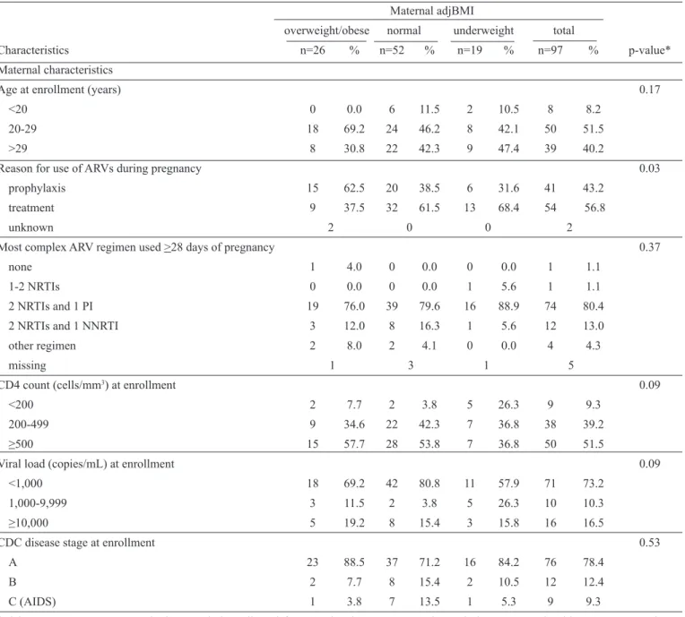
TABLE1 - Characteristics of 97 mother-infant pairs by maternal adjBMI.
Maternal adjBMI
overweight/obese normal underweight total
Characteristics n=26 % n=52 % n=19 % n=97 % p-value*
Maternal characteristics
Age at enrollment (years) 0.17
<20 0 0.0 6 11.5 2 10.5 8 8.2
20-29 18 69.2 24 46.2 8 42.1 50 51.5
>29 8 30.8 22 42.3 9 47.4 39 40.2
Reason for use of ARVs during pregnancy 0.03
prophylaxis 15 62.5 20 38.5 6 31.6 41 43.2
treatment 9 37.5 32 61.5 13 68.4 54 56.8
unknown 2 0 0 2
Most complex ARV regimen used >28 days of pregnancy 0.37
none 1 4.0 0 0.0 0 0.0 1 1.1
1-2 NRTIs 0 0.0 0 0.0 1 5.6 1 1.1
2 NRTIs and 1 PI 19 76.0 39 79.6 16 88.9 74 80.4
2 NRTIs and 1 NNRTI 3 12.0 8 16.3 1 5.6 12 13.0
other regimen 2 8.0 2 4.1 0 0.0 4 4.3
missing 1 3 1 5
CD4 count (cells/mm3) at enrollment 0.09
<200 2 7.7 2 3.8 5 26.3 9 9.3
200-499 9 34.6 22 42.3 7 36.8 38 39.2
≥500 15 57.7 28 53.8 7 36.8 50 51.5
Viral load (copies/mL) at enrollment 0.09
<1,000 18 69.2 42 80.8 11 57.9 71 73.2
1,000-9,999 3 11.5 2 3.8 5 26.3 10 10.3
≥10,000 5 19.2 8 15.4 3 15.8 16 16.5
CDC disease stage at enrollment 0.53
A 23 88.5 37 71.2 16 84.2 76 78.4
B 2 7.7 8 15.4 2 10.5 12 12.4
C (AIDS) 1 3.8 7 13.5 1 5.3 9 9.3
*Fisher’s exact test. adjBMI: body mass index adjusted for gestational age; ARV: antiretroviral; NRTI: nucleoside reverse transcriptase inhibitors; PI: protease inhibitor; NNRTI: non-nucleoside reverse transcriptase inhibitor; CD4: cluster of differentiation 4; CDC: Centers for Disease Control and Prevention.
and infant nutritional variables at the study time points. All p-values were two-sided. The p-values<0.05 were considered statistically signifi cant. Analyses were conducted using SAS version 9.3 statistical software (SAS Institute, Cary, NC, USA).
In total, 123 women that were enrolled in the LILAC protocol at the participating sites provided informed consent to participate in this sub-study, and blood samples were available for 97 mother-infant pairs. Thus, the study population was
composed of 97 mother-infant pairs. All infants were HIV-uninfected.
The characteristics of the 97 mother-infant pairs overall and according to maternal adjBMI at the last study visit
during pregnancy are shown in Table 1. Based on the adjBMI
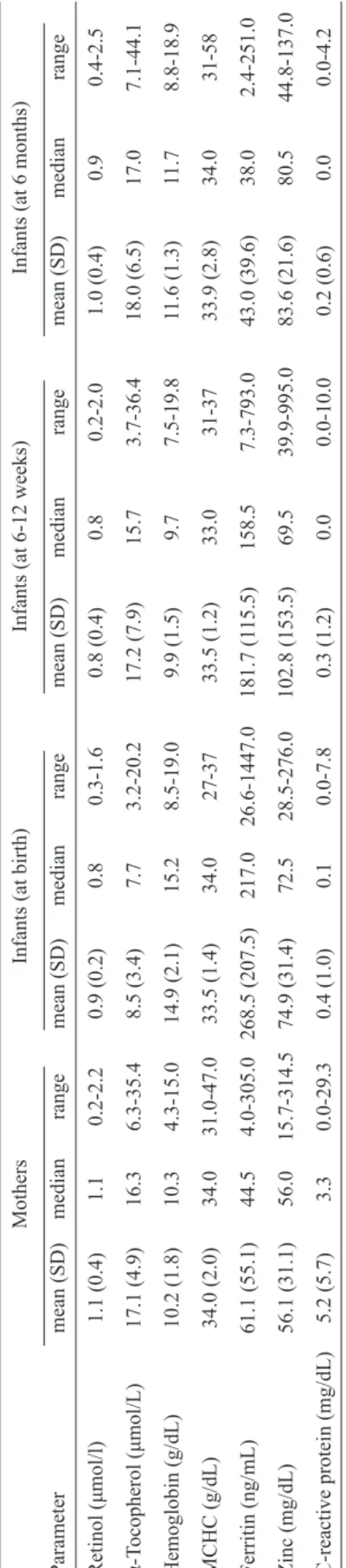
TABLE 2 - Nutritional and infl
ammatory parameters (as continuous variables) among women (after
deliv
ery) and infants (at study visits).
Mothers
Infants (at birth)
Infants (at 6-12 weeks)
Infants (at 6 months)
Parameter mean (SD) median range mean (SD) median range mean (SD) median range mean (SD) median range Retinol (μmol/l) 1.1 (0.4) 1.1 0.2-2.2 0.9 (0.2) 0.8 0.3-1.6 0.8 (0.4) 0.8 0.2-2.0 1.0 (0.4) 0.9 0.4-2.5 α-T ocopherol (μmol/L) 17.1 (4.9) 16.3 6.3-35.4 8.5 (3.4) 7.7 3.2-20.2 17.2 (7.9) 15.7 3.7-36.4 18.0 (6.5) 17.0 7. 1-44.1 Hemoglobin (g/dL) 10.2 (1.8) 10.3 4.3-15.0 14.9 (2.1) 15.2 8.5-19.0 9.9 (1.5) 9.7 7.5-19.8 1 1.6 (1.3) 1 1.7 8.8-18.9 MCHC (g/dL) 34.0 (2.0) 34.0 31.0-47.0 33.5 (1.4) 34.0 27-37 33.5 (1.2) 33.0 31-37 33.9 (2.8) 34.0 31-58 Ferritin (ng/mL) 61.1 (55.1) 44.5 4.0-305.0 268.5 (207.5) 217.0 26.6-1447.0 181.7 (1 15.5) 158.5 7.3-793.0 43.0 (39.6) 38.0 2.4-251.0 Zinc (mg/dL) 56.1 (31.1) 56.0 15.7-314.5 74.9 (31.4) 72.5 28.5-276.0 102.8 (153.5) 69.5 39.9-995.0 83.6 (21.6) 80.5 44.8-137.0
C-reactive protein (mg/dL)
5.2 (5.7) 3.3 0.0-29.3 0.4 (1.0) 0.1 0.0-7.8 0.3 (1.2) 0.0 0.0-10.0 0.2 (0.6) 0.0 0.0-4.2 MCHC:
mean corpuscular hemoglobin concentration.
SD:
standard deviation.
pregnancy compared with normal weight or underweight women. No associations were observed between maternal adjBMI and years of education (p-value=0.45), the number of persons in the household (p-value=0.41), gainful employment outside the home (p-value=0.54), tobacco use during pregnancy (p-value=0.74), alcohol use during pregnancy (p-value=0.71), cocaine use during pregnancy (p-value=0.42) or marijuana use during pregnancy (p-value=0.42). Additionally, maternal adjBMI was not associated with preterm birth (p-value=0.77), low birth weight (p-value=0.28) or infant CD4 count at birth (p-value=0.59). The median gestational age at the time of adjBMI assessment was 33 weeks (minimum, 25 weeks; maximum, 39 weeks). Underweight women were signifi cantly more likely to have low MCHC after delivery than their normal and overweight counterparts (p-value<0.01; data not shown).
Nutritional and infl ammatory marker data for the mothers and infants are summarized [mean, standard deviation (SD)],
median, and range) in Table 2. The test results are categorized as
normal versus defi cient in Table 3. The most frequent maternal defi ciencies were zinc (41.1%), followed by retinol (12.5%), ferritin (6.5%) and α-tocopherol (1%) (Table 3). Additionally, 46.4% of the women had low hemoglobin levels, and the MCHC was <31g/dL in 5.2% of the women. Approximately 54% of the women had high levels of CRP in their blood at hospital discharge following delivery. At birth, 81.1% of the infants were defi cient in α-tocopherol, with the proportion decreasing with age to only 18.5% by six months of age. High proportions of neonates also had defi cient levels of zinc (36.8%) and retinol (29.5%) at birth, and these numbers fell to only 17.5% and 18.5%, respectively, by six months of age. The proportion of infants with low levels of retinol increased to 41.1% by 6-12 weeks of age before decreasing by six months of age. Although none of the infants had low ferritin levels at birth, a sharp increase was observed in the proportion of infants with low ferritin levels at 6-12 weeks of age (58.7%) and at 6 months of age (25.8%). Additionally, low hemoglobin levels were observed in 43.8% and 7.5% of the infants at 6-12 weeks of age and at 6 months of age, respectively. All infants were formula-fed after birth.
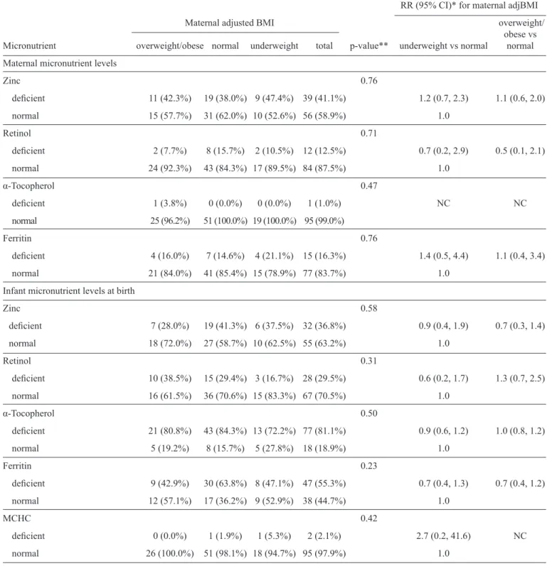
When we compared the underweight adjBMI category with the normal adjBMI category, no association was found between maternal underweight adjBMI during pregnancy and maternal zinc (RR=1.2, 95% CI: 0.7, 2.3), retinol (RR=0.7, 95% CI: 0.2, 2.9), α-tocopherol (RR not calculable), or ferritin (RR=1.4, 95% CI: 0.5, 4.4) defi ciency at delivery, nor was any observed association found between maternal underweight adjBMI during pregnancy and infant zinc (RR=0.9, 95% CI: 0.4, 1.9), retinol (RR=0.6, 95% CI: 0.2, 1.7), α-tocopherol (RR=0.9, 95% CI: 0.6, 1.2), ferritin (RR=0.7, 95% CI: 0.4, 1.3),
or MCHC at birth (RR=2.7, 95% CI: 0.2, 41.6; Table 4). In
TABLE 3 - Nutritional and infl ammatory parameters (as categorical variables) among women (after delivery) and infants (at study visits).
Infants
Category Mothers at birth 6-12 weeks 6 months
Parameter n=97 % n=97 % n=97 % n=97 %
Zinc(mg/dL) normal 56 59.0 55 63.2 34 75.6 33 82.5
defi cient 39 41.1 32 36.8 11 24.4 7 17.5
missing 2 10 52 57
Retinol(μmol/L) normal 84 87.5 67 70.5 56 59.0 75 81.5
defi cient 12 12.5 28 29.5 39 41.1 17 18.5
missing 1 2 2 5
α-Tocopherol(μmol/L) normal 95 99.0 18 19.0 70 73.7 75 81.5
defi cient 1 1.0 77 81.1 25 26.3 17 18.5
missing 1 2 2 5
Ferritin(ng/mL) low 6 6.5 0 54 58.7 16 25.8
normal 77 83.7 38 44.7 34 37.0 45 72.6
high 9 9.8 47 55.3 4 4.4 1 1.6
missing 5 12 5 35
MCHC(g/dL) missing 0 0 1 4
normal 92 94.9 93 95.9 92 95.8 90 96.8
defi cient 5 5.2 4 4.1 4 4.2 3 3.2
Age-adjusted hemoglobin(g/dL) normal 52 53.6 77 79.4 54 56.3 86 92.5
low 45 46.4 20 20.6 42 43.8 7 7.5
missing 0 0 1 4
C-reactive protein(mg/dL) low 18 18.6 0 0.0 0 0.0 0 0.0
normal 27 27.8 77 81.1 83 90.2 79 86.8
high 52 53.6 18 18.9 9 9.8 12 13.2
missing 0 2 5 6
MCHC: mean corpuscular hemoglobin concentration.
acquired immunodefi ciency syndrome (AIDS) were less likely to have normal retinol levels than women with asymptomatic or
mildly symptomatic HIV disease (RR=0.2, 95% CI: 0.1, 0.6;data
not shown). Low maternal hemoglobin was associated only with low maternal CD4+ T-lymphocyte counts at enrollment. Women
who had CD4 cell counts that were <200 cells/mm3 were less
likely to have normal hemoglobin levels than were women with CD4+ T-lymphocyte counts that were ≥ 500 cells/mm3(RR=0.4,
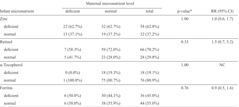
95% CI: 0.3, 0.7; data not shown). Low maternal zinc, ferritin, and hemoglobin were not associated with elevated maternal CRP (p-value>0.8). No observed associations were found between low maternal levels of zinc (RR=1.0, 95%CI: 0.6, 1.7), retinol (RR=1.5, 95%CI: 0.7, 3.2), α-tocopherol (RR not calculable), or ferritin (RR=0.9, 95%CI: 0.5, 1.6) after delivery and corresponding low
levels of these micronutrients in the infants at birth (p-value>0.3;
Table 5). Additionally, linear regression analysis did not reveal
any signifi cant associations between maternal and infant zinc, retinol, α-tocopherol, or ferritin levels at birth when the levels were examined on a continuous scale (p-value>0.15).
TABLE 4 - Association between maternal adjusted BMI (adjBMI) and micronutrient levels.
RR (95% CI)* for maternal adjBMI
Maternal adjusted BMI overweight/ obese vs Micronutrient overweight/obese normal underweight total p-value** underweight vs normal normal
Maternal micronutrient levels
Zinc 0.76
defi cient 11 (42.3%) 19 (38.0%) 9 (47.4%) 39 (41.1%) 1.2 (0.7, 2.3) 1.1 (0.6, 2.0)
normal 15 (57.7%) 31 (62.0%) 10 (52.6%) 56 (58.9%) 1.0
Retinol 0.71
defi cient 2 (7.7%) 8 (15.7%) 2 (10.5%) 12 (12.5%) 0.7 (0.2, 2.9) 0.5 (0.1, 2.1)
normal 24 (92.3%) 43 (84.3%) 17 (89.5%) 84 (87.5%) 1.0
α-Tocopherol 0.47
defi cient 1 (3.8%) 0 (0.0%) 0 (0.0%) 1 (1.0%) NC NC
normal 25 (96.2%) 51 (100.0%) 19 (100.0%) 95 (99.0%)
Ferritin 0.76
defi cient 4 (16.0%) 7 (14.6%) 4 (21.1%) 15 (16.3%) 1.4 (0.5, 4.4) 1.1 (0.4, 3.4)
normal 21 (84.0%) 41 (85.4%) 15 (78.9%) 77 (83.7%) 1.0
Infant micronutrient levels at birth
Zinc 0.58
defi cient 7 (28.0%) 19 (41.3%) 6 (37.5%) 32 (36.8%) 0.9 (0.4, 1.9) 0.7 (0.3, 1.4)
normal 18 (72.0%) 27 (58.7%) 10 (62.5%) 55 (63.2%) 1.0
Retinol 0.31
defi cient 10 (38.5%) 15 (29.4%) 3 (16.7%) 28 (29.5%) 0.6 (0.2, 1.7) 1.3 (0.7, 2.5)
normal 16 (61.5%) 36 (70.6%) 15 (83.3%) 67 (70.5%) 1.0
α-Tocopherol 0.50
defi cient 21 (80.8%) 43 (84.3%) 13 (72.2%) 77 (81.1%) 0.9 (0.6, 1.2) 1.0 (0.8, 1.2)
normal 5 (19.2%) 8 (15.7%) 5 (27.8%) 18 (18.9%) 1.0
Ferritin 0.23
defi cient 9 (42.9%) 30 (63.8%) 8 (47.1%) 47 (55.3%) 0.7 (0.4, 1.3) 0.7 (0.4, 1.2)
normal 12 (57.1%) 17 (36.2%) 9 (52.9%) 38 (44.7%) 1.0
MCHC 0.42
defi cient 0 (0.0%) 1 (1.9%) 1 (5.3%) 2 (2.1%) 2.7 (0.2, 41.6) NC
normal 26 (100.0%) 51 (98.1%) 18 (94.7%) 95 (97.9%) 1.0
*The relative risks (RRs) and 95% confi dence intervals (CIs) for predicting maternal and infant micronutrient defi ciencies as a function
of maternal adjBMI were obtained using log binomial regression models.**p-values were obtained using the Fisher-Freeman-Halton test.
TABLE 5 - Association between maternal and infant micronutrient levels.
Maternal micronutrient level
Infant micronutrient defi cient normal total p-value* RR (95% CI)
Zinc 1.00 1.0 (0.6, 1.7)
defi cient 22 (62.7%) 32 (62.7%) 54 (62.8%)
normal 13 (37.1%) 19 (37.2%) 32 (37.2%)
Retinol 0.33 1.5 (0.7, 3.2)
defi cient 7 (58.3%) 59 (72.0%) 66 (70.2%)
normal 5 (41.7%) 23 (28.0%) 28 (29.8%)
α-Tocopherol 1.00 NC
defi cient 0 (0.0%) 18 (19.3%) 18 (19.1%)
normal 1 (100.0%) 75 (80.7%) 76 (80.9%)
Ferritin 0.76 0.9 (0.5, 1.6)
defi cient 6 (50.0%) 30 (44.1%) 36 (45.0%)
normal 6 (50.0%) 38 (55.9%) 44 (55.0%)
*The p-values were obtained using Fisher’s exact test. NC: value could not be calculated. RR: relative risk; CI: Confi dence interval.
DISCUSSION
Micronutrient defi ciencies are common in HIV-infected pregnant women and their infants in Brazil. The prevalences of most infant micronutrient defi ciencies were lower at six months of age than at birth. Being underweight at the end of pregnancy was not associated with micronutrient defi ciency in HIV-infected women or their infants at birth.
We found that 46.4% of HIV-infected mothers were anemic and that 6.5% had low levels of ferritin immediately postpartum. Few studies have reported iron defi ciency in HIV-infected pregnant women in Latin America; however, some studies have reported an extremely high prevalence of anemia
in HIV-infected pregnant women in other countries28; 78% of
HIV-infected pregnant women in Burkina Faso were reported
to be anemic29, and 83% of HIV-infected pregnant women in
Cote d’Ivoire were reported to be anemic30.
Factors such as poor nutritional status, HIV infection31, ARV
therapy32, and even vitamin D defi ciency33 could explain the high
prevalence of anemia found in the present sub-study. However,
the acute phase response34 was not associated with anemia. Few
studies have examined lipid-soluble vitamins in HIV-infected
pregnant women35–37. In Tanzania38, approximately 35 and 51%
of HIV-infected pregnant women had low levels of vitamins A and E, respectively. In contrast, only 12.5 and 1% of women in our sub-study had vitamin A and E defi ciencies, respectively, although our cut-off for vitamin E defi ciency was lower than that
used in Tanzania38 (vitamin E level<9.7 μmol/L).Zinc defi ciency
has been reported in children39 and in HIV-infected individuals5,40
and is thought to be widely prevalent in pregnant women due to hemodilution, decreased albumin levels and insuffi cient intake41-43.
Infants in our sub-study had lower rates of micronutrient defi ciencies and higher mean levels of vitamin A and vitamin E
than those found by Monteiro et al. in a group of HIV-infected
Latin American children14. Nevertheless, 81% of infants at
birth were defi cient in α-tocopherol, which could partially be explained by increased oxidative stress during delivery. Increased levels of oxidative stress and lipid peroxidation induced by reactive oxygen species (ROS) production play a critical role in the stimulation of HIV replication and immunodefi ciency and can be attenuated by the presence of
vitamin E44. However, vitamin E may facilitate HIV entry into
cells, and higher plasma vitamin E levels have been associated
with adverse outcomes in HIV-infected individuals45. Thus,
lower vitamin E levels may constitute a physiological defense mechanism against HIV in infants during delivery.
A high proportion of infants (41%) had vitamin A defi ciency at 6 to 12 weeks, with mean and SD values of 0.8 and 0.4μmol/L, respectively. Chatterjee A et al. also found vitamin A defi ciency at six weeks of age; The percentages of infants with plasma vitamin A concentrations less than 0.70mmol/L or 0.35mmol/L
were 89% and 22%, respectively, in this sub-study46. These
authors found that higher vitamin A concentrations in plasma at six weeks of age were protective against mortality in children born to HIV-infected women.
Our sub-study had several limitations. Vitamin A defi ciency in this sub-study was defi ned based on WHO recommendations,
which are widely accepted and used28; however, no consensus
represent nutritional defi ciencies during pregnancy. We did not measure serum lipoprotein and cholesterol, which may be important because serum α-tocopherol (vitamin E) concentrations
are highly dependent on serum lipid concentrations2,3. Likewise,
this sub-study did not account for the dietary intake of vitamin A, vitamin E, iron or zinc. All infants were formula-fed and thus received at least some supplementation after birth, although the exact amounts of supplementation are not known. Moreover, the interpretation of serum micronutrient levels is not straightforward due to the wide range of unknown nutrient-nutrient and gene-nutrient-nutrient interactions and physiological and environmental processes involved.
The present sub-study suggests that low serum micronutrient levels are frequently found in HIV-infected pregnant women and their infants and that neither underweight women nor their infants at birth were at increased risk for low serum micronutrient levels. Future studies in different countries that have appropriate designs and that examine genetic and proteomic profi les should be pursued to gain a greater understanding of the interactions among genes, nutrients, and the environment and of micronutrient requirements in infected and
HIV-exposed individuals47–50.
ACKNOWLEDGMENTS
CONFLICT OF INTEREST
FINANCIAL SUPPORT
REFERENCES
We would like to acknowledge all the authors and coauthors, International Site Development Initiative (NISDI) Longitudinal Study in Latin American Countries (LILAC) Study Group and National Institute of Child Health and Human Development (NICHD) for their contribution to this research.
The authors have no competing interests to declare. The fi ndings and conclusions in this report are those of the authors and do not necessarily represent the views of the US National Institutes of Health or the US Department of Health and Human Services.
National Institute of Child Health and Human Development (NICHD) for fi nancial support (Contract # N01-HD-3-3345, 2002-2007, and Contract # HHSN267200800001C, Control # N01-HD-8-0001, 2007-2012).
1. Maternal anthropometry and pregnancy outcomes. A WHO Collaborative Study. Bull World Health Organ 1995; 73 (suppl): S1-S98.
2. Black RE. Micronutrients in pregnancy. (Micronutrients, maternal and child health). Br J Nutr 2001; 85 (suppl):S193-S197.
3. Friis H, Gomo E, Kostel P, Ndhlovu P, Nyazema N, Krarup H, et al. HIV and other predictors of serum beta-carotene and retinol in pregnancy: A cross-sectional study in Zimbabwe. Am J Clin Nutr 2001; 73:1058-1065.
4. Lai H, Lai S, Shor-Posner G, Ma F, Trapido E, Baum MK. Plasma zinc, copper, copper:zinc ratio, and survival in a cohort of
HIV-1-infected homosexual men. J Acquir Immune Defi c Syndr 2001;
27:56-62.
5. Jones CY, Tang AM, Forrester JE, Huang J, Hendricks KM, Knox TA, et al. Micronutrient levels and HIV disease status in HIV-infected patients on highly active antiretroviral therapy in the Nutrition for
Healthy Living cohort. J Acquir Immune Defi c Syndr 2006; 43:
475-482.
6. Wieringa FT, Dijkhuizen MA, Muhilal, Van der Meer JWM.
Maternal micronutrient supplementation with zinc and β-carotene affects morbidity and immune function of infants during the fi rst 6
months of life. Eur J Clin Nutr 2010; 64:1072-1079.
7. Tomkins A. Malnutrition, morbidity and mortality in children and their mothers. Proc Nutr Soc 2000; 59:135-146.
8. Ramalho RA, Flores H, Accioly E, Saunders C. Association between maternal and newborn vitamin A status and economic stratum in Rio de Janeiro, Brazil. Rev Assoc Med Bras 2006; 52:170-175. 9. El Beitune P, Duarte G, Vannucchi H, Quintana SM,
Figueiró-Filho EA, Morais EN, et al. Serum vitamin A during pregnancy and effects on obstetrics and perinatal outcomes in HIV infected pregnant women. Arch Latinoam Nutr 2004; 54:419-427.
10. Allard JP, Aghdassi E, Chau J, Tam C, Kovacs CM, Salit EM, et al. Effects of vitamin E and C supplementation on oxidative stress and viral load in HIV-infected subjects. AIDS 1998; 12:1653-1659. 11. Lee BE, Hong YC, Lee KH, Kim YJ, Kim WK, Chang NS, et al.
Infl uence of maternal serum levels of vitamins C and E during the
second trimester on birth weight and length. Eur J Clin Nutr 2004; 58:1365-1371.
12. Passi S, Morrone A, Picardo M, De Luca C, Bartoli F, Zurlo A, et al. Blood levels of vitamin E, polyunsaturated fatty acids of phospholipids, lipoperoxides, glutathione peroxidase activity and serological screening for syphilis and HIV in immigrants from developing countries. G Ital Dermatol Venereol 1990; 125: 487-491. 13. Tang J, Faustman C, Hoagland TA, Mancini RA, Seyfert M,
Hunt MC. Interactions between mitochondrial lipid oxidation and oxymyoglobin oxidation and the effects of vitamin E. J Agric Food Chem 2005; 53:6073-6079.
14. Monteiro JP, Freimanis-Hance L, Faria LB, Mussi-Pinhata MM,
Korelitz J, Vannucchi H, et al. Both human immunodefi ciency virus-infected and human immunodefi ciency virus-exposed, unvirus-infected
children living in Brazil, Argentina, and Mexico have similar rates of low concentrations of retinol, beta-carotene, and vitamin E. Nutr Res 2009; 29:716-722.
15. Berti C, Biesalski HK, Gärtner R, Lapillonne A, Pietrzik K, Poston L, et al. Micronutrients in pregnancy: Current knowledge and unresolved questions. Clin Nutr 2011; 30: 689-701.
16. Levine AM, Berhane K, Masri-Lavine L, Sanchez M, Young M, Augenbraum M, et al. Prevalence and correlates of anemia in a large cohort of HIV-infected women: Women’s Interagency HIV Study.
J Acquir Immune Defi c Syndr 2001; 26:28-35.
17. Read JS, Duarte G, Hance LF, Pinto J, Gouvea MI, Cohen RA, et al. The NICHD International Site Development Initiative perinatal cohorts (2002-09). Int J Epidemiol 2012; 41:642-649. 18. Arnaud J, Fortis I, Blachier S, Kia D, Favier A. Simultaneous
19. Ferraz IS, Daneluzzi JC, Vannucchi H, Jordão Jr AA, Ricco RG, Del Ciampo LA, et al. Zinc serum levels and their association with
vitamin A defi ciency in preschool children. J Pediatr 2007; 83:512-517.
20. Nutrinfo.com - Comunidad Virtual de Profesionales de la Nutrición [Internet]. [Cited 2014 September 27]. Available at: http://www. nutrinfo.com/.
21. Hotz C, Peerson JM, Brown KH. Suggested lower cutoffs of serum zinc concentrations for assessing zinc status: reanalysis of the second National Health and Nutrition Examination Survey data (1976-1980). Am J Clin Nutr 2003; 78:756-764.
22. Yamini S, West KP, Wu L, Dreyfuss ML, Yang DX, Khatry SK. Circulating levels of retinol, tocopherol and carotenoid in Nepali pregnant and postpartum women following long-term beta-carotene and vitamin A supplementation. Eur J Clin Nutr 2001; 55:252-259. 23. Farrell PM, Levine SL, Murphy MD, Adams AJ. Plasma tocopherol
levels and tocopherol-lipid relationships in a normal population of children as compared to healthy adults. Am J Clin Nutr 1978; 31:1720-1726.
24. Custer, JW, Rau, RE, Johns Hopkins H. The Harriet Lane handbook:
a manual for pediatric house offi cers. 18th Ed. Philadelphia: Mosby/
Elsevier; 2009.
25. Pearson TA, Mensah GA, Alexander RW, Anderson JL, Cannon RO
3rd, Crigui M, et al. Markers of infl ammation and cardiovascular
disease: application to clinical and public health practice: A statement for healthcare professionals from the Centers for Disease Control and Prevention and the American Heart Association. Circulation 2003; 107: 499-511.
26. Hatherill M, Tibby SM, Sykes K, Turner C, Murdoch IA. Diagnostic markers of infection: comparison of procalcitonin with C reactive protein and leucocyte count. Arch Dis Child 1999; 81:417-421.
27. 1993 revised classifi cation system for HIV infection and expanded surveillance case defi nition for AIDS among adolescents and adults.
MMWR Recomm Rep 1992; 41:1-19.
28. World Health Organization (WHO). Global prevalence of vitamin A
defi ciency in populations at risk 1995-2005. World Heal Organ Glob Database Vitam A Defi c 2009; 2009. doi: 9789241598019.
29. Meda N, Dao B, Ouangré A. HIV, maternal anemia and perinatal intervention using zidovudine. DITRAME Study Group (ANRS 049 Clinical Trial). Int J Gynaecol Obstet 1998; 61:65-66.
30. Ramon R, Sawadogo D, Koko FS, Noba V, Likikouët R, Gourvellec G, et al. Haematological characteristics and HIV status of pregnant women in Abidjan, Cote d’Ivoire, 1995-96. Trans R Soc Trop Med Hyg 1999; 93:419-422.
31. Dairo MD, Lawoyin TO, Onadeko MO, Asekun-Olarinmoye EO, Adeniji AO. HIV as an additional risk factors for anaemia in pregnancy: evidence from primary care level in Ibadan, Southwestern Nigeria. Afr J Med Med Sci 2005; 34:275-279.
32. Volberding PA, Baker KR, Levine AM. Human immunodefi ciency
virus hematology. Hematology Am Soc Hematol Educ Program 2003; 2003:294-313.
33. Shin JY, Shim JY. Low vitamin D levels increase anemia risk in Korean women. Clin Chim Acta 2013; 421:177-180.
34. Weiss G, Goodnough LT. Anemia of chronic disease. N Engl J Med 2005; 352:1011-1023.
35. Ahmed F, Azim A, Akhtaruzzaman M. Vitamin A defi ciency in poor, urban, lactating women in Bangladesh: factors infl uencing
vitamin A status. Public Health Nutr 2003; 6:447-452.
36. Keverenge-Ettyang GA, van Marken Lichtenbelt W, Esamai F, Saris W. Maternal nutritional status in pastoral versus farming communities of West Pokot, Kenya: differences in iron and vitamin A status and body composition. Food Nutr Bull 2006; 27:228-235.
37. Zvandasara P, Hargrove JW, Ntozini R, Chidawanika H, Mutasa K, Lliff PJ, et al. Mortality and morbidity among postpartum HIV-positive and HIV-negative women in Zimbabwe: risk factors, causes, and impact of single-dose postpartum vitamin A supplementation.
J Acquir Immune Defi c Syndr 2006; 43:107-116.
38. Mehta S, Spiegelman D, Aboud S, Giovannucci EL, Msanga GI, Hertzmark E, et al. Lipid-soluble vitamins A, D, and E in HIV-infected pregnant women in Tanzania. Eur J Clin Nutr 2010; 64: 808-817.
39. Bhutta ZA, Black RE, Brown KH, Gardner JM, Gore S, Hidayat A, et al. Prevention of diarrhea and pneumonia by zinc supplementation in children in developing countries: pooled analysis of randomized controlled trials. Zinc Investigators’ Collaborative Group. J Pediatr 1999; 135:689-697.
40. Caulfi eld LE, Zavaleta N, Shankar AH, Merialdi M. Potential
contribution of maternal zinc supplementation during pregnancy to maternal and child survival. Am J Clin Nutr 1998; 68 (suppl): S499-S508.
41. Wellinghausen N. Immunobiology of gestational zinc defi ciency.
Br J Nutr 2001; 85(suppl II):81-86.
42. Osendarp SJM, West CE, Black RE. The need for maternal zinc supplementation in developing countries: an unresolved issue. J Nutr 2003; 133:S817-S827.
43. Falcone EL, Mangili A, Tang AM, Jones CY, Woods MN, Polak JF, et al. Micronutrient concentrations and subclinical atherosclerosis in adults with HIV. Am J Clin Nutr 2010; 91:1213-1219.
44. Allard JP, Aghdassi E, Chau J, Salit I, Walmsley S. Oxidative stress and plasma antioxidant micronutrients in humans with HIV infection. Am J Clin Nutr 1998; 67:143-147.
45. Graham SM, Baeten JM, Richardson BA, Bankson DD, Lavreys L, Ndinya-Achola JO, et al. Higher pre-infection vitamin E levels are associated with higher mortality in HIV-1-infected Kenyan women: a prospective study. BMC Infect Dis 2007; 7:63.
46. Chatterjee A, Bosch RJ, Hunter DJ, Manji K, Msamanga GI, Fawzi WW. Vitamin A and vitamin B-12 concentrations in relation to mortality and morbidity among children born to HIV-infected women. J Trop Pediatr 2010; 56:27-35.
47. Killip S, Bennett JM, Chambers MD. Iron defi ciency anemia.
Am Fam Physician 2007; 75: 671-678.
48. Allen LH, Peerson JM, Olney DK. Provision of multiple rather than two or fewer micronutrients more effectively improves growth
and other outcomes in micronutrient-defi cient children and adults.
J Nutr 2009; 139:1022-1030.
49. Irlam JH, Visser MM, Rollins NN, Siegfried N. Micronutrient supplementation in children and adults with HIV infection. Cochrane Database Syst Rev 2010. CD003650.