Medical Radiology · Diagnostic Imaging
Series Editors:
H.-U. Kauczor · H. Hricak · M. Knauth
Imaging of
Complications and
Toxicity following
Tumor Therapy
Hans-Ulrich Kauczor
Medical Radiology
Diagnostic Imaging
Series Editors
Hans-Ulrich Kauczor Hedvig Hricak Michael Knauth
For further volumes:
http://www.springer.com/series/4354
Editorial board
Andy Adam, London Fred Avni, Brussels Richard L. Baron, Chicago Carlo Bartolozzi, Pisa George S. Bisset, Durham A. Mark Davies, Birmingham William P. Dillon, San Francisco D. David Dershaw, New York Sam Sanjiv Gambhir, Stanford Nicolas Grenier, Bordeaux Gertraud Heinz-Peer, Vienna Robert Hermans, Leuven
Hans-Ulrich Kauczor, Heidelberg Theresa McLoud, Boston Konstantin Nikolaou, Munich Caroline Reinhold, Montreal Donald Resnick, San Diego
Hans-Ulrich Kauczor • Tobias Bäuerle
Editors
Imaging of
Complications and
Editors
Hans-Ulrich Kauczor Dept. of Radiology
University Hospital Heidelberg Heidelberg
Germany
Tobias Bäuerle Institute of Radiology University Hospital Erlangen Erlangen
Germany
ISBN 978-3-319-12840-5 ISBN 978-3-319-12841-2 (eBook) DOI 10.1007/978-3-319-12841-2
Library of Congress Control Number: 2015956140
Springer Cham Heidelberg New York Dordrecht London © Springer International Publishing Switzerland 2015 Medical Radiology
This work is subject to copyright. All rights are reserved by the Publisher, whether the whole or part of the material is concerned, specifi cally the rights of translation, reprinting, reuse of illustrations, recitation, broadcasting, reproduction on microfi lms or in any other physical way, and transmission or information storage and retrieval, electronic adaptation, computer software, or by similar or dissimilar methodology now known or hereafter developed.
The use of general descriptive names, registered names, trademarks, service marks, etc. in this publication does not imply, even in the absence of a specifi c statement, that such names are exempt from the relevant protective laws and regulations and therefore free for general use. The publisher, the authors and the editors are safe to assume that the advice and information in this book are believed to be true and accurate at the date of publication. Neither the publisher nor the authors or the editors give a warranty, express or implied, with respect to the material contained herein or for any errors or omissions that may have been made.
Printed on acid-free paper
v
Part I Basics of Toxicity of Tumor Therapies
1 Chemotherapy and Targeted Therapy. . . 3 Florian Lordick and Ulrich Hacker
2 Radiotherapy . . . 17 T. Bostel and F. Sterzing
Part II Brain
3 Brain: Radiotherapy . . . 45 Marco Essig
4 Central Nervous System Complications
in Patients Undergoing Chemotherapy . . . 61 Dimitri Psimaras, D. Leclercq, D. Ricard, and J.Y. Delattre
Part III Head and Neck
5 Therapy-Induced Changes in Head and Neck . . . 95 Michael M. Lell
Part IV Thorax, Lung and Breast
6 Complications and Toxicity of Radiotherapy
for the Breast, Lung and Heart. . . 115 John T. Murchison and Edwin J.R. van Beek
7 Drug-Induced Interstitial Lung Disease
in Oncology Patients . . . 129 Rianne Wittenberg, Santiago Rossi, and Cornelia Schaefer-Prokop
Part V Cardiovascular System
8 Cardiovascular Toxicity and Monitoring Methods
in Oncologic Patients. . . 149 Maxim Avanesov, Andreas Block, and Gunnar K. Lund
vi
Part VI Pediatrics
9 Pediatric Brain Tumors: Imaging of Late Effects
in Pediatric Brain Tumor Survivors. . . 171 G. Tallen, M. Warmuth-Metz, P. Hernáiz Driever,
and Stefan M. Pfi ster
Part VII Pelvis and Genitourinary
10 Imaging of Complications and Toxicity Following
Tumour Therapy: Pelvis and Genitourinary (Male) . . . 195 A. Shah, S.A. Sohaib, and D-M. Koh
11 Female Pelvis: Genital Organs . . . 215 Rosemarie Forstner and Teresa Margarida Cunha
Part VIII Bone Marrow and Spine
12 Radiotherapy Induced Changes in Spine
and Spinal Contents. . . 233 Joana Ramalho and Mauricio Castillo
13 Bone Marrow: Chemotherapy . . . 251 Björn Jobke and Hans Bloem
Part IX Liver and Gastrointestinal
14 Imaging of Gastrointestinal Complications
and Toxicity Following Tumor Therapy . . . 277 Chitra Viswanathan
15 Imaging Liver Complications of Cancer Therapy . . . 287 Sharon Z. Adam, Michal Mauda-Havakuk, Ravit Geva,
and Arye Blachar
Index . . . 305
vii
Sharon Z. Adam Department of Radiology, Tel Aviv Sourasky Medical Center, Tel Aviv, Israel
Department of Diagnostic Radiology, Northwestern University Feinberg School of Medicine, Chicago, IL, USA
Maxim Avanesov Department of Diagnostic and Interventional Radiology, Center for Radiology and Endoscopy, University Medical Center Hamburg-Eppendorf, Hamburg, Germany
Edwin J. R. van Beek Queen’s Medical Research Institute, University of Edinburgh, Edinburgh, UK
Arye Blachar Computed Tomography and Magnetic Resonance Imaging Division, The Tel Aviv University Sackler School of Medicine, Tel Aviv, Israel
Department of Radiology, Tel Aviv Sourasky Medical Center, Tel Aviv, Israel
Andreas Block Department of Internal Medicine II and Clinic (Oncology Center), Center for Oncology, University Medical Center Hamburg-Eppendorf, Hamburg, Germany
Hans Bloem Department of Radiology, Leiden University Medical Center, Leiden, The Netherlands
T. Bostel Department of Radiooncology and Radiation Therapy, Heidelberg University Hospital, Heidelberg, Germany
Mauricio Castillo Division of Neuroradiology, University of North Carolina, Chapel Hill, NC, USA
Teresa Margarida Cunha Department of Radiology, Instituto Português de Oncologia de Lisboa Francisco Gentil, Lisbon, Portugal
J. Y. Delattre AP-HP, Groupe Hospitalier Pitié-Salpêtrière, Service de Neurologie 2-Mazarin, 47 Bd de l’hôpital, Paris, France
Centre OncoNeuroTox, Paris, France
Sorbonne Universités, Sorbonne Universités, UPMC Univ. Paris 06, Inserm, CNRS, UM 75, U 1127, UMR 7225, ICM, F-75013, Paris, France
viii
P. Hernáiz Driever Department of Pediatric Oncology/Hematology, Charité-Universitätsmedizin Berlin, Berlin, Germany
Marco Essig Department of Radiology, University of Manitoba, Winnipeg, MB, Canada
Rosemarie Forstner Department of Radiology, Landeskliniken Salzburg, Paracelsus Medical University, Salzburg, Austria
Ravit Geva Department of Oncology, Tel Aviv Sourasky Medical Center, Tel Aviv, Israel
Ulrich Hacker University Cancer Center Leipzig (UCCL), University Hospital Leipzig, Leipzig, Germany
Björn Jobke Department of Radiology, Deutsches
Krebsforschungszentrum (DKFZ), German Cancer Research Center, Heidelberg, Germany
D-M. Koh Department of Diagnostic Radiology, Royal Marsden Hospital, Sutton, Surrey, UK
D. Leclercq Centre OncoNeuroTox, Paris, France
Sorbonne Universités, Sorbonne Universités, UPMC Univ. Paris 06, Inserm, CNRS, UM 75, U 1127, UMR 7225, ICM, F-75013, Paris, France
Service de Neuroradiologie, Hôpital Salpêtrière, Paris, France
Michael M. Lell Department of Radiology, University Erlangen, Erlangen, Germany
Florian Lordick University Cancer Center Leipzig (UCCL), University Hospital Leipzig, Leipzig, Germany
Gunnar K. Lund Department of Diagnostic and Interventional Radiology, Center for Radiology and Endoscopy, University Medical Center Hamburg-Eppendorf, Hamburg, Germany
Michal Mauda-Havakuk Department of Radiology, Tel Aviv Sourasky Medical Center, Tel Aviv, Israel
John T. Murchison Department of Radiology, Royal Infi rmary of Edinburgh, Edinburgh, UK
Stefan M. Pfi ster Division of Pediatric Neurooncology (B062), Deutsches Krebsforschungszentrum (DKFZ), Heidelberg, Germany
Department of Pediatric Hematology and Oncology, Heidelberg University Hospital, Heidelberg, Germany
Dimitri Psimaras AP-HP, Groupe Hospitalier Pitié-Salpêtrière, Service de Neurologie 2-Mazarin, 47 Bd de l’hôpital, Paris, France
Centre OncoNeuroTox, Paris, France
Sorbonne Universités, Sorbonne Universités, UPMC Univ. Paris 06, Inserm, CNRS, UM 75, U 1127, UMR 7225, ICM, F-75013, Paris, France
ix
Joana Ramalho Division of Neuroradiology, University of North Carolina, Chapel Hill, NC, USA
D. Ricard Centre OncoNeuroTox, Paris, France
Sorbonne Universités, Sorbonne Universités, UPMC Univ. Paris 06, Inserm, CNRS, UM 75, U 1127, UMR 7225, ICM, F-75013, Paris, France
Service de Neurologie, Hôpital d’instruction des armées du Val-de-Grâce, Service de Santé des Armées, Paris, France
Santiago Rossi Centro de Diagnostico Dr Enrique Rossi, Buenos Aires, Argentina
Cornelia Schaefer-Prokop Department of Radiology, Meander Medical Center, Amersfoort, The Netherlands
Department of Radiology, Radboud University, Medical Center, Nijmegen, The Netherlands
A. Shah Department of Diagnostic Radiology, Royal Marsden Hospital, Sutton, Surrey, UK
S. A. Sohaib Department of Diagnostic Radiology, Royal Marsden Hospital, Sutton, Surrey, UK
Department of Diagnostic Radiology, Royal Marsden NHS Foundation Trust, Sutton, Surrey, England, UK
F. Sterzing Department of Radiooncology and Radiation Therapy, Heidelberg University Hospital, Heidelberg, Germany
G. Tallen Department of Pediatric Oncology/Hematology, Charité-Universitätsmedizin Berlin, Berlin, Germany
Department of Pediatrics, Faculty of Medicine, University of Calgary, Calgary, AL, Canada
Chitra Viswanathan Division of Diagnostic Imaging, Department of Diagnostic Radiology, University of Texas MD Anderson Cancer Center, Houston, TX, USA
M. Warmuth-Metz Department of Neuroradiology, Universität Würzburg, Würzburg, Germany
Rianne Wittenberg Department of Radiology, Meander Medical Center, Amersfoort, The Netherlands
Part I
3
Med Radiol Radiat Oncol (2014)
DOI: 10.1007/174_2014_1040, © Springer International Publishing Switzerland Abstract
A precise knowledge of antineoplastic drugs is an indispensable basis for the care of patients with cancer. The mechanisms of action and resistance, cross-resistance pat-terns, pharmacodynamics and pharmacokinet-ics, pharmacological interaction, and last but not least potential adverse effects should be part of this knowledge. As contemporary can-cer care requires interdisciplinary and multi- professional structures, the radiologist is an important and integral part of the oncological treatment team. He has several key roles. Besides the determination of an accurate clini-cal staging which is the basis for all treatment recommendations, he evaluates the response to anticancer treatment and defi nes the remis-sion status following treatment. Importantly, he assesses acute and long-term treatment tox-icities, both having a tremendous impact on patients’ safety and quality of life. This article summarizes the principles of medical antican-cer treatment and outlines the major side effects associated with drug classes and spe-cifi c antineoplastic compounds.
Abbreviations
2-CDA 2-Chlordeoxyadenosine 5-FU 5-Fluorouracil
6-MP 6-Mercaptopurine 6-TG 6-Thioguanine F. Lordick (*) • U. Hacker
University Cancer Center Leipzig (UCCL), University Hospital Leipzig ,
Liebigstr. 20 , Leipzig D- 04103 , Germany e-mail: direktion.uccl@medizin.uni-leipzig.de
Chemotherapy and Targeted
Therapy
Florian Lordick and Ulrich Hacker
Contents
1 Basic Principles of Medical Anticancer
Therapy ... 4
2 Defi nitions of Anticancer Drug Therapy ... 6
2.1 Mono- Versus Combination Therapy ... 6
2.2 Induction Chemotherapy ... 6
2.3 Consolidation Therapy ... 6
2.4 Maintenance Therapy ... 7
2.5 Perioperative (Neoadjuvant and/ or Adjuvant) Chemotherapy ... 7
2.6 Palliative Therapy ... 7
3 Classifi cation of Anticancer Drugs ... 7
4 Classifi cation of Treatment Toxicity ... 8
5 Specifi c Toxicities Associated with Anticancer Treatment ... 10
Conclusions ... 15
4
ACNU Nimustine
ADL Activity of daily living AE Adverse event
ALK Anaplastic lymphoma kinase AMSA Amsacrine
AraC Cytosine arabinoside
ARDS Acute respiratory distress syndrome BCNU Carmustine
bcr/abl Breakpoint cluster region protein/ Abelson murine leukemia viral onco-gene homolog 1
CCDP Cisplatin CCNU Lomustine
CD Cluster of differentiation
c-KIT Hardy-Zuckerman 4 feline sarcoma viral oncogene homolog
CTC Common Toxicity Criteria
CTCAE Common Terminology Criteria for Adverse Events
CTLA-4 Cytotoxic T-lymphocyte-associated protein 4
DNA Deoxyribonucleic acid DTIC Dacarbazine
EGFR Epidermal growth factor receptor EML4 Echinoderm microtubule-associated
protein-like 4 HDAC Histone deacetylase
HER2 Human epidermal growth factor receptor 2
ILD Interstitial lung disease
mTOR Mammalian target of rapamycin MTX Methotrexate
NCI National Cancer Institute NSCLC Non-small cell lung cancer PD-1 Programmed cell death protein 1 PDL-1 Programmed cell death ligand 1 PET Positron emission tomography PlGF Placental growth factor
PRES Progressive reversible encephalopa-thy syndrome
RAF Rapidly accelerated fi brosarcoma SOC System Organ Class
TKI Tyrosine kinase inhibitor
VEGF Vascular endothelial growth factor VEGFR2 Vascular endothelial growth factor
receptor 2 VP-16 Etoposide
WHO World Health Organization
1
Basic Principles of Medical
Anticancer Therapy
Besides the locally active treatment modalities (surgery and radiation therapy), drug therapy is the third important column of anticancer treat-ment. Applied via the bloodstream, medical ther-apy can hit not only the primary tumor but also lymphatic and hematogenous disseminated tumor cells and metastases.
“Cytotoxic drug” denominates a compound that inhibits cell division and kills cells. By its effects on nucleic acid formation, DNA synthesis and repair, and protein synthesis and by the inhi-bition of particular protein functions that are associated with survival, proliferation, and migration, these drugs exert antiproliferative cytostatic effects or cytotoxic effects as pro-grammed cell death (apoptosis), cell destruction (necrosis), and induction of senescence. Of note, all these effects do not only occur in neoplastic tumor cells but can alter also cells of the healthy tissue, depending on the susceptibility of particu-lar organs to the cytotoxic drug effects. Therefore, cancer chemotherapy has transitioned from the use of cytotoxic drugs to the era of agents with an apparent selectivity for a cancer-specifi c target (Phelps and Sparreboom 2014 ). However, targets which are completely specifi c for cancer cells seem to be rare. And even if such characteristics exist, like the Philadelphia chromosome translo-cation in chronic myeloid leukemia coding for the cancer-specifi c bcr/abl tyrosine kinase (Heisterkamp et al. 1985 ), drugs hitting that tar-get do not work absolutely tartar-get specifi c and do have an impact on functional structures of healthy tissue cells as well.
A classifi cation of anticancer treatment into classical cytostatic or cytotoxic chemotherapy, antihormonal therapy, monoclonal antibody treat-ment, or treatment with tyrosine kinase inhibitors has historic reasons and appears arbitrary as the cell biological effects of those therapies are pleio-tropic and have a great overlap. A certain relevance lies in the discrimination of the mostly non-cancer selective classical cytotoxic treatment (“chemo-therapy”) and the so-called selective targeted treat-ment forms like antihormonal therapy, therapeutic
5
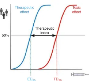
antibodies, and kinase inhibitors. The therapeutic index of classical cytotoxic drugs like alkylating agents is often smaller than that of biologically tar-geted forms of therapy (Fig. 1 ).
Classical cytotoxic drugs have different mech-anisms of action which are outlined in Fig. 2 .
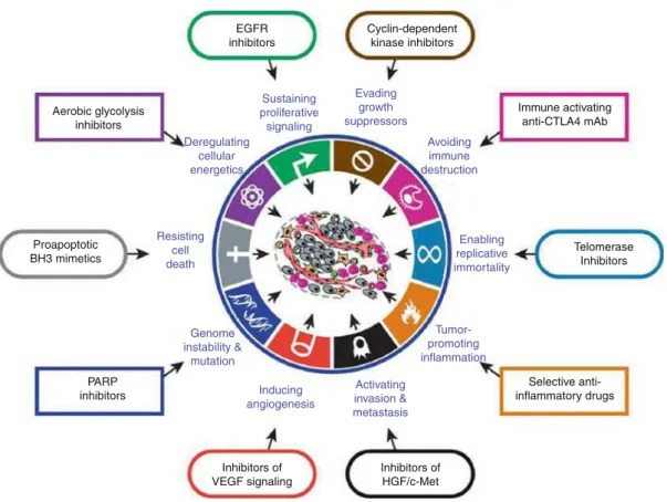
Hanahan and Weinberg described the hall-marks of cancer in a previous landmark article that was updated in 2011. These hallmarks include sustaining proliferative signaling, evad-ing growth suppressors, resistevad-ing cell death, enabling replicative immortality, inducing angio-genesis, and activating invasion and metastasis. Conceptual progress in the last decade has added two emerging hallmarks of potential generality to this list – reprogramming of energy metabolism and evading immune destruction. The “tumor microenvironment” that consists of apparently normal cells adds to the complexity of current tumor characteristics which forms the basis for contemporary drug development and targeted treatment of cancer (Fig. 3 ) (Hanahan and Weinberg 2011 ).
50%
Therapeutic index
Therapeutic effect
ED50 TD50
Toxic effect
Fig. 1 The concept of therapeutic index refers to the rela-tionship between toxic and therapeutic doses. This pharma-codynamic parameter is relevant to clinical practice because it determines how safe or toxic a drug is. Both ED50 and TD50 are calculated from dose-response curves, which rep-resent the frequency with which each dose of drug elicits the desired response or toxic effect in the population. The dose required to cause a therapeutic effect (positive response) in 50 % of a population is the ED50. The dose required to pro-duce a toxic effect in 50 % of the studied population is the TD50 (Redrawn from Craig and Stitzel ( 2003 ))
Nucleic Acids DNA Proteins Mitosis
Purine analogues
6-MP 6-TG MTX
Pyrimidine analogues
5-FU Raltitrexed Pemetrexed MTX
Ribonucleotide reductaseinhibitors
Hydroxyurea
DNA polymerase inhibitor
Cytarabine
DNA alkalyting agent
N-Lost-derivatives Nitrosoureas Oxaphosphorines Platinum compounds Da-/Procarbazine Thiotepa Mitomycine C
Topoisomerase inhibitors
Etoposide Anthracyclines Irinotecan Topotecan
Proteine degradation
L-Asparaginase
Vinca alcaloids
Vincristine Vinblastine Vindesine Vinorelbine
Taxanes
Paclitaxel Docetaxel Cabazitaxel
Fig. 2 Target structures of classical cytotoxic drugs: DNA deoxyribonucleic acid, MTX methotrexate, 5-FU 5- fl uorouracil, 6-MP 6-mercaptopurine, 6-TG 6-thioguanine
6
2
Defi nitions of Anticancer
Drug Therapy
2.1 Mono- Versus Combination Therapy
In principle, combination chemotherapy has advan-tages over monotherapy due to additive or multipli-cative effects of tumor cell kill. Primary or secondary resistant tumor cell clones can be eradicated or sup-pressed by different mechanisms of action. Ideally, combinations have the following features:
• The combined agents are equally effective. • Lack of cross-resistance.
• Different mechanisms of action.
• Additive or synergistic mechanisms of action. • No overlapping toxicities.
For most combinations, this ideal situation does not exist. Especially with regard to side effects, some addition of toxicity must always be accepted when combinations are used.
2.2 Induction Chemotherapy
Induction chemotherapy is used when at the time of diagnosis no acceptable therapeutic alterna-tive exists. Induction chemotherapy shall bring the cancer into a state of better therapeutic options. The goal is “the induction” of an opti-mal remission, which is at best a “complete remission.” High treatment intensities are usu-ally necessary for an optimal induction. Therefore, the probability of inducing adverse effects is usually high.
2.3 Consolidation Therapy
The consolidation therapy shall provide the erad-ication of clinically occult residual tumor. It shall improve the rate of true complete remissions. Thereby, consolidation shall increase the chances of cure or increase the duration of response. EGFR
inhibitors
Cyclin-dependent kinase inhibitors
Telomerase Inhibitors
Inhibitors of HGF/c-Met Inhibitors of
VEGF signaling Sustaining proliferative
signaling
Evading growth suppressors
Avoiding immune destruction
Enabling replicative immortality
PARP inhibitors Aerobic glycolysis
inhibitors
Proapoptotic BH3 mimetics
Immune activating anti-CTLA4 mAb
Selective anti-inflammatory drugs
Tumor-promoting inflammation
Activating invasion & metastasis Inducing
angiogenesis Genome
instability & mutation Resisting
cell death
Deregulating cellular energetics
Fig. 3 The hallmarks of cancer (Redrawn from Hanahan and Weinberg ( 2011 )) are the basis for contemporary drug development and targeted anticancer treatment
7
2.4 Maintenance Therapy
Maintenance therapy, in its classical sense used in the treatment of hematological malignancies like acute leukemia, follows consolidation and shall eradicate or control further residual tumor cells, e.g., those that – due to kinetic resistance – were not yet eradicated by the previous treat-ment. Maintenance therapy can increase the chance of cure or prolong the time interval until further tumor progression. The latter goal is now-adays often chosen in the palliative treatment of solid tumors when a remission has been achieved by a more intensive treatment period preceding maintenance.
2.5 Perioperative (Neoadjuvant and/or Adjuvant)
Chemotherapy
Neoadjuvant (also primary or preoperative) ther-apy is a treatment in patients with localized or locoregional tumor extension in which the appli-cation of local treatment alone (operation or radi-ation therapy) may lead to an unsatisfactory outcome. Neoadjuvant chemotherapy is applied to reduce the extent of surgery (e.g., in breast cancer, where size reduction of large tumors allows for more breast-conserving surgery fol-lowing neoadjuvant chemotherapy) and to increase the chances of cure (like in gastric or muscle invasive bladder cancer). In some cancers (e.g., osteosarcoma and Ewing sarcoma), postop-erative treatment is tailored on the basis of the achieved response during neoadjuvant therapy.
The goal of adjuvant chemotherapy is the eradication of subclinical metastases (“microme-tastases”) following primary local treatment (operation or radiation therapy). The clinical goal of treatment is to increase the cure rate.
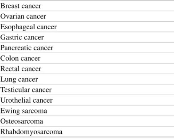
Accepted indications for perioperative che-motherapy are shown in Table 1 . As increased cure rates are the goal of neo-/adjuvant chemo-therapy, optimal dose intensity is necessary and some toxicity must be accepted. On the other hand, treatment safety is of utmost importance as patients may survive with the operation alone. In addition, long-term side effects should be avoided
as they may lead to a signifi cant impairment of quality of life of cancer survivors; alter physical, cognitive, and social functioning; and may even induce secondary diseases (cancers, leukemia, organ dysfunctions, cardiovascular diseases, etc.) leading to a negative impact on life expectancy.
2.6 Palliative Therapy
Palliative chemotherapy is a treatment intended to prolong life, to control symptoms, and to aug-ment quality of life. In case of symptomatic dis-ease, more intensive induction treatment regimens are often applied. For a further stabili-zation of the tumor, most often less intensive monotherapies are regarded as standard of care. Treatment-emergent side effects must be care-fully weighed against potential treatment benefi ts.
3
Classifi cation
of Anticancer Drugs
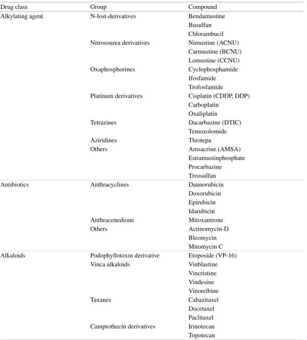
The classifi cation of anticancer drugs can follow different criteria. Traditionally, the World Health Organization (WHO) chose the mechanisms of action (e.g., alkylating agent) and the origin of compounds (e.g., antitumor antibiotics) as their leading criteria for classifi cation. Table 2 groups the compounds predominantly according to their mechanisms of action.
Table 1 Examples for tumors with an established indica-tion for perioperative (neoadjuvant or adjuvant) therapy
Breast cancer Ovarian cancer Esophageal cancer Gastric cancer Pancreatic cancer Colon cancer Rectal cancer Lung cancer Testicular cancer Urothelial cancer Ewing sarcoma Osteosarcoma Rhabdomyosarcoma
8
4
Classifi cation of Treatment
Toxicity
Side effects of medical treatment have been clas-sifi ed according to uniform criteria as long as the drug is applied within a clinical study. Internationally, the so-called Common Toxicity Criteria (CTC) or the newer Common Terminology Criteria for Adverse Events (CTCAE) as developed and published by the
National Cancer Institute (NCI, Bethesda, USA) are most commonly used. Meanwhile, these cri-teria have been well implemented into clinical practice and proved useful. Therefore, thorough oncologists and multidisciplinary teams use it outside of clinical studies in routine cancer care. The current version of CTCAE V4.03 can be downloaded from the Internet ( http://evs.nci.nih. gov/ftp1/CTCAE/CTCAE_4.03_2010-06- 14_ QuickReference_8.5x11.pdf ).
Table 2 Classifi cation of anticancer drugs according to their mechanisms of action and biochemical properties
Drug class Group Compound
Alkylating agent N-lost-derivatives Bendamustine Busulfan Chlorambucil Nitrosourea derivatives Nimustine (ACNU)
Carmustine (BCNU) Lomustine (CCNU) Oxaphosphorines Cyclophosphamide
Ifosfamide Trofosfamide
Platinum derivatives Cisplatin (CDDP, DDP) Carboplatin
Oxaliplatin
Tetrazines Dacarbazine (DTIC)
Temozolomide
Aziridines Thiotepa
Others Amsacrine (AMSA)
Estramustinphosphate Procarbazine Treosulfan
Antibiotics Anthracyclines Daunorubicin
Doxorubicin Epirubicin Idarubicin Anthracenedione Mitoxantrone
Others Actinomycin-D
Bleomycin Mitomycin C Alkaloids Podophyllotoxin derivative Etoposide (VP-16)
Vinca alkaloids Vinblastine Vincristine Vindesine Vinorelbine
Taxanes Cabazitaxel
Docetaxel Paclitaxel Camptothecin derivatives Irinotecan Topotecan
9
Table 2 (continued)
Drug class Group Compound
Antimetabolite Antifolates Methotrexate (MTX)
Pemetrexed
Purine analogues 6-Mercaptopurine (6-MP) 6-Thioguanine (6-TG) Fludarabine
2-Chlordeoxyadenosine (2-CDA) Pyrimidine analogues 5-Fluorouracil (5-FU)
Capecitabine Clofarabine
Cytosine arabinoside (AraC) Gemcitabine
RNR inhibitor Hydroxyurea DNA demethylation Demethylating agents Azacytidine
Decitabine
Protein degradation Enzyme L-asparaginase
Aromatase inhibition Nonsteroidal inhibitors Anastrozole Letrozole Steroidal inhibitor Exemestane Other hormonal therapies Antiandrogens Abiraterone Bicalutamide Flutamide Nilutamide
Antiestrogen Fulvestrant
Gestagens Medroxyprogesterone acetate Megestrol acetate
Selective estrogen receptor modulators
Raloxifene Tamoxifen Immune modulators Cytokines Interferon alpha
Interleukin 2
IMIDs Lenalidomide
Thalidomide Pomalidomide Immune checkpoint inhibitors Ipilimumab
Lambrolizumab Monoclonal antibodies CD20 antibodies Rituximab
Ofatumumab CD30 antibody-toxin conjugate Brentuximab vedotin CD33 antibody Gemtuzumab ozogamicin
CD52 antibody Alemtuzumab
EGFR antibodies Cetuximab Panitumumab HER2 antibodies Trastuzumab
Pertuzumab
HER2 antibody-toxin conjugate Trastuzumab emtansine VEGF antibody Bevacizumab
VEGF recombinant fusion protein Afl ibercept VEGFR2 antibody Ramucirumab
(continued)
10
The NCI Common Terminology Criteria for Adverse Events is a descriptive terminology which can be utilized for adverse event (AE) reporting. A grading (severity) scale is provided for each AE term. System Organ Class (SOC) , the highest level of the reporting hierarchy, is identifi ed by anatomical or physiological system, etiology, or purpose (e.g., SOC Investigations for laboratory test results). Within each SOC, adverse events are listed and accompanied by descrip-tions of severity (grade).
An AE is any unfavorable and unintended sign (including an abnormal laboratory or imaging fi nd-ing), symptom, or disease temporally associated with the use of a medical treatment or procedure that may or may not be considered related to the medical treatment or procedure. An AE is a term that is a unique representation of a specifi c event used for medical documentation and scientifi c analyses.
Grade refers to the severity of the AE. The CTCAE displays grades 1 through 5 with unique clinical descriptions of severity for each AE based on this general guideline (Table 3 ). Not all grades are appropriate for all AEs. Therefore, some AEs are listed with fewer than fi ve options for grade selection.
5
Specifi c Toxicities Associated
with Anticancer Treatment
All organ systems can be subject to treatment- emergent toxicities.
With classical cytotoxic treatment, myelosup-pression (neutropenia, thrombocytopenia, and anemia) is a common side effect. Between 80 and 100 % of all patients undergoing chemotherapy have some grade of myelosuppression leading to
Table 2 (continued)
Drug class Group Compound
Tyrosine kinase inhibitors Bcr/abl Imatinib Dasatinib Nilotinib
cKIT Imatinib
EGFR Afatinib
Erlotinib Gefi tinib
HER2 Lapatinib
Histone deacetylase (HDAC) Romidepsin Vorinostat
mTOR Temsirolimus
Everolimus Multiple kinases Axitinib
Nintedanib Pazopanib Regorafenib Sorafenib Sunitinib
Proteasome Bortezomib
Carfi lzomib
RAF Vemurafenib
Smoothened receptor (hedgehog signaling)
Vismodegib
Somatostatin receptors Octreotide Lanreotide
Compounds are listed with their generic names. Where appropriate, commonly used abbreviations are listed in parentheses
11
alterations of the differential blood counts. Severity and duration depend of course on the applied cytotoxic drug and schedule as well as additional risk factors, like age and general health status. In case of neutropenia, patients are at par-ticular risk of acquiring infections. Febrile neutro-penia is an emergency situation during antineoplastic treatment. It requires immediate clarifi cation and start of empiric antibiotic treat-ment. In most cases (except low-risk neutropenia in otherwise unimpaired and compliant patients), this should be done following hospitalization, and intravenous broad-spectrum antibiotics should be given (Klastersky and Paesmans 2013 ). In more than two thirds of patients, the focus of febrile neutropenia remains unknown, but pulmonary infections, bloodstream infections, urinary infec-tions, infections of the skin and soft tissues, as well as infections of the upper aerodigestive tract should be excluded by appropriate clinical, para-clinical, and radiological diagnostics.
Apart from myelosuppression, non- hematological adverse events are common and
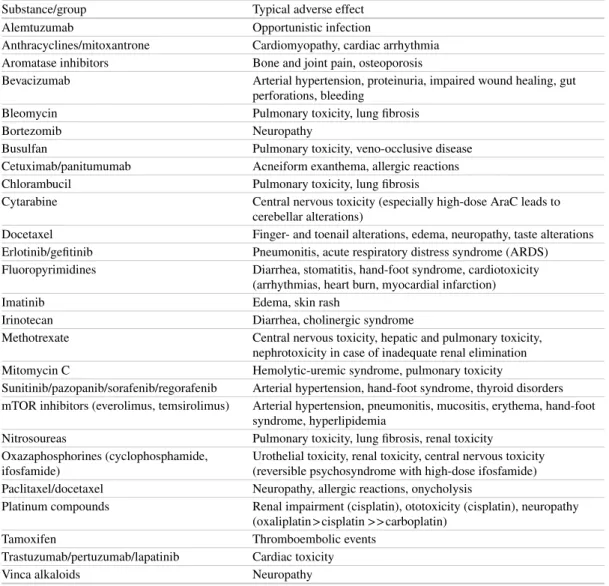
need to be well known by the treatment team. Table 4 outlines a selection of substance- and group-specifi c non-hematological toxicities of anticancer drugs.
Our expectation was that with the introduction of new, more specifi c and biologically targeted drugs, the effi cacy of anticancer treatment would increase, while the side effects would decrease. This hope was desperately disappointed (Niraula et al. 2012 ). International investigators analyzed all randomized controlled trials evaluating agents approved for the treatment of solid tumors by the US Food and Drug Administration between 2000 and 2010. Odds ratios were computed for three end points of safety and tolerability: treatment- related death, treatment discontinuation related to toxicity, and grade 3 or grade 4 adverse events (AEs). These were then pooled in a meta- analysis. Correlations between these end points and the hazard ratios for overall survival and progression- free survival were also assessed. The investiga-tors came to the conclusion that new anticancer agents that lead to improvements in time-to-event end points also increase morbidity and treatment- related mortality. The balance between effi cacy and toxicity may be less favorable in clinical practice because of selection of fewer patients with good performance status and limited comor-bidities. Patients’ baseline health characteristics should be considered when choosing therapy.
With the use of targeted therapies, novel side effects have emerged that are closely related to the specifi c mechanisms of action of the respec-tive drug. Targeted therapies in general block cer-tain signaling pathways that play important roles in promoting tumor cell survival and proliferation or interfere with stromal cells like vascular endo-thelial cells to inhibit tumor angiogenesis or with immune cells to modify antitumor immune responses. Monoclonal antibodies and tyrosine kinase inhibitors (TKI) represent the drug classes that are most commonly used for targeted cancer therapy. Furthermore, specifi c intracellular sig-naling checkpoints can be blocked by chemical compounds (i.e., mTOR inhibitors). Another group of drugs targets immune function to improve host anticancer immunity. CTLA-4 anti-bodies are used to enhance T-cell co-stimulation,
Table 3 Toxicity grades according to the “Common Terminology Criteria for Adverse Events” (CTCAE) reporting system provided by the National Cancer Institute, Bethesda, USA
Grade Severity Grade
1
Mild; asymptomatic or mild symptoms; clinical or diagnostic observations only; intervention not indicated
Grade 2
Moderate; minimal, local, or noninvasive intervention indicated; limiting age- appropriate instrumental activity of daily living (ADL) a
Grade 3
Severe or medically signifi cant but not immediately life-threatening; hospitalization or prolongation of hospitalization indicated; disabling; limiting self-care ADL b
Grade 4
Life-threatening consequences; urgent intervention indicated
Grade 5
Death related to an adverse event
A semicolon indicates “or” within the description of the grade
a Instrumental ADL refer to preparing meals, shopping for
groceries or clothes, using the telephone, managing money, etc.
b Self-care ADL refer to bathing, dressing and undressing,
feeding self, using the toilet, taking medications, and not bedridden
12
Table 4 Selection of substance and group-specifi c non-hematological toxicities of anticancer drugs
Substance/group Typical adverse effect
Alemtuzumab Opportunistic infection
Anthracyclines/mitoxantrone Cardiomyopathy, cardiac arrhythmia Aromatase inhibitors Bone and joint pain, osteoporosis
Bevacizumab Arterial hypertension, proteinuria, impaired wound healing, gut perforations, bleeding
Bleomycin Pulmonary toxicity, lung fi brosis
Bortezomib Neuropathy
Busulfan Pulmonary toxicity, veno-occlusive disease Cetuximab/panitumumab Acneiform exanthema, allergic reactions Chlorambucil Pulmonary toxicity, lung fi brosis
Cytarabine Central nervous toxicity (especially high-dose AraC leads to cerebellar alterations)
Docetaxel Finger- and toenail alterations, edema, neuropathy, taste alterations Erlotinib/gefi tinib Pneumonitis, acute respiratory distress syndrome (ARDS) Fluoropyrimidines Diarrhea, stomatitis, hand-foot syndrome, cardiotoxicity
(arrhythmias, heart burn, myocardial infarction)
Imatinib Edema, skin rash
Irinotecan Diarrhea, cholinergic syndrome
Methotrexate Central nervous toxicity, hepatic and pulmonary toxicity, nephrotoxicity in case of inadequate renal elimination Mitomycin C Hemolytic-uremic syndrome, pulmonary toxicity
Sunitinib/pazopanib/sorafenib/regorafenib Arterial hypertension, hand-foot syndrome, thyroid disorders mTOR inhibitors (everolimus, temsirolimus) Arterial hypertension, pneumonitis, mucositis, erythema, hand-foot
syndrome, hyperlipidemia
Nitrosoureas Pulmonary toxicity, lung fi brosis, renal toxicity Oxazaphosphorines (cyclophosphamide,
ifosfamide)
Urothelial toxicity, renal toxicity, central nervous toxicity (reversible psychosyndrome with high-dose ifosfamide) Paclitaxel/docetaxel Neuropathy, allergic reactions, onycholysis
Platinum compounds Renal impairment (cisplatin), ototoxicity (cisplatin), neuropathy (oxaliplatin > cisplatin > > carboplatin)
Tamoxifen Thromboembolic events
Trastuzumab/pertuzumab/lapatinib Cardiac toxicity
Vinca alkaloids Neuropathy
and drugs targeting the PD-1/PD-L1 pathway have been developed to block inhibitory immune checkpoints.
An overview of key side effects can be found in Table 2 . Specifi c side effects resulting in path-ological radipath-ological fi ndings are shortly summa-rized in the following section.
Agents Targeting the Epidermal Growth Factor Receptor (EGFR): The monoclonal anti-bodies (cetuximab, panitumumab) are used for the treatment of RAS wild-type metastatic colorectal cancer, while TKI (gefi tinib, erlotinib, afatinib) represent a standard of care in the
treat-ment of EGFR-mutated non-small cell lung can-cer (NSCLC) patients. Skin toxicities occur with high frequency in both groups of drugs. In con-trast, interstitial lung disease (ILD) represents a rare complication, and the mechanism is not fully understood. Disruption of the alveolar epithelial function however may play a role. Based on this, the frequency of ILD is higher in smokers and in patients with preexisting lung disease (Ando et al. 2006 ).
Agents Targeting Her-2: Chemotherapy com-bined with monoclonal antibodies (trastuzumab, pertuzumab) represents a treatment standard in
13
Her2-positive breast cancer and in Her2 gastric cancer (trastuzumab). The TKI lapatinib targeting EGFR and Her2neu is approved for the treatment of breast cancer. An important side effect of this class of drugs is cardiotoxicity that is related to the expression of Her2 on cardiomyocytes. Mechanistically, Her2 signaling results in sarco-mere stability and initiates repair processes that are important to counteract toxic stress (Tocchetti et al. 2012 ).
Agents Targeting Tumor Angiogenesis: The monoclonal antibody bevacizumab binds vascu-lar endothelial growth factor (VEGF), and the fusion construct afl ibercept binds VEGF and pla-cental growth factor (PlGF). Both drugs are used in combination with chemotherapy for the treat-ment of metastatic colorectal cancer. Additionally, a large number of TKI targeting VEGF receptors and other receptors are in clinical use for the treatment of a wide variety of cancer types (Table 2 ). Hypertension and proteinuria represent common side effects of VEGF-targeting therapy. Furthermore, the rate of thromboembolic compli-cations is increased. Other side effects are related to impaired tissue repair capacity and comprise gastrointestinal pneumatosis perforations and the formation of fi stulas (Shinagare et al. 2012 ). Overall, bleeding is a rare side effect. However, frequent bleeding complications have resulted in the exclusion of the use of bevacizumab in squa-mous cell carcinoma of the lung. Progressive reversible encephalopathy syndrome (PRES) is a very rare (≤0.1 %) but severe neurological com-plication that has been reported in patient treat-ment with bevacizumab or afl ibercept (Seet and Rabinstein 2012 ). The disruption of cerebrovas-cular endothelial cell signaling is related to the disruption of cerebrovascular autoregulation preferentially in the posterior circulation of the brain. Finally, pancreatitis (sunitinib, sorafenib, pazopanib) and acalculous cholecystitis (suni-tinib) have been reported in the literature on a casuistic basis.
Anaplastic Lymphoma Kinase (ALK) Inhibitors: ALK inhibitors are used for the treat-ment of NSCLC harboring specifi c genomic rear-rangements (EML4-ALK). Pneumonitis has been
reported with the use of the ALK inhibitor crizo-tinib and symptoms started within two months of treatment. The underlying mechanisms are not yet clarifi ed.
RAF-Targeting Agents: RAF-targeting agents include the multi-TKI sorafenib for the treatment of renal cell and hepatocellular cancer as well as vemurafenib and dabrafenib, which are used for the treatment of melanoma harboring the B-Raf mutation V600E and other B-Raf mutations. An increase in the occurrence of cutaneous squamous cell carcinomas has been reported, and nodular panniculitis (Monfort et al. 2012 ) may result in increased radiotracer uptake during 18F- FDG positron emission tomography (PET).
Agents Targeting Mammalian Target of Rapamycin (mTOR) and Targeted Immune Modulators: These agents (everolimus, temsiroli-mus) are used for the treatment of breast and renal cancers and pancreatic neuroendocrine tumors. Mucositis and aphthous mucosal lesions are common side effects. Additionally, interstitial pneumonitis is an important side effect of this class of drugs with up to 36 % of patients show-ing any pulmonary abnormalities durshow-ing treat-ment (Duran et al. 2014 ).
Ipilimumab is a novel targeted immune modu-lator that interacts with CLTA-4, thus fostering co-stimulatory function to improve host antitu-mor immune response. Due to immune function deregulation, autoimmune-related side effects like enterocolitis and hypophysitis may occur. Additionally, unspecifi c lymph node enlargement and soft tissue changes like myositis or fasciitis as well as retroperitoneal fat opacities due to lymphocyte infi ltration may interfere with treat-ment response assesstreat-ment (Bronstein et al. 2011 ). As examples of “new toxicities” emerging from biologically selective targeted drugs, Fig. 4 dis-plays a perforation at the rectosigmoid level that occurred during treatment of metastatic colorectal cancer with the anti-VEGF antibody bevacizumab. Another patient who was also treated for meta-static colorectal cancer received the monoclonal anti-EGFR antibody cetuximab plus chemother-apy and developed a grade 3 skin rash during weeks 4–6 of this combined treatment (Fig. 5 ).
14
a b
Fig. 4 Gut perforation leading to an ileus and peritonitis, emerging from a pararectal abscess in a patient with colorectal cancer with simultaneous liver and lung metas-tases. ( a ) Is illustrating the coronary section through the abdomen; ( b ) is illustrating a transversal section through
the pelvis. The two white arrows in b are highlighting the formation of a pararectal abscedation. This patient was treated with the anti-VEGF monoclonal antibody bevaci-zumab in combination with chemotherapy
a b
Fig. 5 ( a , b ) Patient who developed severe (grade 3 according to CTCAE V4.03) skin rash during weeks 4–6 of che-motherapy combined with the anti-EGFR-directed monoclonal antibody cetuximab
15
Conclusions
For clinical practice, we have to state that medical anticancer treatment is more demand-ing than ever, as toxicities are very common, polymorphic and allotropic. They may lead to severe impairment of the patients’ safety and quality of life. All members of the treatment team, including the radiologist, need to do their best to support patients during anticancer treatment. Treatment- emergent as well as tumor-related complications may not be missed, and the severity of events must be appropriately classifi ed. In addition, for drug development it has been advocated to move “Toward Patient-Centered Drug Development in Oncology” (Basch 2013 ).
References
Ando M, Okamoto I, Yamamoto N, Takeda K, Tamura K, Seto T, Ariyoshi Y, Fukuoka M (2006) Predictive fac-tors for interstitial lung disease, antitumor response, and survival in non-small-cell lung cancer patients treated with gefi tinib. J Clin Oncol 24(16):2549–2556. doi: 10.1200/JCO2005.04.9866
Basch E (2013) Toward patient-centered drug develop-ment in oncology. N Engl J Med 369(5):397–400. doi: 10.1056/NEJMp1114649
Bronstein Y, Ng CS, Hwu P, Hwu WJ (2011) Radiologic manifestations of immune-related adverse events in patients with metastatic melanoma undergoing anti-CTLA- 4 antibody therapy. AJR Am J Roentgenol 197(6):W992–W1000. doi: 10.2214/AJR.10.6198 Craig CR, Stitzel CR (2003) Modern pharmacology with
clinical applications, 6th edn. Lippincott, Williams & Wilkins, Philadelphia
Duran I, Goebell PJ, Papazisis K, Ravaud A, Weichhart T, Rodriguez-Portal JA, Budde K (2014) Drug-induced
pneumonitis in cancer patients treated with mTOR inhibitors: management and insights into possible mechanisms. Expert Opin Drug Saf 13(3):361–372. doi: 10.1517/14740338.2014.888056
Hanahan D, Weinberg RA (2011) Hallmarks of cancer: the next generation. Cell 144(5):646–674. doi: 10.1016/j.cell.2011.02.013
Heisterkamp N, Stam K, Groffen J, de Klein A, Grosveld G (1985) Structural organization of the bcr gene and its role in the Ph′ translocation. Nature 315(6022):758–761 Klastersky J, Paesmans M (2013) The Multinational
Association for Supportive Care in Cancer (MASCC) risk index score: 10 years of use for identifying low- risk febrile neutropenic cancer patients. Support Care Cancer 21(5):1487–1495. doi: 10.1007/s00520-013-1758-y Monfort JB, Pages C, Schneider P, Neyns B, Comte C,
Bagot M, Vignon-Pennamen MD, Viguier M, Lebbe C (2012) Vemurafenib-induced neutrophilic panniculi-tis. Melanoma Res 22(5):399–401. doi: 10.1097/ CMR.0b013e3283570792
Niraula S, Seruga B, Ocana A, Shao T, Goldstein R, Tannock IF, Amir E (2012) The price we pay for prog-ress: a meta-analysis of harms of newly approved anti-cancer drugs. J Clin Oncol 30(24):3012–3019. doi: 10.1200/JCO.2011.40.3824
Phelps MA, Sparreboom A (2014) A snapshot of challenges and solutions in cancer drug development and therapy. Clin Pharmacol Ther 95(4):341–346. doi: 10.1038/clpt.2014.15 Seet RC, Rabinstein AA (2012) Clinical features and out-comes of posterior reversible encephalopathy syn-drome following bevacizumab treatment. QJM 105(1):69–75. doi: 10.1093/qjmed/hcr139
Shinagare AB, Howard SA, Krajewski KM, Zukotynski KA, Jagannathan JP, Ramaiya NH (2012) Pneumatosis intes-tinalis and bowel perforation associated with molecular targeted therapy: an emerging problem and the role of radiologists in its management. ARJ Am J Roentgenol 199(6):1259–1265. doi: 10.2214/AJR.12.8782
Tocchetti CG, Ragone G, Coppola C, Rea D, Piscopo G, Scala S, De Lorenzo C, Iaffaioli RV, Arra C, Maurea N (2012) Detection, monitoring, and management of trastuzumab-induced left ventricular dysfunction: an actual challenge. Eur J Heart Fail 14(2):130–37. doi: 10.1093/eurjhf/hfr165
17
Med Radiol Diagn Imaging (2014)
DOI 10.1007/174_2014_1041, © Springer International Publishing Switzerland
Abstract
The focus of this chapter lies on the description of the general basics of early and late radiation effects and the translation of these pathogenetic processes into imaging; furthermore, a few short clinical examples including imaging patterns of those underlying pathogenetic normal tissue reactions are given to provide a better under-standing. In addition, the margin concepts used in radiotherapy as well as the important radiation techniques are summarized, as it is very impor-tant for diagnostic radiologists to correlate post-therapeutic tissue and organ changes in follow-up examinations with dose characteristics of a cer-tain treatment to achieve a higher degree of reli-ability in image interpretation. Furthermore, for T. Bostel • F. Sterzing (*)
Department of Radiooncology and Radiation Therapy , Heidelberg University Hospital ,
Im Neuenheimer Feld 400 , Heidelberg 69120 , Germany
e-mail: bostel.tilmann@med.uni-heidelberg.de;
sterzing.fl orian@med.uni-heidelberg.de
Radiotherapy
T. Bostel and F. Sterzing
Contents
1 Introduction ... 18
2 Radiation Delivery Techniques ... 20 2.1 Traditional External-Beam Radiation
Therapy (EBRT) ... 20 2.2 Conformal Radiation Therapy ... 20 2.3 Intensity-Modulated Radiation
Therapy (IMRT) ... 21 2.4 Stereotactic Body Radiation
Therapy (SBRT) ... 22 2.5 Particle Therapy ... 23 2.6 Brachytherapy ... 24
3 Radiation Biology: A Refresher... 25
4 Basics of Radiation Effects of Normal
Tissues ... 26 4.1 Classifi cation of Radiation Effects ... 26 4.2 Radiobiological Characteristics of Early
and Late Radiation Effects ... 26 4.3 Consequential Late Effects (CLE) ... 27 4.4 Cellular Basis of Radiation Effects ... 27 4.5 Tolerance Dose Concept ... 27 4.6 Classifi cation Systems ... 28
5 Early Radiation Effects ... 29 5.1 Pattern of Cell Divisions in Early-Reacting
Tissues ... 29 5.2 Pathogenesis of Early Radiation Reactions .... 30
6 Chronic Radiation Effects... 31 6.1 Concepts of Radiation Pathophysiology ... 31
6.2 General Pathogenesis of Chronic
Radiation Effects ... 32 6.3 Dose Dependency of the Latency Period ... 32 6.4 Chronic Radiation Effects
in the Vascular System ... 33 6.5 Chronic Radiation Effects
in the Mesenchymal Tissues ... 34 6.6 General Chronic Radiation Effects
in the Epithelia and Organ Parenchyma ... 35 6.7 Modulation of the Immune System ... 37
7 Radiation-Induced Cancers ... 38 7.1 Secondary Cancer Rate ... 38 7.2 Secondary Cancers in Adults ... 38 7.3 Secondary Cancers in Children ... 39 7.4 Development and Manifestation
of Secondary Tumors ... 40
Conclusion ... 40
18
a better understanding of the cellular basis of the various radiogenic tissue effects, a short refresher about the underlying radiobiological principles is given. The detailed description of specifi c radiation effects and imaging patterns of clini-cally relevant organs and tissues, however, fol-lows in the specifi c organ chapters in order to avoid redundancy.
Abbreviations
CLE Consequential late effects COX-2 Cyclooxygenase-2 CT Computed tomography CTV Clinical target volume 3D Three dimensional 4D Four dimensional DNA Deoxyribonucleic acid
EBRT External-beam radiation therapy e.g. Exempli gratia
GTV Gross tumor volume Gy Gray
i.e. Id est
IL-1a Interleukin-1 alpha
iNOS Inducible nitric oxide synthase IGRT Image-guided radiotherapy
IMRT Intensity-modulated radiation therapy MRI Magnetic resonance imaging
mRNA Messenger ribonucleic acid
NTCP Normal tissue complication probability OAR Organs at risk
PET Positron emission tomography PTV Planning target volume
RBE Relative biological effectiveness RR Relative risk
SBRT Stereotactic body radiation therapy TD Tolerance dose
TGF-ß Transforming growth factor-ß TNF-a Tumor necrosis factor alpha VOD Veno-occlusive disease
1
Introduction
Radiotherapy plays a vital role in the oncological treatment concept besides surgical and systemic therapies. Moreover, it is an effective local cancer
therapy like surgery, but beyond that it offers the chance for regional high-volume treatments of microscopic tumor deposits or lymphatic path-ways as transition to systemic treatments. This pivotal role of radiotherapy is also supported by epidemiological data: More than half of all can-cer patients can be cured nowadays, owing to improved effi cacy of advanced and mostly multi-modal cancer therapies, and around half of these patients receive either radiotherapy alone or radiotherapy in combination with other cancer treatments. Moreover, about two thirds of cancer patients gain valuable palliation by radiation to alleviate the symptoms and to improve the qual-ity of life in the course of their advanced disease.
In recent years, substantial advances in radio-logical imaging as well as computer hardware and software along with improved design of medical linear accelerators have contributed sig-nifi cantly to the development in radiation ther-apy. Nowadays, existing modern radiation techniques enable the delivery of conformal dose distributions with steep dose gradients between the tumor and adjacent normal tissue structures. Thus, intensifi cation of the radiation dose to the tumor and reduction of high-dose irradiation of sensitive organs and normal tissues are possible resulting in higher curing rates and lower rates of side effects (i.e., increased therapeutic ratio). However, despite these advances, modern radio-therapy still leaves signifi cant proportions of healthy tissue structures exposed to relatively high doses. This is in part caused by the margin concepts used in radiotherapy. In general, the determination of the planning target volume (PTV) necessarily requires the inclusion of the visible or palpable extent of tumor (i.e., gross tumor volume, GTV) as well as an additional sur-rounding area without visible branches of the tumor in order to take microscopic disease into account (i.e., clinical target volume, CTV). Furthermore, a patient-specifi c safety margin is added, if necessary, to account for the range of target motion related to breathing, pulsations, or intestinal peristalsis (e.g., lung or liver lesions) that is often based on four-dimensional (4D) imaging information derived from the planning
19
CT. And fi nally, a margin to encompass variabil-ity in patient positioning (setup) and mechanical uncertainty is added to create the fi nal PTV. This PTV concept accounts for all available radiation techniques, even though modern approaches such as stereotactic radiation therapy or intensity- modulated radiation therapy enable to adapt the dose distribution more precisely to the tumor boundaries than traditional radiation techniques, which in turn helps to spare the adjacent healthy organs and tissues. On the other hand, PTV can encompass extended areas of normal tissues, dependent on the tumor and disease stage, for example, irradiations of the whole body, whole brain, spinal column, or breast with or without the supraclavicular lymph nodes after breast- conserving surgical treatment.
In the follow-up care of cancer patients, how-ever, it is very important that side effects after therapeutic irradiations are not in general regarded as an indicator for medical malpractice. Moreover, it is an indicator for the best-possible treatment and maximum cure probability when these radio-genic effects manifest with only a defi ned low incidence of sequelae of defi ned severity in cured patients (Dorr 2009 ) (Fig. 1 ). Regarding the eval-uation of side effects, it has also to be mentioned that radiotherapy is increasingly combined with other local and systemic therapeutic approaches such as operation, chemotherapy, or molecular targeting which may lead not only to additive but also to synergistic effects for the tumor response and for organ- specifi c injuries (Dische et al. 1989 ; Pedersen et al. 1994 ). Furthermore, it has to be considered that a certain number of pathological conditions may be triggered by other reasons than specifi c tumor therapies, such as comorbidities, the tumor itself, or other non-oncological treat-ments, for example, obstipation due to analgesia with opioids.
As a consequence of increased numbers of cancer survivors and prolongation of survival times, late radiation sequelae as well as secondary cancers are more frequently seen than in the past. This subject has therefore gained more relevance in oncological studies as well as clinical follow-up examinations in recent years. Therefore, it is of utmost importance that radiologists are familiar
with the imaging patterns of these therapy-related tissue reactions as they may both mimic and obscure tumor relapses. Beyond interpretation of posttreatment imaging, diagnostic specialists can make further valuable contributions to increase the therapeutic ratio preceding the radiation treat-ment process (Terezakis et al. 2011 ). First of all, every cancer treatment, particularly radiotherapy, heavily relies on accurate staging of the cancerous disease in order to select an appropriate treatment regimen for each patient. It is obvious that misdi-agnosis in staging may have fatal consequences for the patient with regard to therapy-associated complications and treatment outcome. For exam-ple, unrecognized local tumor extension in an early stage of the disease may result in an insuffi -cient local therapy with persistence of residual tumor cells that trigger the further course of the disease, either with new local symptoms or with propagation of systemic spread of these cells. Similar devastating consequences may result from overtreatment, for example, through radio-therapy, with avoidable early and late therapy effects and worsening of the patient’s general con-dition. This is especially important, since late sequelae of any oncological treatment are thera-peutically diffi cult to infl uence and characterized by a progressive pattern in many cases (see below). Taken together, accurate staging is an essential precondition for a successful treatment
Probability
Dose
7,500 75,000
10 20
0 30 40 50 60 70 80 90 100%
Local tumor control Complication
Therapeutic window Toleranz überscnritten
Fig. 1 Dose dependency of tumor control ( green sigmoid curve) and side effect ( red sigmoid curve ) probability (according to Holthusen): Due to the fact that both curves overlap, there is no chance for complete tumor destruction through radiotherapy without any risk of normal tissue complications – the third blue curve depicts the therapeutic window (Figure provided by courtesy of Dr. Dr. Thieke)
20
of cancer patients. Further input of diagnostic spe-cialists may be provided during the routine radia-tion oncology workfl ow: Delineation of the macroscopic tumor (GTV) often requires addi-tional advanced imaging modalities such as MRI or PET/CT and reaches beyond the normal ana-tomic information. As tumor imaging has been increasing in both the amount and the complexity of information, an in-depth knowledge of onco-logic radiology has become more and more cru-cial in recent times. Furthermore, tumor tissue is often diffi cult to distinguish from normal tissue changes, for example, due to prior treatments or stromal reactions seen in infi ltrating cancers, radiologic input may add to the precision in delin-eating the GTV. In summary, radiation therapy has become increasingly based on multimodal imaging, and oncologically trained diagnostic radiologists are increasingly important for the successful application of modern radiotherapy treatments (Terezakis et al. 2011 ).
2
Radiation Delivery
Techniques
For radiologists it is important to consider not only the delivered overall dose for image interpre-tation of normal tissue changes but also the used treatment technique. This means that depending on the irradiation technique, a given specifi c over-all dose may be distributed in normal tissues in completely different ways, and thus the organ exposure can vary signifi cantly with consecutive different image presentations in the follow-up. Furthermore, it would be extremely helpful for radiologists if dose overlays from treatment plan-ning software could be integrated into PACS workstations in the near future to achieve a higher degree of safety in image interpretation.
2.1 Traditional External-Beam Radiation Therapy (EBRT)
First, therapeutic applications of ionizing radia-tion started early after their discovery by Conrad Roentgen in 1895. For many decades, irradiation of cancerous tissues was performed with X-ray
devices, which allowed only relatively low energy doses with peak doses near the entrance site of the beam. Thus, major drawbacks were dose-limiting radiation effects in skin and epider-mis and the rapid decline of the depth-dose curve being unfavorable especially for the treatment of deep-seated local tumors.
It was only in the 1950s until high-energy lin-ear accelerators were developed – a milestone for the specialty of radiation oncology. From the 1950s to the 1980s, radiation treatment was administered by the use of planar radiographs in two dimensions, which visualized osseous land-marks. These bony landmarks were used for delineation of radiation portals and localization of therapeutic targets. Depending on the tumor site, the number of beams used for radiotherapy ranged from two to six. However, treatment plan-ning was limited by poor tumor visualization of mainly X-ray-based imaging methods and tech-niques available for radiation delivery (Purdy 2008 ; Bortfeld and Jeraj 2011 ).
2.2 Conformal Radiation Therapy
In the 1980s, cross-sectional imaging procedures (i.e., CT and MRI) entered clinical routine, which were essential for a more accurate delineation of cancerous tissues and risk structures. These advances in radiological imaging were fundamental for further progress in radiation oncology with development of computerized treatment planning and delivery systems that enabled an exquisite tai-loring of 3D radiation dose distributions to the can-cerous tissues (Bortfeld and Jeraj 2011 ). These 3D conformal dose applications were reached by the use of a larger number of lower-dose radiation beams aimed at the target volume from different directions (up to 10 beams) (Fig. 2 ). As a conse-quence, the low-dose exposition of healthy tissues was increased, but the amount of tissues receiving high doses was signifi cantly decreased (Bortfeld and Jeraj 2011 ); thus, the dose in the tumor could be escalated, while the surrounding healthy tissues and organs at risk could be protected better than with traditional EBRT helping to increase the therapeutic ratio. Furthermore, dynamic multileaf collimators were developed and clinically established for more
21
precise shaping of radiation beams compared to the previously used lead blocks (Purdy 2008 ).
Modern conformal radiation therapy plans may also include intensity-modulated radiation therapy (i.e., IMRT) and stereotactic body radia-tion therapy (i.e., SBRT), which are described in the next two sections.
2.3 Intensity-Modulated Radiation Therapy (IMRT)
The mathematical basis of IMRT was developed in the early 1980s to address the problem of irradiation of complex-shaped tumors in close
proximity to or within risk structures, for exam-ple, paraspinal tumors (Fig. 3 ). But it still took a while until theoretical knowledge was put into practice, with fi rst IMRT treatments applied to patients in 1997. The concept of IMRT is based on two decisive pillars, which are inverse treat-ment planning and nonuniform photon intensities across each of several radiation beams – usually 5–9 in modern treatment plans (Brahme et al. 1982 ). In the pre-IMRT era, physical dose distri-bution was calculated by trial and error; this means by trying out different intensities and directions of radiation beams. IMRT, on the other hand, takes the abovementioned path of inverse treatment planning, that is, dose distribution is tailored exactly to the target volume at the begin-ning of the planbegin-ning process. Subsequent model-ing of the direction, contour, and intensity of each treatment beam follows this by computerized treatment planning systems. For this purpose, radiation beams are subdivided into many seg-ments and subsegseg-ments (i.e., often more than 100), in which intensities can be specifi ed inde-pendently of each other by the use of multiple overlapping fi eld segments or moving collimator leaves. This enables reduction of the dose for a certain beam direction, if risk structures are included in the beam. However, this approach would result in underdosing of the target volume, if conventional radiation techniques were used (Fig. 4 ). In IMRT plans, the lack of dose in the target volume is compensated by additional dose through another beam (Sterzing et al. 2009 ; Paumier et al. 2011 ).
Fig. 2 Dose distribution for primary irradiation of an NSCLC in the right upper lobe. The purple- and red - colored inner region represents the high-dose region. The yellow , green , and blue areas represent decreasing isodose lines towards the periphery
a b
Fig. 3 Presentation of a mass along the dorso-cranial thoracic wall left sided with infi ltration of the paraverte-bral space, upper part of the thoracic spine and spinal canal in the planning CT (status post-laminectomy) ( a ). In
IMRT plan ( b ) with depiction of steep dose gradients to the surrounding normal tissues and the myelon ( red area represent high-dose area; yellow , green , and blue areas represent decreasing isodoses)
22
Taken together, accurate computation of an optimized dose distribution in IMRT makes it possible to apply a highly conformal radiation dose to tumors of complex shapes in the immedi-ate vicinity of high-risk organs such as the optic nerve, the brain stem or spinal cord, the intestine, or the lungs without damaging healthy surround-ing tissues. This implies that the high-dose region is smaller and the low-dose region is larger at IMRT than at 3D conformal radiation therapy (Purdy 2008 ; Paumier et al. 2011 ). However, the highly conformal nature of IMRT makes it more sensitive to geometric error, which was the ratio-nale for development of image-guided radiation therapy (i.e., IGRT) techniques in order to ensure that radiation dose is delivered as planned (Perks et al. 2008; Boda-Heggemann et al. 2011 ; Sterzing et al. 2011 ).
2.4 Stereotactic Body Radiation Therapy (SBRT)
SBRT was pioneered in the 1980s and represents a special form of 3D conformal radiotherapy,
which enables precise delivery of large single doses (in general, more than 3 Gy) in one or a just a few fractions to a confi ned area. Compared with other conformal radiation techniques, the advan-tage of SBRT lies mainly in maximization of tumor cell killing while minimizing the dose to the surrounding normal tissues (Kavanagh et al. 2011). Another advantage is shortening of the overall treatment time, which is more convenient for the patients.
However, safety and effi ciency of this approach strongly depend on several factors such as adequate and very often multimodal treatment planning, accurate dose delivery, rigid immobili-zation, and/or regular image-guidance and dynamic-motion compensation methods.
The use of highly dose-intense or ablative treatment regimens imposes tough requirements on target delineation and defi nition of organs/ structures at risk; thus, besides planning CT other imaging modalities like MRI or PET/CT are very often included into the planning process. Highly conformal dose distributions are achieved by the use of a large number of beams from various directions which are usually more narrowly
a b
Fig. 4 IMRT plan with 9 beams for irradiation of an advanced nasopharyngeal cancer with infi ltration of the skull – note the purple area of dose distribution, which indi-cates the high-dose region encompassing the primary tumor and the steep dose gradients, which enables an excellent sparing of the adjacent brain stem ( a ). Same patient showing
the integrated boost concept, that is, the primary tumor and lymph node metastases receive the boost ( purple-colored area ) and the cervical lymphatic pathways a slightly lower dose ( red-colored area ) to treat potential microscopic tumor deposits ( b ). The yellow , green and blue coloured areas indi-cate the decreasing isodoses towards the periphery