Interictal rhythmical midline theta differentiates frontal
from temporal lobe epilepsies
1
Pedro Beleza, O
¨ zgu¨r Bilgin, and Soheyl Noachtar
Epilepsy Center, Department of Neurology, University of Munich, Munich, Germany
S
UMMARYPurpose:We evaluated the role of interictal rhyth-mical midline theta (RMT) in the identification of frontal lobe epilepsy (FLE) and its differentiation from temporal lobe epilepsy (TLE) and nonepilep-tic controls.
Methods:We included 162 individuals in the study: 54 FLE patients, 54 TLE patients, and 54 nonepi-leptic controls. Continuous electroencephalo-graphic (EEG)-video monitoring was performed in all individuals. Interictal RMT was included only if it occurred during definite awake states. RMT associated with drowsiness or mental activation and ictal RMT was excluded.
Results: We identified RMT significantly more frequently in FLE patients (48.1%, 26 of 54) than in TLE patients (3.7%, 2 of 54) (p < 0.01), and not in
the control group. The average frequency was 6 Hz (range 5–7 Hz), and the average RMT bursts lasted 8 s (3–12 s). Interestingly, all mesial FLE patients (n = 4) (as established by invasive EEG recordings) showed RMT, whereas this was less frequently the case in the other FLE patients (44%, 22 of 50) (p = 0.03). Thirteen of our 54 patients with FLE (24%) did not have any interictal epilepti-form discharges (IEDs), but RMT was observed in the majority of these patients (62%, 8 of 13). Conclusion: Interictal RMT is common and has a localizing value in patients with FLE, provided that conditions such as drowsiness and mental activa-tion as confounding factors for RMT are excluded. RMT should be included in the evaluation of patients considered for resective epilepsy surgery. KEY WORDS:Epileptogenic zone, Epilepsy moni-toring, EEG, Epilepsy surgery.
The localization of the epileptogenic zone in focal epilepsy is typically based on seizure semiology, inter-ictal and inter-ictal electroencephalographic (EEG) findings, and magnetic resonance imaging (MRI) lesions (Rosenow & Luders, 2001). In select patients, functional neuroimag-ing tools such as fluorodeoxyglucose-positron emission tomography (FDG-PET) and ictal single-photon emission computed tomography (SPECT) may add localizing infor-mation (Duncan, 1997). Interictal epileptiform discharges (IEDs) are more frequently found in patients with tempo-ral lobe epilepsy (TLE) than in those with frontal lobe epilepsy (FLE) (Blume et al., 1993; Salanova et al., 1993; Bautista et al., 1998). The highly localizing value of
uni-lateral anterior temporal spikes in repeated electroenceph-alograms in TLE has already been reported by pioneers in the field of electroencephalography (Gibbs & Gibbs, 1952) and has been confirmed by later studies (Morris et al., 1989; Blume et al., 1993). Ictal EEG seizure patterns such as the rhythmic theta or localized repetitive epilepti-form activity accurately localize temporal lobe and lateral frontal lobe epilepsy, respectively (Foldvary et al., 2001). IEDs consistently arising from one temporal lobe have an excellent prognosis for surgical treatment of TLE (Schulz et al., 2000). In FLE, IEDs may be less frequently recorded but may still allow differentiation between mesial and lateral frontal epileptogenic zones (Bautista et al., 1998).
The role of interictal or postictal regional slowing in the localization of the epileptogenic zone is less well estab-lished, but it seems that postictal slowing and interictal slowing, particularly if rhythmical and localized to the temporal region, may add to the localization of the epilep-togenic zone (Geyer et al., 1999; Jan et al., 2001). We aimed to evaluate the role of interictal rhythmical fronto-central midline theta in FLEs, the second most common
Accepted July 8, 2008; Early View publication September 17, 2008. Address correspondence to Prof. Dr. Soheyl Noachtar, Epilepsy Center, Department of Neurology, Klinikum Grosshadern, University of Munich, Marchioninistr. 15, 81337 Munich, Germany. E-mail: noa@ med.uni-muenchen.de
1
Current address: Department of Neurology, S¼o Marcos Hospital, Braga, Portugal.
Wiley Periodicals, Inc.
ª2008 International League Against Epilepsy
epilepsy syndrome amenable to resective epilepsy surgery. The results were compared with TLE patients and a control group of nonepileptic patients.
Methods
For the purpose of this study, we performed a data search to identify FLE patients in the database of the epi-lepsy monitoring unit (EMU) at the University of Munich. We included a total of 162 patients in this study. Fifty-four FLE patients with noninvasive EEG monitoring were first identified. The mean age of the patients was 35 years (range 14–65 years), with a slight male predominance (33 male vs. 21 female patients).We then matched a group of 54 patients with TLE selected from the same database according to exact age and sex. These two groups of epi-lepsy patients had refractory epilepsies and underwent a standardized presurgical evaluation, including EEG-video monitoring, high resolution magnetic resonance imaging (MRI), neuropsychological testing, and 99m Tc-ethyl-cysteinate-dimer single photon emission computerized tomography (ECD-SPECT) ECD-SPECT (n = 86) and FDG-PET in select patients (n = 97). The localization of the epileptogenic zone was defined in a patient manage-ment meeting attended by epileptologists, neuroradiolo-gists, neurosurgeons, and neuropsycholoneuroradiolo-gists, according to the criteria depicted in Tables 1 and 2. The control group of nonepileptic patients (n = 54) consisted of exactly age-matched patients, who were admitted to our EMU for differential diagnosis of epileptic versus nonepi-leptic conditions. Only two of these patients could not be sex-matched. EEG was normal in all individuals of
the control group. Most individuals in the control group (n = 35; 64.8%) had no previous remarkable illnesses, and the paroxysmal events were considered to be non-epileptic in nature. Eight patients (15%) had psychiatric disorders (dissociative disorder, n = 3; depressive disor-der, n = 2; pavor nocturnus, n = 2; anxiety disordisor-der, n = 1), seven patients (13%) had general medical disorders (cardiogenic syncope, n = 2; arterial hypertension, n = 2; familiar adenomatosis, n = 1; Down syndrome, n = 1; hemophilia A and HIV, n = 1), and four patients (7%) had neurologic disorders (trauma, n = 2; paroxysmal dystonia, n = 1; tumor, n = 1).
EEG monitoring
All 162 patients underwent at least 3 days of continuous noninvasive EEG-video monitoring with closely spaced surface electrodes using the international 10–10 electrode system with 32–64 channel EEG machines (Vanguard, Cleveland⁄Ohio; XLTEK, Oakville, Ontario⁄Canada).
A total of 26 patients, including 15 (28%) with FLE and 11 (20%) with TLE, underwent invasive EEG recordings in addition to noninvasive electroencephalography. Inva-sive EEG recordings included stereotactically implanted depth electrodes (n = 15) and subdural strip and grid elec-trodes (n = 26).
A review of the continuous EEG recording of all patients as done in real time was performed by one EEG reviewer who was blinded to the results of the EEG-video monitoring, for the occurrence and characterization of interictal rhythmical midline theta (RMT). RMT was included only if it lasted at least 3 s (Westmoreland & Klass, 1986) and occurred during definite awake states
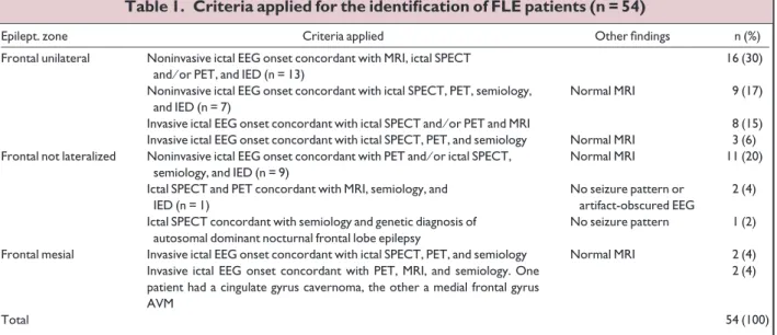
Table 1. Criteria applied for the identification of FLE patients (n = 54)
Epilept. zone Criteria applied Other findings n (%)
Frontal unilateral Noninvasive ictal EEG onset concordant with MRI, ictal SPECT
and⁄or PET, and IED (n = 13)
16 (30)
Noninvasive ictal EEG onset concordant with ictal SPECT, PET, semiology, and IED (n = 7)
Normal MRI 9 (17)
Invasive ictal EEG onset concordant with ictal SPECT and⁄or PET and MRI 8 (15)
Invasive ictal EEG onset concordant with ictal SPECT, PET, and semiology Normal MRI 3 (6)
Frontal not lateralized Noninvasive ictal EEG onset concordant with PET and⁄or ictal SPECT,
semiology, and IED (n = 9)
Normal MRI 11 (20)
Ictal SPECT and PET concordant with MRI, semiology, and IED (n = 1)
No seizure pattern or artifact-obscured EEG
2 (4)
Ictal SPECT concordant with semiology and genetic diagnosis of autosomal dominant nocturnal frontal lobe epilepsy
No seizure pattern 1 (2)
Frontal mesial Invasive ictal EEG onset concordant with ictal SPECT, PET, and semiology Normal MRI 2 (4)
Invasive ictal EEG onset concordant with PET, MRI, and semiology. One patient had a cingulate gyrus cavernoma, the other a medial frontal gyrus AVM
2 (4)
Total 54 (100)
characterized by predominance of occipital alpha background activity, eye blinking, and lack of any drowsy pattern. Therefore, RMT associated with drowsiness was excluded for the purpose of this study. In addition, RMT associated with mental activation and occurring during clinical seizures was not included. Mental activation was excluded by observing the video. The EEG studies of the patients were included for further analysis only if another EEG reviewer, who reviewed solely the most representa-tive EEG periods considered by the first EEG reviewer, agreed on the result of RMT and the above-mentioned conditions were excluded. The concordance between both reviewers who were blinded to each other’s opinion was total.
Statistical analysis
Chi-square analysis was used to evaluate the signifi-cance of association of RMT between the different groups.
Results
RMT was found significantly more frequently in FLE patients (48.1%, 26 of 54) than in TLE patients (3,7%, 2 of 54) (p < 0.01). In individual patients, RMT occurred daily for five times per day on average (range from 2–10). A typical example of RMT in an FLE patient is given in Fig. 1. RMT was not observed in the control group of nonepileptic patients. Moreover, there was no statistically significant difference between RMT occurrence in patients with TLE and the control group.
RMT was located in the vertex (electrode Cz) in 22 of 28 patients (79%) and in the frontocentral midline (electrode Fz) in the remaining 6 patients (21%). The average frequency seen was 6 Hz (range 5–7 Hz). Average RMT bursts lasted 8 s (range 3–12 s).
When comparing the intralobar localization, it is inter-esting to note that all (100%) mesial FLE patients (n = 4) (as established by mesial frontal subdural electrodes) showed RMT, whereas this was less frequently the case in the other FLE patients (44%, n = 22) (p = 0.03). The epi-leptic pathology among these 22 FLE patients may also include the mesial frontal structures, but we did not estab-lish an exclusive mesial frontal epileptogenic zone in these 22 patients, of whom 3 underwent invasive EEG studies.
Thirteen of our 54 patients with FLE (24%) did not have any IED. RMT was present in 8 of these 13 patients (62%).
Discussion
This study demonstrates that RMT is common in patients with FLE (48%) and may help to differentiate FLE from TLE in patients with refractory epilepsies con-sidered for resective epilepsy surgery. The frequency of RMT may have been underestimated in this study, as methodologically the second EEG reviewer, reviewed only a select sample of the continuous EEG-video moni-toring in preselected patients. RMT was originally attrib-uted to TLE on the basis of electroclinical data before the imaging era (Ciganek, 1961). Others confirmed that RMT is more frequent in epilepsy patients than nonepileptic
Table 2. Criteria applied for the identification of TLE patients (n = 54)
Epilept. zone Criteria applied Other findings n (%)
Temporal unilateral Noninvasive ictal EEG onset concordant with MRI, ictal SPECT
and⁄or PET, semiology, and IED
29 (54)
Noninvasive ictal EEG onset concordant with ictal SPECT, PET, semiology, and IED
Normal MRI 7 (13)
Invasive ictal EEG onset concordant with PET, semiology, and IED (n = 2)
Normal MRI or bitemporal lesions
4 (7)
Invasive ictal EEG onset concordant with PET, MRI, and IED (n = 1) 2 (4)
Temporal mesial Invasive ictal EEG onset concordant with ictal SPECT and⁄or PET,
semiology, and IED
Normal MRI or bilateral hippocampal sclerosis
4 (7)
Noninvasive ictal EEG onset concordant with MRI, ictal SPECT, PET, semiology, and IED
2 (4)
Temporal bilateral Noninvasive ictal EEG onset concordant with ictal SPECT, PET,
semiology, and IED
Normal MRI 3 (6)
Noninvasive ictal EEG onset concordant with MRI, ictal SPECT, PET, and IED
2 (4)
Invasive ictal EEG onset concordant with PET, MRI, and semiology 1 (2)
Total 54 (100)
patients (Mokran et al., 1971; Westmoreland & Klass, 1986). However, 22% or 39% of patients showing RMT, depending on the series, did not have epilepsy, but had other conditions such as headache, head trauma, stroke, multiple sclerosis and brain abscess, labyrinthine disease, and syncope (Mokran et al., 1971; Westmoreland & Klass, 1986). RMT was described in the EEG studies of 2% of the patients in a large series of patients (n = 1,250) with various neurologic conditions (Mokran et al., 1971). The association with epilepsy has been questioned, since RMT was recorded in approximately 35% of normal adults and considered a drowsy pattern (Maulsby, 1968). RMT also appears in nonepileptic persons while they are performing working memory tasks (Gevins et al., 1997). Therefore, there is uncertainty about the prevalence of RMT and its clinical significance. The available studies show discrep-ant results, which may be related to the lack of imaging data in the epilepsy studies and the difficulty in excluding RMT as a normal drowsy pattern and as a product of
mental activation. According to previous studies, one would expect to find RMT in 35% (12 of 54 of our controls) of nonepileptic individuals (Maulsby, 1968). This was not the case in our study, most probably because we meticulously excluded RMT during drowsiness and during mental activation. Therefore, no RMT was observed in the control group of nonepileptics.
RMT in normal subjects is most likely generated in the medial prefrontal cortices, including the anterior cingulate cortex, as demonstrated by dipole modeling and magneto-encephalography (MEG) (Gevins et al., 1997; Ishii et al., 1999). Electrical stimulation of the human anterior cingu-late cortex elicited a regular 3–8 Hz rhythm, which can be recorded noninvasively at the vertex (Talairach et al., 1973). In view of this experience, it is interesting to note that, although few, all of our invasively proven mesial FLE patients showed RMT in the scalp EEG study, which makes it likely that RMT in these patients reflects mesial frontal lobe abnormality. RMT was described in 4 of 10
Figure 1.
Interictal EEG in transverse (A) and longitudinal (B) bipolar montage of a 19-year-old female patient with a right FLE caused by focal cortical dysplasia. This electroencephalogram shows interictal rhythmical midline theta (RMT) local-ized to the vertex (electrode Cz). Muscle artifacts and eye blinks indicate the awake state of the patient. There was no mental activation during this period of EEG study.
epileptic patients (40%) with interictal midline parasagit-tal epileptogenic discharges, but no icparasagit-tal EEG was avail-able in this study (Pedley et al., 1981).
The clinical significance of RMT may increase because about 30–40% of FLE patients may lack IED (Morris et al., 1988; Salanova et al., 1993), or show predominantly temporal IED (Williamson & Spencer, 1986). In our ser-ies, 24% (n = 13) of the FLE patients lacked IED. In a most of these patients (62%; n = 8), RMT provided additional interictal localizing information to suggest a frontal epi-leptogenic zone. The lack of IED in our FLE patients, which is smaller than in other studies (Morris et al., 1988; Salanova et al., 1993), is possibly explained by the fact that we visually inspected the continuous long-term EEG recording, which may have a significantly higher yield (Kellinghaus & Luders, 2004).
Both TLE patients with RMT presented predominant unilateral temporal lobe IED. Therefore, RMT is not nec-essarily an interictal element of FLE, and may coexist with unilateral temporal IED, perhaps reflecting a more wide-spread abnormality. Furthermore, IEDs were present in all TLE patients. Only one of our TLE patients showed addi-tional extratemporal IED, namely unilateral frontal, but no RMT. Consequently, although our study did not specif-ically address this issue, one could speculate that when RMT occurs coupled with frontal IED, it emphasizes a frontal lobe epileptogenic area.
We would consider RMT as an EEG correlate of dysfunction rather than an epileptogenic abnormality, because it is a nonspecific pattern seen in a heteroge-neous group of neurologic and normal conditions, not correlated with epilepsy unless the normal drowsy variants and RMT associated with mental activation are excluded. The similarity of RMT to frontal inter-mittent rhythmical delta activity (FIRDA) may reflect common cortical dysfunction (frontal mesial) com-bined with abnormal thalamocortical circuits (Schaul, 1990). It is known that the mesial prefrontal cortex, including the anterior cingulate cortex, has functional connections with the thalamus (Hsu & Shyu, 1997; Masterman & Cummings, 1997). Thus pacemakers of the thalamus possibly work as generators of RMT, and thalamocortical volleys may act to trigger mal-functioning cortical neurons. Accordingly, experimen-tal studies in rats showed that theta rhythm was more commonly seen in the cortical stroke group (10 of 10) than in the control group (5 of 9) (Kharlamov et al., 2003).
In conclusion, to make use of the additional information that RMT may provide as a common finding in FLE, one should ensure that the normal conditions, such as drowsi-ness or mental activation, which are associated with RMT are excluded. Under this precondition, RMT is able to dif-ferentiate FLE from TLE, and should be included in the evaluation of patients considered for resective epilepsy
surgery. On the basis of the hypothesis that RMT reflects a dysfunctional abnormality in epilepsy, further studies should address the correlation of RMT on scalp recording with simultaneous invasive monitoring and also whether RMT disappears postoperatively in patients, who were rendered seizure free.
Acknowledgments
The authors thank E. Sincini, R. Grossmann, E. Scherbaum, R. Tschackert, and O. Klein for technical assistance in the EEG-video monitoring unit and K. Ogston for copyediting the manuscript.
We confirm that we have read the Journal’s position on issues involved in ethical publication and affirm that this report is consistent with those guidelines.
Conflict of interest: None of the authors has any conflicts of interest to disclose.
References
Bautista RE, Spencer DD, Spencer SS. (1998) EEG findings in frontal lobe epilepsies.Neurology50:1765–1771.
Blume WT, Borghesi JL, Lemieux JF. (1993) Interictal indices of tempo-ral seizure origin.Ann Neurol34:703–709.
Ciganek L. (1961) Theta-discharges in the middle-line-EEG symptom of temporal lobe epilepsy. Electreoencephaolgr Clin Neurophysiol 13:669–673.
Duncan JS. (1997) Imaging and epilepsy.Brain120:339–377.
Foldvary N, Klem G, Hammel J, Bingaman W, Najm I, Luders H. (2001) The localizing value of ictal EEG in focal epilepsy. Neurology 57:2022–2028.
Gevins A, Smith ME, McEvoy L, Yu D. (1997) High-resolution EEG mapping of cortical activation related to working memory: effects of task difficulty, type of processing, and practice.Cereb Cortex7:374– 385.
Geyer JD, Bilir E, Faught RE, Kuzniecky R, Gilliam F. (1999) Significance of interictal temporal lobe delta activity for localization of the primary epileptogenic region.Neurology52:202– 205.
Gibbs FA, Gibbs EL. (1952)Atlas of electroencephalography. Addison-Wesley, Cambridge, MA.
Hsu MM, Shyu BC. (1997) Electrophysiological study of the connection between medial thalamus and anterior cingulate cortex in the rat. Neuroreport8:2701–2707.
Ishii R, Shinosaki K, Ukai S, Inouye T, Ishihara T, Yoshimine T, Hirabu-ki N, Asada H, Kihara T, Robinson SE, Takeda M. (1999) Medial pre-frontal cortex generates pre-frontal midline theta rhythm.Neuroreport 10:675–679.
Jan MM, Sadler M, Rahey SR. (2001) Lateralized postictal EEG delta predicts the side of seizure surgery in temporal lobe epilepsy. Epilep-sia42:402–405.
Kellinghaus C, Luders HO. (2004) Frontal lobe epilepsy. Epileptic Disord6:223–239.
Kharlamov EA, Jukkola PI, Schmitt KL, Kelly KM. (2003) Electrobe-havioral characteristics of epileptic rats following photothrombotic brain infarction.Epilepsy Res56:185–203.
Masterman DL, Cummings JL. (1997) Frontal-subcortical circuits: the anatomic basis of executive, social and motivated behaviors. J Psychopharmacol11:107–114.
Maulsby R. (1968) The normative electroencephalographic data reference library. Final report, contract NAS 9-1200. National Aeronautics and Space Administration, Washington, DC.
Mokran V, Ciganek L, Kabatnik Z. (1971) Electroencephalographic theta discharges in the midline.Eur Neurol5:288–293.
Morris HH 3rd, Kanner A, Luders H, Murphy D, Dinner DS, Wyllie E, Kotagal P. (1989) Can sharp waves localized at the sphenoidal electrode accurately identify a mesio-temporal epileptogenic focus? Epilepsia30:532–539.
Pedley TA, Tharp BR, Herman K. (1981) Clinical and electroencephalo-graphic characteristics of midline parasagittal foci. Ann Neurol 9:142–149.
Rosenow F, Luders H. (2001) Presurgical evaluation of epilepsy.Brain 124:1683–1700.
Salanova V, Morris HH 3rd, Van Ness PC, Luders H, Dinner D, Wyllie E. (1993) Comparison of scalp electroencephalogram with subdural electrocorticogram recordings and functional mapping in frontal lobe epilepsy.Arch Neurol50:294–299.
Schaul N. (1990) Pathogenesis and significance of abnormal nonepilepti-form rhythms in the EEG.J Clin Neurophysiol7:229–248.
Schulz R, Luders HO, Hoppe M, Tuxhorn I, May T, Ebner A. (2000) Inte-rictal EEG and ictal scalp EEG propagation are highly predictive of surgical outcome in mesial temporal lobe epilepsy. Epilepsia 41:564–570.
Talairach J, Bancaud J, Geier S, Bordas-Ferrer M, Bonis A, Szikla G, Rusu M. (1973) The cingulate gyrus and human behaviour. Electro-encephalogr Clin Neurophysiol34:45–52.
Westmoreland BF, Klass DW. (1986) Midline theta rhythm.Arch Neurol 43:139–141.