Review
Frontal lobe epilepsy
Pedro Beleza
⇑, João Pinho
Epilepsy Group, Department of Neurology, Braga Hospital, Largo Carlos Amarante, Apartado 2242, Braga 4701-965, Portugal
a r t i c l e
i n f o
Article history:
Received 13 January 2010 Accepted 7 August 2010
Keywords:
EEG
Frontal lobe epilepsy Refractory epilepsy SPECT
Surgery Treatment
a b s t r a c t
About one-quarter of patients with refractory focal epilepsies have frontal lobe epilepsy (FLE). The typical seizure semiology for FLE includes unilateral clonic, tonic asymmetric or hypermotor seizures. Interictal electroencephalograms (EEG) usually reveal interictal epileptiform discharges and rhythmical midline theta, which has localizing value. The usefulness of ictal EEG recordings is limited by frequent muscle artifacts in motor seizures and because a large portion of the frontal lobe cortex is ‘‘hidden’’ to scalp elec-trodes. Ictal single photon emission CT and positron emission tomography are able to localize FLE in about one-third of patients only. A pre-surgical evaluation should include, whenever possible, a subclas-sification of FLE as dorsolateral frontal, mesial frontal or basal frontal lobe epilepsy to allow a minimal cortical resection. A review of the typical findings of seizure semiology, interictal and ictal EEG regarding the different FLE subtypes is given. Etiology, medical treatment and surgery are also discussed.
Ó2010 Elsevier Ltd. All rights reserved.
1. Introduction
Refractory epilepsy is diagnosed when there is inadequate sei-zure control despite use of potentially effective antiepileptic drugs (AED) at tolerable levels for 1–2 years. Once refractoriness is estab-lished, surgical treatment must be considered.1Of all patients with
refractory focal epilepsies referred to epilepsy surgery, 25% have frontal lobe epilepsy (FLE).2 The objective of resective surgery is
the removal of the entire epileptogenic zone (EZ) without causing permanent neurological deficits. Given this objective, localization of the EZ is of paramount importance. This can be achieved by combining seizure semiology, interictal and ictal electroencephalo-gram (EEG) findings, as well as fluorodeoxyglucose (FDG)-positron emission tomography (PET), single photon emission CT (SPECT) and MRI.3
Unilateral clonic seizures,4tonic asymmetric seizures with
pre-served consciousness5and hypermotor seizures,4while not
patho-gnomonic, are specific for FLE. Even though abdominal auras may occur in FLE, the evolution of an abdominal aura into an automotor seizure is typical of temporal lobe epilepsy (TLE), which allows its differentiation from FLE.6The presence of a visual aura strongly ar-gues against an FLE, since visual auras are associated with parietal, temporal or occipital lobe epilepsy.7Given that an ictal EEG from a
patient with FLE is characterized by frequent false negatives and frequent muscle artifacts,8the analysis of ictal semiology is crucial
for the differential diagnosis between FLE and psychogenic non-epileptic seizures (PNES), the most frequent (90%) condition
misdiagnosed as epilepsy.9 Certain characteristics of the motor
phenomena are strongly associated with PNES, including a very gradual onset or termination, pseudosleep, discontinuous (stop-and-go) and irregular or asynchronous (out-of-phase) activity, side-to-side head shaking, opisthotonic posturing, stuttering and weeping.10
Interictal epileptiform discharges (IED) occur in 60% to 80% of FLE and are considered to be of less localizing value than in TLE be-cause they can be bilateral, multilobar or even generalized.11
Inter-ictal rhythmical midline theta (RMT) is common (50% of FLE
patients) and has localizing value in patients with FLE, provided that it can be distinguished from normal variants occurring with drowsiness and mental activation tasks.12Ictal EEG is often
gener-alized and locgener-alized patterns are observed in fewer than one-third of patients (Fig. 1).13
Localization of seizure onset with ictal SPECT in adults is possi-ble in only 30% to 43% of patients with FLE.14With the use of
FDG-PET, it is possible to localize a hypometabolic region in about 75% of patients with unilateral FLE and abnormal MRI,15but in only 29%
to 45% of patients with unilateral FLE and normal MRI.16
FLE should be, whenever possible, further classified as dorsolat-eral frontal, mesial frontal or basal to allow minimal cortical resection.
2. Dorsolateral frontal lobe epilepsy
Dorsolateral FLE may be further subdivided into central, premo-tor and prefrontal lobe epilepsy. The central lobe is sometimes de-scribed as the region formed by the primary motor cortex and the sensory cortex (Brodmann areas 4 and 3) (Fig. 2). The border
be-0967-5868/$ - see front matterÓ2010 Elsevier Ltd. All rights reserved. doi:10.1016/j.jocn.2010.08.018
⇑ Corresponding author. Tel.: +35 1253209000.
E-mail address:[email protected](P. Beleza).
Journal of Clinical Neuroscience 18 (2011) 593–600
Contents lists available atScienceDirect
Journal of Clinical Neuroscience
tween these motor and sensory areas was thought to be the central sulcus, but recent studies showed both motor (tonic, clonic or
mo-tor arrest) and sensory responses after electrical stimulation of the gyrus precentralis and gyrus postcentralis.17Functionally, the
pre-motor cortex (Fig. 2) includes the secondary motor area (posterior parts of the frontal gyri), the frontal eye field (intersection of sulcus precentralis and superior frontal sulcus) and Broca’s language area (opercular and triangular parts of the inferior frontal gyrus in the dominant hemisphere). The premotor cortex projects to the pri-mary motor cortex and, less extensively, to the motor systems of the spinal cord, and there is evidence in animal studies supporting its role in motor preparation and motor learning.18Extensive
fron-tal lobe resections up to the precentral sulcus, sparing the supple-mentary motor area, do not lead to any permanent or even transient motor disturbance.19The prefrontal cortex (Fig. 2) is in-volved in emotion processing, moral behaviour, executive control, monitoring in working memory, learning and temporal structuring of behavior by context.20Even though some hypotheses propose that individualized tasks are carried out by the prefrontal cortex, this brain region might be responsible for the coordination of infor-mation processing and transfer, required for occurrence of multiple high-level cognitive operations.21
2.1. Seizure semiology
2.1.1. Central lobe
Although non-specific auras occur in most patients with FLE, fo-cal somatosensory auras, more commonly unilateral parasthesias (‘‘tingling’’, ‘‘numbness’’ or ‘‘strange feeling’’ sensations) restricted to the hand, the face/tongue or the foot, are specific to contralateral
Fig. 1.Ictal electroencephalogram (EEG) in longitudinal bipolar montage of a 16-year-old female with a ring chromosome 20 syndrome. The EEG shows a predominantly right frontal seizure pattern occuring during a dyscognitive seizure.
involvement of the central lobe.4Likewise, unilateral myoclonic or
clonic seizures, more frequently affecting distal segments of the body (such as the face or tongue), are generally also the expression of the epileptic activation of the contralateral central lobe. As for electrical stimulation of the primary motor area, it usually does not cause tonic contractions, but rather clonic twitching of the af-fected muscles. The pathogenesis of clonic seizures, which consists primarily of repetitive myoclonic jerks, is probably very similar to that of myoclonic seizures.22Typical seizure evolution includes: (i) focal clonic seizures with Jacksonian march without secondary generalization, usually accompanied by ipsilateral head version and followed by postictal paresis; and (ii) somatosensory aura of-ten followed by tonic posturing and head version or clonic sei-zures; automatisms and vocalization are rare.4
2.1.2. Premotor cortex
Typical seizure evolution associated with lesions of the premo-tor cortex includes early versive seizure, frequently followed by other motor manifestations such as automatisms.
Versive seizures, characterized by lateral deviation of the eyes (tonic or saccadic), version of the head and, frequently, also of the trunk, especially when followed by a secondary generalized to-nic–clonic seizure, indicate an epileptic activation of the frontal eye field contralateral to the side of eye deviation.23Aphasic
sei-zures may occur if Broca’s language area is involved. Long-lasting postictal aphasia is seen in >90% of seizures starting in the frontal lobe of the dominant side that spreads to the ipsilateral temporal lobe.24
2.1.3. Prefrontal cortex
Hypermotor seizures were defined by Lüders et al.25as complex
movements involving trunk and proximal limb segments, usually with the preservation of consciousness, and are considered specific for FLE, in close association with frontopolar and orbitofrontal cor-tical lesions.4This type of seizure is frequently preceded by an aura (fear, ill-defined feelings, and somatosensory phenomena) and in-cludes bizarre gestures, repetitive movements, bicycle peddling, pelvic thrusting and shouting, often charged with emotional and aggressive features. Hypermotor seizures are usually short and tend to occur during sleep. Unlike seizures involving the central lobe, the complex semiology of prefrontal seizures may be caused by disruption of neuronal synchrony between different brain re-gions rather than by excitation of single areas of the cortex.26
2.2. Interictal EEG
A concordant EZ and irritative zone was found in 72% of patients with dorsolateral FLE compared to 33% with mesial FLE (Fig. 3).27
Possible reasons for this difference are the smaller distance be-tween lateral cortex and scalp electrodes and that the dipoles tan-gential to the scalp in mesial FLE cannot be detected by EEG. The sensitivity of interictal EEG is higher in intracranial subdural than in scalp recordings. Due to the closer distance to the cortex, sub-dural electrodes may reveal a smaller irritative zone in some pa-tients, when compared to surfaces electrodes. However, a sampling bias remains in invasive monitoring studies.28
2.3. Ictal EEG
Ictal scalp EEG in 127 seizures of 15 patients with dorsolateral FLE showed correct localization of the EZ in 65% of patients, while 26% of seizures started with generalized EEG activity and 3% were mislateralized in EEG analysis.29In this study only 1.5% of the
sei-zures was obscured by artifacts or did not show EEG changes. The most frequent EEG patterns at seizure onset were repetitive epilep-tiform activity (36%), rhythmic delta (26%) and EEG suppression
(14%), in contrast to rhythmical theta activity, the most frequent seizure pattern in TLE, which was seen in only 9% of the 127 sei-zures. A study comparing medial (n= 5) with dorsolateral (n= 4) patients with FLE found that absence of focal electrographic sei-zure activity excluded the possibility of dorsolateral frontal lobe seizures with a negative predictive value of 93%, but this conclu-sion may be misleading because of the small number of study par-ticipants.13 Several authors have reported that, although scalp
electrodes showed widespread seizure onset and MRI was normal or non-localizing, the use of subdural grid electrodes that exten-sively covered the frontal areas was able to localize the seizure on-set zone in >90% of patients.30
3. Mesial frontal epilepsy
The mesial surface of the frontal lobe includes primary sensory and motor cortex for the lower limb, the supplementary sensori-motor area (SSMA), the anterior cingulate cortex and the prefrontal cortex31(Fig. 2). The SSMA extends anteriorly approximately to the
level of the genu of the corpus callosum. SSMA stimulation results in usually bilateral and proximal tonic posturing and motor re-sponses, but frequently show predominance on the contralateral side. Additionally, contralateral sensory phenomena may occur. The SSMA has a somatotopic distribution: the head and upper limbs are represented at the anterior and the lower limbs at the posterior surface of the interhemispheric region. Stimulation of the anterior portion of the SSMA results in arrest or slowing of vol-untary motor activity. Furthermore, stimulation of the cingulate gyrus near the SSMA leads to motor responses that overlap those occurring in the SSMA,32but automatisms, namely oro-alimentary,
have also been described.33
3.1. Seizure semiology
A somatosensory aura consisting of ‘‘tingling’’ or a feeling of tension, pulling or heaviness in a limb or the impression of impending movement of the limb may precede the tonic seizure. The sensation may be relatively focal, involving a portion of a limb, lateralized with both upper and lower limbs involved simulta-neously, or a poorly defined bilateral sensation in the head or body.34The symptoms may arise from the sensory representation
within the SSMA32or may be the awareness of tension developing
in muscle groups involved in the tonic contraction. Bilateral asym-metric tonic seizures are characterized by an abrupt onset of tonic posturing maintained for 10 s to 40s and absence of any postictal stupor or confusion.35Penfield and Jasper described ‘‘the arm being
raised and the head and eyes turned as though to look at the hand’’, which is called the ‘‘fencing posture’’.36Moreover, Ajmone-Marsan
and Ralston created the term ‘‘M2e’’ to describe tonic abduction and external rotation of the shoulder with flexion of the elbow. They described SSMA involvement if M2e posturing occurred with-out loss of consciousness and withwith-out progression into a secondar-ily generalized tonic–clonic seizure.37Although asymmetric tonic
seizure is typically associated with mesial FLE, it is not specific.38
Tonic seizures arising from the SSMA preferentially affect muscle groups on both sides of the body, yet, they more often predominate in the contralateral musculature.39In most patients with focal
epi-lepsy, consciousness remains unclouded during tonic seizures, at least at the onset of seizures.39 Strictly unilateral tonic seizures
have a highly lateralizing significance, pointing to a contralateral seizure onset.39Other distinct semiologies may also be associated
dorsolateral frontal,11frontopolar and opercular–insular41seizure
onset have all been reported. While seizure onset in several frontal regions may produce this seizure semiology, the anterior cingulate region has been frequently proposed as the cortical region respon-sible for the clinical signs and symptoms. Dialeptic seizures, as de-fined by Lüders et al.,25 consist of episodes with loss of
consciousness, during which a patient cannot react or reacts only to a limited extent to external stimuli and shows minimal motor activity. Dialeptic seizures are rare in patients with FLE and were termed ‘‘frontal absences’’ due to their resemblance to dialeptic seizures in patients with generalized epilepsies (‘‘absence’’). In contrast to childhood absences, patients with frontal lobe absences may have subtle repetitive vocalizations, rocking movements, small degrees of head and eye turning, report awareness of a motor arrest without loss of consciousness and have brief postictal confu-sion.41Staring may evolve into a generalized tonic–clonic seizure
via version of the head and eyes, focal tonic posturing of an upper limb or bilateral tonic posturing. Patients with dialeptic seizures in FLE seem to have a more anterior EZ than those with bilateral asymmetric tonic seizures.41 This clinical semiology has been
as-cribed to bilateral cingulate gyrus involvement via the callosal route.42 Negative myoclonic seizure consists of short phases of
muscle atonia (30–400 ms), which are preceded by epileptiform discharges in the central region (20–30 ms). Generalized and focal negative myoclonic seizures have also been reported.43Several
re-ports indicate that these seizures are caused by the sudden inhibi-tion of tonic inervainhibi-tion of motor neurons, as evidenced by the silent electromyelogram (EMG) period. Recent studies showed that
SSMA stimulation induces silent periods only, regardless of the stimulus intensity, whereas occurrence of silent periods following stimulation of the premotor cortex, primary motor cortex or pri-mary somatosensory area depended mainly on the intensity of stimulation.44Gelastic seizures are seizures characterized by ictal
laughing, sometimes accompanied by mirth, that frequently occur in patients with hypothalamic hamartomas.45 The anteromesial
superior frontal gyrus and anterior cingulate gyrus have been de-scribed as involved in motor aspects of laughter,46while the
tem-poral lobes, particularly the basal regions, seem to be mainly involved in the processing of mirth.47
3.2. Interictal EEG
The interictal EEG in patients with mesial FLE generally shows either abundance of non-lateralised epileptiform activity or none at all.48Focal IED at or adjacent to the midline have been reported
in patients with tonic postural seizures.35 Blume and Oliver49
found that about 50% of patients (n= 13) with ‘‘supplementary mo-tor area epilepsy’’ show midline (Fz, Cz) (five patients) or frontal (F4, F3) (two patients) spike foci. EEG analysis with transverse montages and using midline electrodes Fz, Cz and Pz is essential, as maximal discharges at these electrodes may have a limited field.49 In addition, bilateral frontal synchronous discharges are
characteristic, but not specific, of mesial FLE.40 A recent study
showed that all mesial FLE patients (n= 4, established by invasive EEG recordings) were characterised by interictal RMT, but this finding was less frequent in other FLE patients (44%, 22 of 50),
viding evidence that RMT may be a neurophysiological marker for mesial frontal lobe abnormality.12 Nevertheless, replication of
these results by further studies with a larger cohort of patients is needed. RMT is seen in patients with bilateral asymmetric tonic seizures and in patients with midline parasagittal epileptic discharges.50
3.3. Ictal EEG
Muscle activity is prominent from the onset of bilateral asym-metric tonic seizures and the EEG is frequently contaminated with considerable EMG and movement artifacts. Seizure patterns may still be evident at the vertex, where EMG activity is minimal. Ab-sence of any ictal or immediate postictal EEG slowing has been re-ported in patients with mesial FLE.13In the study by Foldvary et al.,
just over 50% of the seizures analyzed were obscured or showed no EEG change in the mesial frontal lobe epilepsy patients, an uncom-mon occurrence in the other focal epilepsy groups.29Characteristic
findings include an initial high amplitude slow wave transient or midline sharp wave, followed by bilateral frontocentral low volt-age fast activity or electrodecrement.51Accordingly, one study
re-ported that seizures from the mesial frontal lobe more frequently showed paroxysmal fast activity (33%) or electrodecrement (29%) as the initial ictal pattern.29Electrodecrement will usually evolve
into low voltage fast activity and then bilateral frontocentral or generalized rhythmic theta slowing.52The low voltage fast activity
and the rhythmic slow activity may be either localized in the ver-tex or be more diffuse. Subtle lateralization of these rhythms may occur but, in general, the lateralizing information from ictal EEG is minimal. Indeed, it has been shown that only 25% of mesial FLE sei-zures correctly localized or lateralized on EEG and 75% had non-lateralised patterns.29When indicated, intracranial EEG with depth
electrodes, usually placed bilaterally, provides greater accuracy for lateralization and localization, but also carries a significant risk of parenchymal hemorrhage.
4. Basal frontal lobe epilepsy
On the basal (orbital) surface of the frontal lobes, five gyri can be identified: lateral orbital gyrus, anterior and posterior orbital gyri, medial orbital gyrus and gyrus rectus19(Fig. 2). The posterior
part of the orbitofrontal region is continuous with the rostral part of the agranular insula, and is hence regarded as more ‘‘limbic’’ in character. However, the rostrally placed isocortical orbitofrontal region has features of granular isocortex and blends into the dorso-lateral heteromodal components of the prefrontal cortex.53
4.1. Seizure semiology
Olfactory auras, usually described by patients as unpleasant, are frequently an expression of epileptic activation of the orbitofrontal part of the gyrus rectus.54Autonomic seizures may also occur, and
consist of ictal events during which the main abnormality is an objective autonomic change including cardiovascular (tachycardia, bradycardia, asystole, arrhythmia), respiratory (hyperventilation, apnea, dyspnea and stridor), gastrointestinal (epigastric aura, vom-iting, spitting, defecation), cutaneous (piloerection, pallor, flush-ing), papillary (mydriasis, miosis) or urogenital (urinary urge, sexual/orgasmic aura, genital aura, sexual and genital automa-tisms) manifestations. To document its epileptic nature, a simulta-neously recorded EEG seizure pattern is usually required. Ictal ‘‘vegetative’’ manifestations are thought to result from activation of the orbitofrontal and opercular insular regions.41 Hypermotor
seizures are also common, although not specifically associated with this region.
4.2. Interictal and ictal EEG
Typically, abnormalities detected by scalp EEG do not allow for topographic localization of foci residing in the basal frontal lobe, mostly due to the inaccessibility of the basal frontal surface to scalp electrodes. When present, IED may have a regional distribu-tion or appear generalized as a result of secondary bilateral syn-chrony. Case reports by Ludwig and co-workers highlighted the occurrence of bilaterally synchronous epileptiform discharges, with a bifrontal or frontopolar maximum, as well as discharges involving one anterior quadrant, with or without evidence of addi-tional temporal lobe involvement.55In a single patient with
orbito-frontal epilepsy described by Chang et al., the use of invasive recordings showed that sphenoidal recordings were able to lateral-ize the EZ, and the infraorbital scalp electrodes added to the scalp EEG revealed that the observed bisynchronous discharges had a more basal distribution with a maximum in the infraorbital re-gions.55 False localization to the anterior temporal region is not
uncommon in patients with basal frontal epilepsies.56 Occasion-ally, propagated epileptiform activity can be present over central or frontolateral regions. Moreover, epileptiform abnormalities may have a misleadingly widespread appearance because of the large distance and intervening cortical area that separates the EZ from the scalp EEG electrodes.54
5. Etiology
In a study of 68 patients with FLE who underwent frontal lesionectomy, the histopathological findings were: tumors in 24 patients (35%) (glioneural tumors, 8; astrocytic tumors, 15; and osteoma, 1), dysgenetic lesions in 18 patients (26%) (glioneural hamartoma, 15; cortical dysplasia, 1; cortical subcortical disorga-nization, 1; ectopical cortical neurons, 1), gliosis in 14 patients (21%), vascular malformations in 10 patients (15%) (cavernomas, 6; arteriovenous malformations, 4), encephalitis in one patient (1.5%) and necrosis in one patient (1.5%).57The Cleveland Clinic
series included 70 patients with FLE who underwent a frontal lobectomy between 1995 and 2003. Based on MRI and surgical pathology, patients were divided into the following etiological sub-groups: (i) malformation of cortical development (MCD) with abnormal MRI (41% of patients); (ii) MCD with normal high-resolu-tion MRI (17%); (iii) tumor (19%); (iv) vascular malformahigh-resolu-tion (3%); (v) cryptogenic with normal MRI and histology (10%); and (vi) encephalomalacia following stroke or trauma (10%).58
MRI-nega-tive MCD as a disease etiology proved to be an independent predic-tor of seizure recurrence after frontal lobectomy.58Another study
found that of a total of 21 patients with refractory nocturnal FLE submitted to surgery, 20 (95%) patients had focal cortical dysplasia detected on histological examination (including one patient with familial pedigree suggestive of autosomal dominant nocturnal FLE) and only 11 (52%) patients showed frontal lobe abnormalities on MRI. Invasive recording by stereo-EEG was performed in 18 (86%) patients.59The main genetic cause of FLE is autosomal
dom-inant nocturnal FLE (ADNFLE), a channelopathy inherited with incomplete (70%) penetrance resulting from mutations in genes coding for subunits of the nicotinic acetylcholine receptor.60
Clini-cally available molecular genetic testing reveals mutations in CHRNA4 or CHRNB2 in approximately 10% to 20% of individuals with a positive family history and in fewer than 5% of individuals with a negative family history of ADNFLE.60Ring chromosome 20
should be suspected in patients with recurrent frontal status and normal MRI.61Slight mental retardation or dysmorphism may also
be found.62A recent report described a patient with a ring
chromo-some 17 who presented with an epileptic syndrome similar to the
ring chromosome 20 syndrome, raising the question of overlap of ring chromosome epileptic syndromes.63
6. Additional and experimental methods
Despite its low spatial resolution, MR spectroscopy may help to lateralize and even to localize epileptogenic frontal and central lobe lesions by detection of reduced N-acetylaspartate levels.64
The area of decreased N-acetylaspartate concentration frequently exceeds the epileptogenic lesion as seen in MRI.65Diffusion tensor imaging may be helpful for detection of the epileptogenic lesion in patients without structural changes on conventional MRI, espe-cially in patients with focal cortical dysplasia.66Furthermore, mul-tiplanar reconstruction and curvilinear reformatting have been shown to improve the localization of focal cortical dysplasias in the frontal lobe.67
7. Treatment of refractory FLE
7.1. Surgery
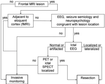
The algorithm used in our institution for pre-surgical evaluation of patients with FLE is outlined inFig. 4. A FLE patient with a lesion not adjacent to the eloquent cortex, with a congruent EEG (ictal and interictal), seizure semiology, and neuropsychology evaluation may be submitted to resective surgery without the need for inva-sive monitoring if: (i) ictal EEG reveals a lateralized or localized sei-zure pattern; or (ii) ictal EEG is normal or contains artifacts but PET or ictal SPECT is localized. Invasive monitoring is recommended when there is: FLE without a lesion; a lesion adjacent to an elo-quent cortex; no congruence between the different zones; or con-gruence but the ictal EEG is normal and the PET and ictal SPECT are not localized.3
Extratemporal lobe surgery for focal epilepsy accounts for less than 50% of all epilepsy surgeries.68In FLE surgery the probability
of becoming seizure-free is 55.7% at 1 year, 45.1% at 3 years, and 30.1% at 5 years.58 Mesial temporal lobe epilepsy (MTLE)
associ-ated with hippocampal sclerosis is the most common form of focal epilepsy, with around 60% of patients having anterior temporal lobe resections, of whom 60–70% are seizure free at 1–2 years of follow-up69 but only 58% are seizure free at 10 years.70 Patients
with FLE and favorable prognostic factors (MRI lesion restricted
to one frontal lobe, complete resection, regional or lateralized ictal scalp EEG pattern) show a seizure-free outcome comparable to MTLE patients after temporal lobectomy, with 50% to 60% being seizure free at 3 years. Regarding etiology, patients with low-grade tumors have the best outcome (62%) followed by patients with MRI visible malformations of cortical development (52%).
7.2. Palliative interventions
Complete seizure control is virtually unachievable for some pa-tients, but useful palliation can sometimes be achieved with tech-niques such as vagal nerve stimulation or multiple subpial transections.
Vagal nerve stimulation is indicated in adults with focal epilep-sies who are not surgical candidates or who have had surgery per-formed without success. On average, a 50% reduction of seizure frequency has been reported in about one-third of patients, the same range of expected benefit if a new AED is added, with the advantage of lower adverse effects. However, seizure freedom is rare.71
Multiple subpial transections use radially oriented incisions in the grey matter at 4-mm intervals to limit propagation of epileptic activity within eloquent cortex and to reduce seizure spread with-out disturbing functional integrity. A significant seizure reduction has been reported.72
A ketogenic diet, high in fat and low in carbohydrate, is mainly used in pediatric patients (due to questions of tolerability) as a sec-ond line treatment in focal non-surgical refractory epilepsy. A re-cent randomized controlled trial showed a reduction of seizure frequency of more than 50% in 38% of children with drug-resistant epilepsy.73
In chronic epilepsies (more than 5 years) the addition of a pre-viously unused AED provided seizure freedom in 17% and a 50% to 99% seizure reduction for 25%. For non-responders to the first trial, a similar benefit might be expected for at least two more trials. At the end, 28% of patients were seizure free.74 Zonisamide (100–
400 mg id), levetiracetam (1000–3000 mg id), lamotrigine (300– 500 mg id), topiramate (300–1000 mg id) and gabapentine (600– 1800 mg id) have demonstrated efficacy (evidence level A) as add-on therapy in patients with refractory focal epilepsy.75 Even
though the methodology was similar for all studies, a direct com-parison between outcomes does not allow determination of the relative efficacy, because populations differed and some drugs were not used in maximum doses, whereas others appear to have been administered above the ideal dose. For essentially all drugs, efficacy as well as side effects increased with increasing doses.75 In refractory epilepsy it is convenient to manage AED by: (i) increasing the dosage up to the maximum tolerable dose; (ii) if the patient is non-responsive, then replace the AED; if the patient responds partially, then add on an AED chosen according to the mechanism of action of the first AED (e.g. lamotrigine and valpro-ate are synergistic), efficacy and adverse effects.76
8. Conclusion
FLE is an important cause of refractory focal epilepsy and repre-sents a substantial group of patients referred for epilepsy surgery. Seizure semiology, MRI, ictal EEG, interictal EEG and PET/SPECT should be judiciously analyzed to further classify the FLE as central, premotor, prefrontal, frontal mesial or frontal basal epilepsy. Accu-rate localization of the EZ and recognition of prognostic factors fur-ther contribute to the success in FLE surgery. Antiepileptic drugs, vagal nerve stimulation, a ketogenic diet and multiple subpial tran-sections are beneficial in patients not eligible for resective surgery.
Acknowledgement
The authors acknowledge Dr. A. S. Costa for helpful English lan-guage editing assistance.
References
1. Beleza P. Refractory epilepsy: a clinically oriented review. Eur Neurol
2009;62:65–71.
2. Rasmussen T. Tailoring of cortical excisions for frontal lobe epilepsy.Can J Neurol Sci1991;18:606–10.
3. Rosenow F, Lüders H. Presurgical evaluation of epilepsy. Brain
2001;124:1683–700.
4. Manford M, Fish DR, Shorvon SD. An analysis of clinical seizure patterns and their localizing value in frontal and temporal lobe epilepsies. Brain
1996;119:17–40.
5. Quesney LF, Constain M, Fish DR, et al. The clinical differentiation of seizures arising in the parasagittal and anterolaterodorsal frontal convexities. Arch Neurol1990;47:677–9.
6. Henkel A, Noachtar S, Pfander M, et al. The localizing value of the abdominal aura and its evolution: a study in focal epilepsies.Neurology2002;58:271–6. 7. Noachtar S, Peters AS. Semiology of epileptic seizures: a critical review.Epilepsy
Behav2009;15:2–9.
8. Lee SK, Kim JY, Hong KS, et al. The clinical usefulness of ictal surface EEG in neocortical epilepsy.Epilepsia2000;41:1450–5.
9. Benbadis SR, O’Neill E, Tatum WO, et al. Outcome of prolonged video-EEG monitoring at a typical referral epilepsy center.Epilepsia2004;45:1150–3. 10. Benbadis S. The differential diagnosis of epilepsy: a critical review.Epilepsy
Behav2009;15:15–21.
11. Laskowitz DT, Sperling MR, French JA, et al. The syndrome of frontal lobe epilepsy: characteristics and surgical management.Neurology1995;45:780–7. 12. Beleza P, Bilgin O, Noachtar S. Interictal rhythmical midline theta differentiates
frontal from temporal lobe epilepsies.Epilepsia2009;50:550–5.
13. Bautista RE, Spencer DD, Spencer SS. EEG findings in frontal lobe epilepsies.
Neurology1998;50:1765–71.
14. Lee SK, Lee SY, Yun CH, et al. Ictal SPECT in neocortical epilepsies: clinical usefulness and factors affecting the pattern of hyperperfusion.Neuroradiology
2006;48:678–84.
15. Ryvlin P, Bouvard S, Le Bars D, et al. Clinical utility of flumazenil-PET versus [18F]fluorodeoxyglucose-PET and MRI in refractory partial epilepsy. A prospective study in 100 patients.Brain1998;121:2067–81.
16. Hamer HM, Morris HH, Mascha EJ, et al. Complications of invasive video-EEG monitoring with subdural grid electrodes.Neurology2002;58:97–103. 17. Nii Y, Uematsu S, Lesser RP, et al. Does the central sulcus divide motor and
sensory functions? Cortical mapping of human hand areas as revealed by electrical stimulation through subdural grid electrodes. Neurology
1996;46:360–7.
18. Kandel ER, Schwartz JH, Jessell TM.Principles of neuroscience. 3rd ed. New York: McGraw-Hill; 2000.
19. Tamraz JC, Comair YG. Atlas of regional anatomy of the brain using MRI. Berlin: Springer; 2006.
20. Bechara A, Damasio H, Tranel D, et al. Deciding advantageously before knowing the advantageous strategy.Science1997;275:1293–5.
21. Ramnani N, Owen AM. Anterior prefrontal cortex: insights into function from anatomy and neuroimaging.Nat Rev Neurosci2004;5:184–94.
22. Noachtar S, Luders HO. Classification of epileptic seizures and epileptic syndromes. In: Gildenberg PL, Tasker RR, editors.Textbook of stereotactic and functional neurosurgery. New York: McGraw-Hill; 1997. p. 1763–74. 23. Rheims S, Demarquay G, Isnard J, et al. Ipsilateral head deviation in frontal lobe
seizures.Epilepsia2005;46:1750–3.
24. Goldberg-Stern H, Gadoth N, Cahill W, et al. Language dysfunction after frontal lobe partial seizures.Neurology2004;62:1637–8.
25. Lüders H, Acharya J, Baumgartner C, et al. A new epileptic seizure classification based exclusively on ictal semiology.Acta Neurol Scand1999;99:137–41. 26. Bartolomei F, Trebuchon A, Gavaret M, et al. Acute alteration of emotional
behaviour in epileptic seizures is related to transient desynchrony in emotion-regulation networks.Clin Neurophysiol2005;116:2473–9.
27. Vadlamudi L, So EL, Worrell GA, et al. Factors underlying scalp-EEG interictal epileptiform discharges in intractable frontal lobe epilepsy.Epileptic Disord
2004;6:89–95.
28. Salanova V, Morris 3rd HH, Van Ness PC, et al. Comparison of scalp electroencephalogram with subdural electrocorticogram recordings and functional mapping in frontal lobe epilepsy.Arch Neurol1993;50:294–9. 29. Foldvary N, Klem G, Hammel J, et al. The localizing value of ictal EEG in focal
epilepsy.Neurology2001;57:2022–8.
30. Cukiert A, Buratini JA, Machado E, et al. Results of surgery in patients with refractory extratemporal epilepsy with normal or nonlocalizing magnetic resonance findings investigated with subdural grids.Epilepsia2001;42:889–94. 31. Donoghue JP, Sanes JN. Motor areas of the cerebral cortex.J Clin Neurophysiol
1994;11:382–96.
32. Lim SH, Dinner DS, Pillay PK, et al. Functional anatomy of the human supplementary sensorimotor area: results of extraoperative electrical stimulation.Electroencephalogr Clin Neurophysiol1994;91:179–93.
33. Unnwongse K, Lachhwani D, Tang-Wai R, et al. Oral automatisms induced by stimulation of the mesial frontal cortex.Epilepsia2009;50:1620–3.
34. Bleasel AF, Morris HH. Supplementary sensorimotor area epilepsy in adults. In: Lüders HO, editor.Advances in neurology. Philadelphia: Lippincott-Raven; 1996. p. 271–84.
35. Morris 3rd HH, Dinner DS, Lüders H, et al. Supplementary motor seizures: clinical and electroencephalographic findings.Neurology1988;38:1075–82. 36. Penfield W, Jasper H. Epilepsy and the functional anatomy of the human
brain. Boston: Little Brown; 1954.
37. Ajmone-Marsan C, Ralston BL.The epileptic seizure. Its functional morphology and diagnostic significance. Springfield, IL, USA: Charles C. Thomas; 1957. 38. Williamson PD. Frontal lobe epilepsy. Some clinical characteristics.Adv Neurol
1995;66:127–52.
39. Werhahn KJ, Noachtar S, Arnold S, et al. Tonic seizures: their significance for lateralization and frequency in different focal epileptic syndromes.Epilepsia
2000;41:1153–61.
40. Harvey AS, Hopkins IJ, Bowe JM, et al. Frontal lobe epilepsy: clinical seizure characteristics and localization with ictal 99mTc-HMPAO SPECT. Neurology
1993;43:1966–80.
41. Bancaud J, Talairach J. Clinical semiology of frontal lobe seizures. In: Chauvel P, Delgado-Escueta AV, Halgren E, Bancaud J, editors.Frontal lobe seizures and epilepsies. New York: Raven Press; 1992. p. 3–58.
42. Huck FR, Radvany J, Avila JO, et al. Anterior callosotomy in epileptics with multiform seizures and bilateral synchronous spike and wave EEG pattern.Acta Neurochir Suppl (Wien)1980;30:127–35.
43. Werhahn KJ, Noachtar S. Epileptic negative myoclonus. In: Lüders HO, Noachtar S, editors. Epileptic seizures: pathophysiology and clinical semiology. New York: Churchill Livingstone; 2000. p. 473–83.
44. Rubboli G, Mai R, Meletti S, et al. Negative myoclonus induced by cortical electrical stimulation in epileptic patients.Brain2006;129:65–81.
45. Arroyo S, Santamaria J, Sanmarti F, et al. Ictal laughter associated with paroxysmal hypothalamopituitary dysfunction.Epilepsia1997;38:114–7. 46. Unnwongse K, Wehner T, Bingaman W, et al. Gelastic seizures and the
anteromesial frontal lobe: A case report and review of intracranial EEG recording and electrocortical stimulation case studies. Epilepsia
2010;51:2195–8.
47. Arroyo S, Lesser RP, Gordon B, et al. Mirth, laughter and gelastic seizures.Brain
1993;116:757–80.
48. Williamson PD, Spencer DD, Spencer SS, et al. Complex partial seizures of frontal lobe origin.Ann Neurol1985;18:497–504.
49. Blume WT, Oliver L. Noninvasive electroencephalography in the evaluation of supplementary sensoriomotor area epilepsy.Adv Neurol1996;70:309–17. 50. Pedley TA, Tharp BR, Herman K. Clinical and electroencephalographic
characteristics of midline parasagittal foci.Ann Neurol1981;9:142–9. 51. Jobst BC, Siegel AM, Thadani VM, et al. Intractable seizures of frontal lobe
origin: clinical characteristics, localizing signs, and results of surgery.Epilepsia
2000;41:1139–52.
52. Bleasel A. Studies in electroencephalography. Sydney: University of Sydney; 1997. p. 169–74.
53. Cavada C, Company T, Tejedor J, et al. The anatomical connections of the macaque monkey orbitofrontal cortex. A review.Cereb Cortex2000;10:220–42. 54. Ebner A. Epilepsy syndromes in adults. In: Lüders HO, editor. Epilepsy: comprehensive review and case discussions. London: Martin Dunitz; 2001. p. 169–79.
55. Chang CN, Ojemann LM, Ojemann GA, et al. Seizures of fronto-orbital origin: a proven case.Epilepsia1991;32:487–91.
56. Walczak T. Interictal EEG. In: Engel Jr J, Pedley TA, editors. Epilepsy: a comprehensive textbook, vol. 1. Philadelphia, USA: Lippincott-Raven; 1998. p. 831–48.
57. Schramm J, Kral T, Kurthen M, et al. Surgery to treat focal frontal lobe epilepsy in adults.Neurosurgery2002;51:644–55.
58. Jeha LE, Najm I, Bingaman W, et al. Surgical outcome and prognostic factors of frontal lobe epilepsy surgery.Brain2007;130:574–84.
59. Nobili L, Francione S, Mai R, et al. Surgical treatment of drug-resistant nocturnal frontal lobe epilepsy.Brain2007;130:561–73.
60. Combi R, Dalpra L, Tenchini ML, et al. Autosomal dominant nocturnal frontal lobe epilepsy – a critical overview.J Neurol2004;251:923–34.
61. Inoue Y, Fujiwara T, Matsuda K, et al. Ring chromosome 20 and nonconvulsive status epilepticus. A new epileptic syndrome.Brain1997;120:939–53. 62. Garcia DM, Ortiz R, Gomez A, et al. Ring 20 chromosome syndrome with
epilepsy and dysmorphic features: a case report.Epilepsia2001;42:1607–10. 63. Ricard-Mousnier B, N’Guyen S, Dubas F, et al. Ring chromosome 17 epilepsy
may resemble that of ring chromosome 20 syndrome. Epileptic Disord
2007;9:327–31.
64. Lundbom N, Gaily E, Vuori K, et al. Proton spectroscopic imaging shows abnormalities in glial and neuronal cell pools in frontal lobe epilepsy.Epilepsia
2001;42:1507–14.
65. Li LM, Cendes F, Andermann F, et al. Spatial extent of neuronal metabolic dysfunction measured by proton MR spectroscopic imaging in patients with localization-related epilepsy.Epilepsia2000;41:666–74.
66. Walczak TS, Radtke RA, Lewis DV. Accuracy and interobserver reliability of scalp ictal EEG.Neurology1992;42:2279–85.
67. Dlugos DJ, Sperling MR. Propagation of neocortical extratemporal seizures.Adv Neurol2000;84:287–97.
68. Mosewich RK, So EL, O’Brien TJ, et al. Factors predictive of the outcome of frontal lobe epilepsy surgery.Epilepsia2000;41:843–9.
69. Wiebe S, Blume WT, Girvin JP, et al. A randomized, controlled trial of surgery for temporal-lobe epilepsy.N Engl J Med2001;345:311–8.
70. Jeha LE, Najm IM, Bingaman WE, et al. Predictors of outcome after temporal lobectomy for the treatment of intractable epilepsy. Neurology 2006;66: 1938–40.
71. Morris 3rd GL, Mueller WM. Long-term treatment with vagus nerve stimulation in patients with refractory epilepsy. The Vagus Nerve Stimulation Study Group E01-E05.Neurology1999;53:1731–5.
72. Spencer S, Huh L. Outcomes of epilepsy surgery in adults and children.Lancet Neurol2008;7:525–37.
73. Neal EG, Chaffe H, Schwartz RH, et al. The ketogenic diet for the treatment of childhood epilepsy: a randomised controlled trial.Lancet Neurol2008;7:500–6.
74. Luciano AL, Shorvon SD. Results of treatment changes in patients with apparently drug-resistant chronic epilepsy. Ann Neurol 2007;62: 375–81.
75. French JA, Kanner AM, Bautista J, et al. Efficacy and tolerability of the new antiepileptic drugs II: treatment of refractory epilepsy: report of the Therapeutics and Technology Assessment Subcommittee and Quality Standards Subcommittee of the American Academy of Neurology and the American Epilepsy Society.Neurology2004;62:1261–73.