FERNANDES IL, SANTANA LO, JÚNIOR JBDS, MOTTA MMD, MOuRA AR, PRuDENTE ACL, TORRES NETO JR. Anus neoplasm: study of a case series. Rev bras Coloproct, 2011;31(3): 285-290.
AbstRACt: Anus neoplasm accounts for 2 to 4% of colorectal tumors, being more prevalent around the seventh and the eighth decades. Females are mostly affected, and the ratio is 3:1. Its increased prevalence amongst the population in the past years is prob-ably related to the higher number of people that are affected by sexually transmitted diseases, mainly human papillomavirus (types
16 and 18, mostly) and/or the human immunodeiciency virus. Diagnosis is based on clinical indings and anatomopathological tests.
the treatment of choice is radiochemotherapy, and the rescue surgery with abdominoperineal resection is used for recurrence and persistence cases. A retrospective and prospective longitudinal observational study was performed with 11 patients diagnosed with
anal neoplasm from 2004 to 2010. Six (54.5%) were females and ive (45.5%) were males. The incidence was higher in the sixth decade,
at the mean age of 54.45 years. the most frequent histological type observed was the epidermoid carcinoma, and the most frequent cell differentiation type was the moderately differentiated. Chemotharapy associated with radiotherapy was used in 81.9% of the patients, and abdominoperineal resection was necessary as a rescue surgery in 18.2% of the patients.
Keywords: anus neoplasms; diagnosis; chemotherapy; radiotherapy; surgery.
Anus neoplasm: study of a case series
IGOR LIMA FERNANDES1, LARICE OLIVEIRA SANTANA1, JOSÉ BATISTA DA SILVA JÚNIOR1, MARCEL
MACHADO DA MOTTA1, ALEx RODRIGuES MOuRA2 ANA CAROLINA LISBôA PRuDENTE3, JuVENAL DA
ROCHA TORRES NETO4
1Medical Student at Universidade Federal de Sergipe (UFS) – Aracaju (SE), Brazil. 2Resident of Coloproctology at the
University Hospital of UFS - Aracaju (SE), Brazil. 3Coloproctologist at the University Hospital of UFS – Aracaju (SE), Brazil. 4Professor and Head of the Coloproctology Service of UFS – Aracaju (SE), Brazil.
Study carried out at the Coloproctology Service of the University Hospital of Universidade Federal de Sergipe (HU-UFS), Aracaju (SE), Brazil. Financing source: none.
Conlict of interest: nothing to declare.
Submitted on: 05/04/2011 Approved on: 02/05/2011
INTRODUCTION
The anus neoplasm is not prevalent in the popu-lation, and corresponds to 2 to 4% of the neoplasms that affect the large intestine1.
It is more prevalent after the sixth decade of life, and some of the risk factors associated with the ne-oplasm are: infection by the human papillomavirus (HPV), subtypes 16 and 18, smoking and immuno-suppression; it is also more prevalent among patients
who have the human immunodeiciency virus (HIV)
around the third and fourth decades of life, due to the immunologic depression2-8.
In a study conducted in Sergipe, which involved almost all databases of pathology laboratories in the State, the incidence rate ranged from 0.18 to 0.83 per 100,000 people, with mean age of 63.8 years; also, there is the proportion of 3.3 women for each man who have the anal carcinoma3.
Even if some patients are asymptomatic, more than half of the anus neoplasm cases present with
ble-eding. Besides, other indings may be present, such
as pruritus, mucus discharge, changes in bowel habit, anal pain and changes in stool diameter9,10.
been radiotherapy associated with chemotherapy as the initial treatment, with the use of mitomycin and
5-luorouracil; cisplatin was the alternative when mi -tomycim was being tested2,3,7,9,11-13. The surgical treat-ment with abdominoperineal resection can be used as rescue therapy2,9,11.
This paper aims to study a case series of anus
neoplasm with the objective to deine the proile of
patients who have this condition, focusing on the diag-nosis and follow-up, and also showing determined tests and treatments.
SAMPLE AND METHOD
sample
It is a retrospective and prospective longitudinal observational study of the case series based on two groups. Group A was comprised of all the patients who had been diagnosed with and treated for anus neoplasm from 2004 to 2008 at the Coloproctology Service of the university Hospital of Universidade Federal de Sergipe (Hu/uFS); Group B was prospec-tive and comprised of all the patients who had been diagnosed and followed-up from 2009 to 2010 in the same service.
Exclusion criteria
Patients who abandon the treatment/follow-up or those who could not be contacted were excluded from the study.
Method
Patients in group A were analyzed by the medical records of patients who died. The living patients were contacted for a periodical review and evaluation of the studied variables.
Patients in group B were prospectively analyzed with the same variables.
a. studied variables: demographic data: (age, gen-der, provenance); family history, data related to the tumor (histological type, topographic loca-tion, tumor differentialoca-tion, lymphatic, neural and vascular invasion, staging); data related to the
treatment (type, eficacy, recurrence, complica -tions, treating the complications); and mortality rates.
b. Ethical considerations: the project was submit-ted to the Research Ethics Committee of uFS.
All patients were enlightened as to the objectives of the research and signed the informed consent form. This research offered no risks or extra ex-penses to the subject of study. The participants remained anonymous, and his right not to answer the questionnaire was assured.
c. statistical analysis: the collected data were in-serted in a database system, thus being submitted to statistical analysis with the software Statistics Package of the Social Science (SPSS/PC+), ver-sion 9.0 (1998). In order to characterize the sam-ple, simple frequency tables were used, as well
the deinition of median, mean and standard de -viation.
RESULTS
Eleven patients diagnosed with anus neoplasm were analyzed. Six (54.5%) belonged to Group A, and
were diagnosed before 2009; ive (45.5%) belonged
to Group B, and were diagnosed after 2009. Out of
these, ive (45.5%) were males, and six (54.5%) were
females.
The age of the patients ranged from 36 to 89 ye-ars, with mean age of 54.45 years median of 55 years and standard deviation ±14.45. Two (18.2%) patients were in the fourth decade of life, two (18.2%) were in
the ifth decade, ive (45.5%) were in the sixth deca -de, one (9.1%) were in the seventh deca-de, and one (9.1%) in the ninth decade.
Mean age among females was 61 years, ranging from 48 to 89 years and standard deviation ±14.63. As to males, mean age was 50.6 years, ranging from 36 to 59 years, and standard deviation ±10.83.
As to provenance, six (54.5%) patients lived in
the capital, and ive (45.5%) lived in the countryside.
Eight patients (72.7%) had epidermoid carcino-ma, 2 (18.2%) patients had cloacogenic tumor, and one (9.1%) of squamous verrucous carcinoma.
Seven (63.6%) patients had anal canal neoplasm, and four (36.4%) had anal margin cancer. There were four (36.4%) cases of well differentiated carcinoma, six cases (54.5%) of moderately differentiated carci-noma and we could not obtain such information from one patient (9.1%).
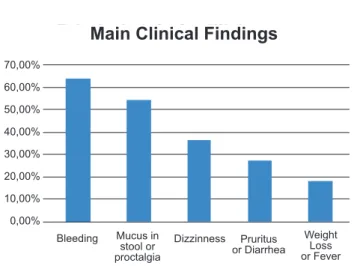
proctalgia was present in six patients (54.5%); dizzi-ness was observed in four patients (36.4%), pruritus, in three (27.3%), diarrhea, in three (27.3%), weight loss, in two (18.2%), and fever, in two (18.2%). Syn-cope, anal incontinence, purulent discharge, anemia, abdominal distention, anal mass sensation and consti-pation corresponded to one case each (9.1%). All pa-tients (100%) presented with at least two symptoms each (Graphic 1).
Anuscopy was used as the diagnostic method for all patients. ulcerative lesion was present in four me-dical records (36.4%), and tumor was found in seven records (63.7%).
Three patients (27.3%) had T1 tumors, four (36.3%) had T2 tumors (27.3%), one (9.1%) had T3 tumor, one (9.1%) had T4 tumor, and two (18.2%) pa-tients had no data concerning the size of the tumor. Six patients had no lymph node compromise, one (9.1%) were in N2, one (9.1%) was in N1 and three (27.3%) had no data. One patient (9.1%) had lung metastasis. Thus, one (9.1%) was in stage I, three (27.3%) were in stage II, one (9.1%) was in stage IIIA, and one patient (9.1%) was in stage IV. Two patients (18.2%) still had not been submitted to imaging examinations, and one had T4NxMx. Two patients (18.2%) had no informa-tion concerning staging.
Nine patients (81.9%) underwent radiochemothera-py. The other two patients (18.2%) are under oncologic analysis until the present time, thus not being treated.
Four patients (36.4%) presented with tumor re-currence at follow-up biopsy; one of them (9.1%) had not initially adhered to the previously established tre-atment. Two (18.2%) underwent abdominoperineal resection for the rescue surgery, and one (9.1%) had another series of chemoradiotherapy sessions.
Three patients (27.3%) with epidermoid carcino-ma presented recurrence, and one (9.1%) died. There was one case of recurrence in a patient with squamous verrucous carcinoma, and recurrence was not obser-ved in any patient with basaloid tumor (Table 1).
One of the patients (9.1%) died and two patients (18.2%) were no longer followed-up.
DISCUSSION
Anal canal neoplasm is not frequent in the gas-trointestinal tract, corresponding to 1 to 2% of large
intestine cancers, and 3 to 3.5% of anorectal tumors. In Sergipe, its incidence it 0.18 to 0.83 per 100,000 people/year3,10. In this study, this corresponded to 11 diagnosed patients who were followed-up at the colo-proctology service of Hu/uFS, which reinforces the idea that this is the least frequent tumor in the digesti-ve tract. The low sample was a result of the low inci-dence and prevalence of the neoplasm, which made it
dificult to effectively make the statistical analysis.
The anal carcinoma is more prevalent among wo-men, at a proportion of 1.6:1 to 5:1. In our study, data
analysis conirmed the prevalence of anal carcinoma
among females (54.5%), at a proportion of 1.2:12,3,14. The anal cancer is more frequent at the seventh and eighth decades of life, and the mean age reported is 63.8 years3. The most affected age group is compri-sed of people in the sixth decade of life. Mean age of female patients was 61 years, from 48 to 89, and
stan-Table 1. Distribution as to number of recurrences per histological subtype.
staging Follow-up biopsy Recurrence No recurrence
Squamous cell
carcinoma 3 5
Basaloid carcinoma 0 2
Squamous verrucous
carcinoma 1 0
Total 4 7
Graphic 1. Graphic showing the prevalence of clinical indings in
patients with anus neoplams.
70,00%
60,00%
50,00%
40,00%
30,00%
20,00%
10,00%
0,00%
Main Clinical Findings
Bleeding Mucus in stool or proctalgia
Dizzinness Pruritus or Diarrhea
dard deviation of ±14.63. As to males, mean age was 50.6 years, from 36 to 59 years, and standard deviation of ±10.83. The three patients (27.3%) aged less than 50 years were males. There are reports regarding the increased prevalence of anus neoplasm in homosexual male patients, maybe due to the association with HPV. This fact could be the cause for a higher prevalence of anus neoplasm among men than young women3,5.
The increased incidence of anus neoplasm has been reported in urban centers, which was also found in our study, in which 54.5% of the patients lived in the capital10.
The epidermoid carcinoma was the most
com-mon histopathological inding (72.7%), followed by
two patients (18.2%) with cloacogenic tumor, and one (9.1%) with the rare variable of squamous verrucous carcinoma. This is in accordance with literature, in which the epidermoid type is more prevalent, follo-wed by the cloacogenic tumor3,14. It is important to emphasize that we observed the squamous verrucous carcinoma in our series, which is a rare variable of the epidermoid carcinoma that can manifest as a mass that
resembles a caulilower, pale rose, at the anal margin
or anal canal. Histopathological examination shows a well differentiated lesion that can invade subjacent tis-sues, despite being apparently benign9. The squamous verrucous carcinoma in the series was well differen-tiated, as demonstrated in literature. The anal canal is the most affected site, which is also in accordance with our study10.
About 20% of the patients affected by the anal canal tumor are asymptomatic, and the most common
clinical inding is bleeding2,10. The mass sensation and/or rectal pain correspond to 30% of the patients
with neoplasm. Other indings include pruritus, anal
incontinence, changes in bowel habit and mucus dis-charge4. No patient was asymptomatic at diagnosis. Thus, bleeding was present in 63.7% of the patients,
proving to be the most common inding. Mass and/or
anal pain were observed in 36.4% of the patients, as described in the review. Such symptoms and signs are
also present in other conditions, such as issure and
hemorrhoid, which are important for the differential diagnosis of anal neoplasm9,10.
The anus neoplasm diagnosis is based on anam-nesis and physical examination. Anuscopy is essential
for the diagnosis, to be conirmed by the lesion biopsy
that will deine histological type and level of differen -tiation10. This routine was adopted to diagnose the le-sions of all patients in the present series. Anuscopy not
only deines if the lesion is anal canal or anal margin,
but also describes the aspect of the lesion, which was ulcerous in four cases, and its size, which is important for staging.
The size of the tumor is an important prognos-tic factor10. Despite the few patients in this series, the only one who had a T4 tumor was the same person who had the worst prognosis, developing recurrence after radiochemotherapy, thus being submitted to
ab-dominoperineal resection to rescue, conirming this
observation.
About 50 to 60% of the patients have T1 and T2 tumors, with survival rates of 80 to 90%. In our study, 63.3% of the cases corresponded to these sizes4.
The patient in stage IV (9.1%) with distant me-tastasis showed recurrence to radiochemotherapy, with posterior abdominoperineal resection, which shows the prognostic importance of staging10. Five of them (45.5%) presented incomplete data as to TNM grading, which is partly due to lack of information in medical records (27.3%), and also because (18.2%) these patients were still being clinically investigated.
As to staging, the thoracic x-ray and the tompu-ted tomography of abdomen and pelvis were used. Re-cently, the importance of the endorectal ultrasound has
been increasing, since it enables to deine the level of
invasion in the anal wall and to assess perirectal
gan-glion to deine gangan-glion metastasis. At the moment,
we still do not dispose of this examination for semio-tic complement10.
In spite of the liver being the most common loca-tion of distant metastasis, according to literature, the lung was the only location (9.1%) affected by dissemi-nation in our study4.
There are other important factors in prognosis, such as histological type, anemia, irradiation dose and gender. Epidermoid carcinomas have more chances of therapy failure and causes more obits than the
cloa-cogenic subtype, which is conirmed in our study, in
which three patients with squamous cell carcinoma had recurrence, and no cloacogenic presented therapy failure10.
Since Nigro’s study from 1974, in which
RESUMO: Neoplasias do ânus correspondem de 2 a 4% dos tumores de intestino grosso, sendo predominante nas sétima e oitava décadas. A maior prevalência é em gênero feminino, com proporção de 3:1. O aumento da prevalência na população nos últimos anos provavelmente está relacionado ao número maior de pessoas com doenças sexualmente transmissíveis, principalmente o
papiloma-vírus humano (tipos 16 e 18, mais comumente) e/ou o papiloma-vírus da imunodeiciência humana. O diagnóstico é feito a partir de achados clínicos somados ao exame anatomopatológico. O tratamento de escolha baseia-se na radioquimioterapia, sendo a cirurgia de resgate
com amputação abdominoperineal utilizada para casos de recidiva ou persistência. Foi feito um estudo observacional longitudinal re-trospectivo e prospectivo, com 11 pacientes diagnosticados com neoplasia anal no período de 2004 a 2010. seis (54,5%) eram do gênero
feminino e 5 (45,5%) do masculino. O pico de incidência foi em sexta década, com média de idade de 54,45 anos. O tipo histológico
mais encontrado foi o carcinoma epidermoide (72,7%), sendo o moderadamente diferenciado o mais frequente grau de diferenciação. A quimioterapia associada à radioterapia foi instituída em 81,9% dos pacientes, sendo necessária a cirurgia de amputação abdomino-perineal como terapia de resgate em 18,2% dos pacientes.
Palavras-chave: neoplasias do ânus; diagnóstico; quimioterapia; radioterapia; cirurgia.
therapy for the abdominoperineal resection, until there was no residual lesion in the response, the treatment
with 5-luorouracil and mitomycim with radiotherapy
became essential to resolve anal neoplasm, thus going through changes with time as to doses and number of radiochemotherapy sessions9,10,12,13. Such therapy was used for 9 (81.9%) patients, thus becoming the treat-ment of choice. The other two cases (18.2%) are still in oncologic evaluation.
The role of the rescue surgery for a recurrent or persistent disease is still relevant, besides the pallia-tive care for the anus neoplasm. Two (18.2%) out of the three patients who presented with recurrence un-derwent abdominoperineal resection2,11.
Predisposing factors for anal tumor are usually infection by HPV subtypes 16 and 184,5,8. Thus, de-tecting and eliminating HPV clinical and subclinical lesions may provide better prognosis and prevent the tumor from becoming malignant15.
Genetic studies may be used in the future to tra-ck patients who are more prone to anal canal and anal margin neoplasm. Studies have tried to establish the relation between genetic changes and anus neoplasm. Such studies showed the increased expression of 14-3-3σ, inhibitor regulator of p53 in its association with anus neoplasm. However, there is no relation betwe-en this protein and p53, or the association of the neo-plasm with p1616,17.
A signiicant part of the medical records had in -complete data as to staging, although the patients had been properly staged at diagnosis, undergoing radio-chemotherapy. The need to computerize the informa-tion of medical records is urgent, so that important
data concerning the patients’ follow-up cannot be lost. Since we are dealing with a university hospital, such
fact makes it dificult to access the information and conduct scientiic work. The American Joint Commit
-tee on Cancer (AJCC) observes that “the classiication
and the staging of cancer enable doctors and cancer registers to stratify the patients, which will lead to bet-ter decisions as to treatment and the development of a common language”; this organizes the assistance to patients and the therapy of choice, analyzing progno-sis, enabling the professionals to change experiences and assess therapy results18.
CONCLUSION
• Seven patients were diagnosed with anal canal
câncer, and four were diagnosed with anal mar-gin câncer;
• It was more prevalent among females in relation
to males;
• The incidence peak was during the sixth decade
of life;
• The most frequent histological type was epider -moid carcinoma, followed by the cloacogenic carcinoma. The rare squamous verrucous carci-noma was found;
• The moderately differentiated level was present
in 54.5% of the cases, and the well differentiated level was found in 36.4% of the cases;
REFERENCEs
1. Nadal SR, Calore EE, Cruz SHA, Horta SHC, Manzione CR, Bin FC, et al. Comparação das contagens das células de Langerhans de tecidos contendo carcinoma anal em doentes com e sem infecção pelo HIV. Rev Bras Coloproct 2006;26(3):269-74.
2. Santos Jr JCM. Câncer ano-retal-cólico - aspectos atuais: I - câncer anal. Rev Bras Coloproct 2007;27(2):219-23. 3. Torres-Neto JR, Prudente ACL, Santos RL. Estudo
demográico do câncer de canal anal e ânus no estado de
Sergipe. Rev Bras Coloproct 2007;27(2):190-5.
4. uronis HE, Bendell CJ. Anal cancer: an overview. Oncologist 2007;12(5):524-34.
5. Frisch M, Glimelius B, Van den Brule AJ, Wohlfah J, Meijer CJ, Wallboomers JM, et al. Sexually transmitted infection as a cause of anal cancer. N Engl J Med 1997;337(19):1350-8. 6. Ryan DP, Compton CC, Mayer RJ. Carcinoma of the anal
canal. N Engl J Med 2000;342(11):792-800.
7. Dallan LAP, Cruz SHA, da Rosa DL, Bin FC, Nadal SR, Capelhuchnik P, et al. Avaliação dos resultados do tratamento de 14 doentes de carcinoma espinocelular anal. Rev Bras Coloproct 2005;26(1):34-40.
8. Nakamura RA, Ferrigno R, Salvajoli JV, Nishimoto IN, David Filho WJ, Lopes A. Tratamento conservador do carcinoma do canal anal. Rev Col Bras Cir 2005;32:23-31.
9. Keighley M, Williams N. Cirurgia do ânus, reto e colo. 1st ed. São Paulo: Manole, 1998.
10. Soares WGP. Radioquimioterapia no câncer de canal anal: avaliação de 12 pacientes. Sergipe, 2002.
11. Rodrigues MRS, Magi JC, Corrêa RS, Guerra GMLSR, Souza
HFS, Fonseca MFM, et al. Cirurgia de resgate no carcinoma de canal anal. Rev Bras Coloproct 2004;24(2):137-9. 12. Nigro ND, Vaitkevicius VK, Considine BJR. Combined
therapy for cancer of the anal canal: a preliminary report. Dis Colon Rectum 1974;17(3):354-6.
13. Charnley N, Chouldhury A, Chesser P, Cooper RA,
Sebag-Monteiore D. Effective treatment of anal cancer in the
elderly with low-dose chemoradiotherapy. Br J Cancer 2005;92(7):1221-5.
14. Larangeira LLS, Andrade SKV. Incidência do carcinoma de canal anal na Regional de Saúde de Londrina (PR). Rev Bras Coloproct 2004;24(3):240–6.
15. Nadal SR, Manzione CR. Papilomavirus humano e o câncer anal. Rev Bras Coloproct 2006;26(2):204-7.
16. Roma AA, Goldblum JR, Fazio V, Yang B. Expression of
14-3-3σ, p16 and p53 proteins in anal squamous intraepithelial
neoplasm and squamous cell carcinoma. Int J Clin Exp Pathol 2008;1(5):419-25.
17. Contu SS, Agnes G, Damin AP, Contu PC, Rosito MA, Alexandre CO, et al. Lack of correlation between p53 codon 72 polymorphism and anal cancer risk. World J Gastroenterol 2009;15(36):4566-70.
18. Greene FL. Cancer staging handbook from the AJCC cancer staging manual. 6th ed. Springer-Verlag (NY): Springer Science + Business Media; 2004.
Correspondence to:
Juvenal da Rocha Torres Neto
Rua Ananias Azevedo Nº 100, apto. 902, Praia 13 de Julho CEP: 49020-080 – Aracaju (SE), Brazil.