Key words:
Penile Neoplasms; Humans; Prevalence; Lymphatic Metastasis; Biological Markers
Int Braz J Urol. 2013; 39: 542-50
__________________ Submitted for publication: November 11, 2012
__________________ Accepted after revision: May 25, 2013
Objectives: To evaluate the prevalence, distribution and association of HPV with histolo-gical pattern of worse prognosis of penile cancer, in order to evaluate its predictive value of inguinal metastasis, as well as evaluation of other previous reported prognostic factors.
Material and Methods: Tumor samples of 82 patients with penile carcinoma were tested in order to establish the prevalence and distribution of genotypic HPV using PCR. HPV status was correlated to histopathological factors and the presence of inguinal mestas-tasis. The influence of several histological characteristics was also correlated to inguinal disease-free survival.
Results: Follow-up varied from 1 to 71 months (median 22 months). HPV DNA was iden-tified in 60.9% of sample, with higher prevalence of types 11 and 6 (64% and 32%, res-pectively). There was no significant correlation of the histological characteristics of worse prognosis of penile cancer with HPV status. Inguinal disease-free survival in 5 years did also not show HPV status influence (p = 0.45). The only independent pathologic factors of inguinal metastasis were: stage T ≥ T1b-T4 (p = 0.02), lymphovascular invasion (p = 0.04) and infiltrative invasion (p = 0.03).
Conclusions: HPV status and distribution had shown no correlation with worse prognosis of histological aspects, or predictive value for lymphatic metastasis in penile carcinoma.
INTRODUCTION
Epidermoid carcinoma of penis (ECP) is a rare neoplasm, but with high incidence in under-developed countries (1). In Brazil, the incidence varies from 2.9 to 6.8 cases/100.000 inhabitants/ year (2). In an epidemiological study in Pará Sta-te, the observed incidence was 5.7 cases/100.000 inhabitants/year (3).
Its etiology is associated with several risk factors, including presence of prepuce, bad hygie-nic habits, chrohygie-nic dermatitis and smoking.
Howe-ver, the most extensively studied factor is human papilloma virus infection (HPV) (4).
HPV has more than 100 subtypes that can be classified as with low (HPVlow) or high risk (HPVhigh) oncogenic potential (5). Its role in cer-vical uterine cancer is well established, but its as-sociation with ECP is still being discussed.
In patients with ECP, the presence of HPV va-ries from 15% to 80% suggesting that only a subgroup of these tumors is associated with HPV infection (6).
Natural history of the disease is characteri-zed by progressive invasion of surrounding penile
Human Papilloma Virus: Prevalence, distribution and
predictive value to lymphatic metastasis in penile
carcinoma
_______________________________________________
Aluizio Gonçalves da Fonseca, Fernando Augusto Soares, Rommel Rodriguez Burbano, Rodrigo
Vellasco Silvestre, Luis Otávio Amaral Duarte Pinto
Urology Department, Hospital Ophyr Loyola, Belem, PA, Brazil
ABSTRACT
ARTICLE
INFO
tissues, and posteriorly metastatic dissemination to inguinal lymph nodes (6). Lymph node involve-ment is the prognostic factor of worst impact on prognosis of disease-specific survival (6) and the analysis of this involvement is very important in order to determine the cure of the disease.
Nowadays, the available methods for sta-ging regional lymph node involvement have low accuracy. For example, Gonzaga-Silva et al., using gamma probe in the sentinel lymph node, showed sensitivity of only 25% and 42.8% of false negati-ve results (7).
The physical exam of inguinal region (ins-pection and palpation) is also inconclusive, since half the cases with lymph node involvement does not present tumor. Also, 20% of patients without lymph node detected during physical exam pre-sent local micrometastasis (8).
Inguinal lymph node involvement is the most accurate method of staging to diagnose oc-cult metastasis. However, it has significant morbi-dity and does not benefit a relevant subgroup of patients without metastasis (8).
In literature, several histological factors of the primary lesion were identified with predictive value of inguinal metastasis. These include: diffe-rentiation grade, tumor staging, and presence of neurovascular and lymph vascular invasion, with independent prognostic importance (9).
Although HPV infection is associated with the most aggressive histological subtypes, its va-lue as a predictive factor of lymph node metastasis and prognosis is still debated (10).
The present study was conducted in a group of patients from Northern region of Brazil (Pará State), in order to evaluate the prevalence, distribution and association of HPV infection with histological pattern of worse prognosis in ECP and to determine its possible predictive value of lymph node metastasis.
MATERIALS AND METHODS
Patients
This study, as well as its consent inform, was approved by the Local Ethical Committee. Eighty--two patients with ECP were treated at the Hospital Ophir Loyola, Belém - Pará, from 2001 to 2008. The
patients were submitted to penile amputation and the analysis of the lymph nodes followed a pro-tocol proposed by the Urologic Department of the Hospital. All patients were prospectively evaluated, by follow-up appointments, medical files charts or telephonic contact of relatives.
Treatment protocol
Lymph node resection was performed after 4 to 6 weeks of the penile surgery. Du-ring this interval patients received antibiotico-therapy with fluoroquinolones. For the patients with palpable inguinal lymph nodes after the antibiotic treatment it was performed bilateral superficial lymph node resection with freezing histological samples. Patients with T2-T4 sta-ging and/or G2-G3 differentiation grade (accor-ding to TNM 2009 UICC System of Staging and Pathology) were also submitted to superficial bilateral lymph node resection.
In the presence of tumor involvement of superficial lymph nodes, radical lymphadenec-tomy was performed. Pelvic lymphadeneclymphadenec-tomy was performed in all cases with deep inguinal lymph node invasion.
The surgery was not performed in patients with Tis or T1, G1 staging, without lymph vascular invasion or palpable lymph nodes. These patients followed a careful ambulatory schedule of sur-veillance for at least five years.
Pathological Analysis
All samples were revised blindly by a sin-gle pathologist. The tumors were retrospectively staged according to 2009 TNM System.
Lymph vascular invasion (LVI) was defi-ned as the presence of tumor cells in the interior of lymphatic veins, blood veins or arteries. Perineu-ral invasion (PNI) was determined as the presence of tumor tissue next to peripheral nerve branches. As for invasion, the tumor was considered “in-filtrative”, when it was observed the presence of branches of malignant cells within the stroma, or “pushing”, when it was observed an interface be-tween the tumor and the host cells.
HPV Analysis
This part of the study was developed by the Papilloma Virus Laboratory of Instituto Evandro Chagas, a HPV referential laboratory of Brazilian Ministry of Health.
Parafin blocks were serially cut by microto-me and the cuts were used for viral DNA detection.
Samples were submitted to polimerase chain reaction (PCR) using initially human beta globin G73 - 5´GAA GAG GGA AGG ACA GGT AC 3´ e G74 - 5´ CAA CTT CAT CCA CGT TCA CC 3´ (PCR).
Viral gene specific reaction used the initia-tors GP5+ - 5´ TTT GTT ACT GTG GTA GAT ACT AC 3´ e GP6+ - 5´ GAA AAA TAA ACT GTA AAT CAT ATT C 3´, that were able to amplify a 150pb fragment of the L1 gene, that corresponds to a well preserved region of the virus genome.
After PCR, the identification of the virus was carried out using direct sequencing of the PCR pro-duct, using a capillary sequencer ABI 3130 Genetic analyser, Hitachi©.
The presence of HPV was classified in three subgroups:
HPVhigh - infection by one or more high risk virus
HPVlow - infection by one or more low risk virus
HPVmult - presence of more than one subtype of virus in the sample, whether of high or low risk.
Follow-up and statistical analysis
Follow-up of patients was carried out throu-gh periodic ambulatory evaluations, quarterly in the first two years after surgery, and then semiannually.
The presence or absence of HPV infection, as well the presence of subgroups HPVhigh, HPVlow and HPVmult were related to several clinical and pa-thological variables, using the Chi-Square method or Fisher exact test.
The presence of inguinal metastasis was correlated to: pT, G, LVI, PNI, Infiltratative, Pushing, histological subtype and HPV status, using univa-riate and multivauniva-riate analysis by Cox proportional hazard method.
The analysis of inguinal disease-free interval (IDFI) in five years used the Kaplan-Meier method
and the log Rank test. SPSS® 13.0 was used for all analyses, and the result was considered significant if p < 0.05.
RESULTS
Median age of patients was 58 years (22-91 years) and follow-up ranged from 1 to 71 months (median 20 months).
Presence of HPV DNA
HPV DNA was detected in 60.9% (50/82) fixed samples in paraffin. Viral distribution was: HPV11 (64%), HPV6 (32%), HPV16 (30%), HPV53 (18%), HPV33 (4%) and subtypes 18, 68, 45, 51, 52 and 58 (2%).
Subgroups HPVhigh, HPVlow and HPV-mult were identified in 12/50 (24%), 25/50 (50%) and 25/50 (50%) respectively. Viral types that pre-dominated in each group were 16, 11 and 11.
Histological characteristics of subgroups HPV po-sitive (HPVhigh, HPVlow and HPVmult) and HPV negative
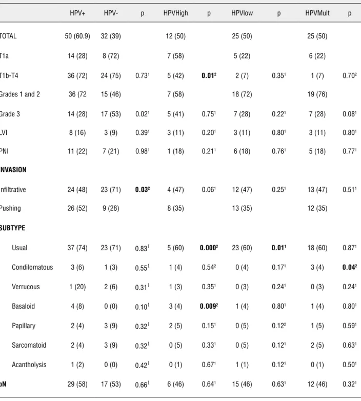
Patients without HPV infection had predomi-nantly infiltrative invasion. HPV positive group was correlated to more differentiated tumors (Table-1).
HPVhigh was associated more frequently to basaloid subtype, and rarely was present in patients with usual pattern. There was also a low prevalence of HVPhigh in patients with more advanced penile carcinoma (Table-1).
Most patients with usual pattern were in-fected by HPVlow. Condylomatous type was as-sociated more frequently to HPVmult infection (Table-1).
Multiple logistic regression analysis did not show any histological variable independently rela-ted to status or HPV subgroup.
Table 1 - Histopathological characteristics of 82 patients with penile cancer according to HPV positive (n = 50) or negative (n = 32), HPVhigh (n = 12), HPVlow (n = 25) and HPVmult (n = 25) groups.
DNA-HPV
HPV+ HPV- p HPVHigh p HPVlow p HPVMult p
TOTAL 50 (60.9) 32 (39) 12 (50) 25 (50) 25 (50)
T1a 14 (28) 8 (72) 7 (58) 5 (22) 6 (22)
T1b-T4 36 (72) 24 (75) 0.731 5 (42) 0.012 2 (7) 0.351 1 (7) 0.702
Grades 1 and 2 36 (72 15 (46) 7 (58) 18 (72) 19 (76)
Grade 3 14 (28) 17 (53) 0.021 5 (41) 0.751 7 (28) 0.221 7 (28) 0.081
LVI 8 (16) 3 (9) 0.391 3 (11) 0.201 3 (11) 0.801 3 (11) 0.801
PNI 11 (22) 7 (21) 0.981 1 (18) 0.211 6 (18) 0.761 5 (18) 0.771
INVASION
Infiltrative 24 (48) 23 (71) 0.032 4 (47) 0.061 12 (47) 0.251 13 (47) 0.511
Pushing 26 (52) 9 (28) 8 (35) 13 (35) 12 (35)
SUBTYPE
Usual 37 (74) 23 (71) 0.831 5 (60) 0.0002 23 (60) 0.011 18 (60) 0.871
Condilomatous 3 (6) 1 (3) 0.551 1 (4) 0.542 0 (4) 0.171 3 (4) 0.042
Verrucous 1 (20) 2 (6) 0.311 1 (3) 0.351 0 (3) 0.241 0 (3) 0.241
Basaloid 4 (8) 0 (0) 0.101 3 (4) 0.0092 1 (4) 0.801 1 (4) 0.801
Papillary 2 (4) 3 (9) 0.321 2 (5) 0.151 0 (5) 0.122 1 (5) 0.591
Sarcomatoid 2 (4) 3 (9) 0.321 0 (5) 0.331 0 (5) 0.121 2 (5) 0.631
Acantholysis 1 (2) 0 (0) 0.421 0 (1) 0.671 1 (1) 0.121 0 (1) 0.501
pN 29 (58) 17 (53) 0.661 6 (46) 0.641 15 (46) 0.631 12 (46) 0.321
The values in parenthesis indicate percentage 1: Chi square test;
Likewise, there was no statistical significant difference of IMFS among the groups HPVhigh, HPVlow and HOVmult (Table-2).
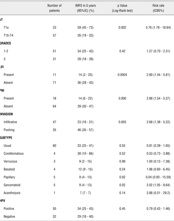
Univariate analysis of IMFS in five years, according to histopatological patterns, showed that stage T ≥ T1b-T4 (HR = 5.76), LVI (HR = 2.90), PNI (HR = 2.88), and infiltrative invasion (HR = 2.68) were, decreasingly, strong predictors of inguinal metastasis, respectively 82.6% (38/52), 100% (11/11), 94.4% (17/18) and 70.2% (33/47) (Table-2).
Only sarcomatoid subtype was related to regional metastasis (4/5). Papillary subtype showed negative correlation to metastasis in 100% (0/5).
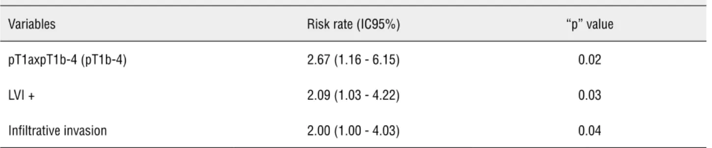
Multivariated analysis showed that T ≥ T1b--T4, LVI and infiltrative invasion were strong inde-pendent predictors of inguinal metastasis (Table-3).
DISCUSSION
The present study is the first scientific report of the presence of HPV and its genotype variants correlated to ECP in a northern state of Brazil, a region with high prevalence of the disease.
In this sample, HPV prevalence in patients with ECP was 60.9%, in accordance to literature. It is reported a variation of 14% to 100% (11).
Miral-les-Guri et al. metanalysis in 2009, comprasing 30 studies and 1266 patients with ECP reported a HPV DNA prevalence of 47.9%. In South America, viral DNA was specifically found in 40.7% of tumors (12).
As for the genotype distribution, in our sam-ple there was a clear prevalence of types 11 and 6, contrary to the previous report of Bezerra et al., that reported an absolute prevalence of type 16 (12). We also registered infection by multiple HPV genotypes in 30.4% (25/50), contrary to the study by Krustrup et al., that did not show multiple viral infections in their sample (13).
Variations of the reported prevalence of DNA HPV are mainly due to different techniques of sampling, studied population, detection methods, tumoral tissue storage and inclusion of multiple his-tological subtypes (14).
The role of HPV in epithelium carcinogene-sis of uterine cervix is well established. In more than 95% of patients it is observed several high risk sub-types of virus infection (15); however, in ECP, the vi-ral infection is not mandatory and there are sevevi-ral other causes not related to HPV infection (15). Re-lation of genital tumors and HPVhigh is stimulating research and interest of the biological potential viral interference on metastasis and disease prognosis.
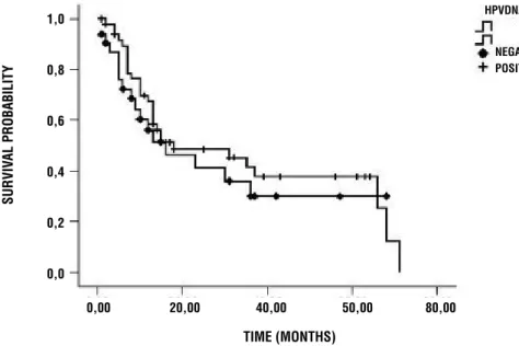
Figure 1 - Inguinal metastasis-free survival after treatment of the primary tumor showed no significant differences between hpv positive tumors (n50/82) or negative hpv tumors (n32/82), p = 0.45.
TIME (MONTHS) 1,0
0,8
0,6
0,4
0,2
0,0
0,00 20,00 40,00 50,00 80,00
NEGATIVE POSITIVE HPVDNA
SURVIV
Table 2 - Inguinal metastasis-free survival index in 5 years and risk rate according to clinical and pathological variables – univariate analysis
Number of patients
IMFS in 5 years (95%IC) (%)
p Value (Log-Rank test)
Risk rate (IC95%)
pT
T1a 23 59 (45 - 73) 0.002 5.76 (1.78 - 18.64)
T1b-T4 57 26 (19 - 33)
GRADES
1-2 51 34 (25 - 43) 0.42 1.27 (0.70 - 2.31)
3 31 28 (18 - 39)
LVI
Present 11 14 (3 - 25) 0.0004 2.90 (1.44 - 5.81)
Absent 71 36 (28 - 43)
PNI
Present 18 14 (6 - 22) 0.000 2.88 (1.54 - 5.37)
Absent 64 39 (30 - 47)
INVASION
Infiltrative 47 23 (16 - 31) 0.003 2.68 (1.38 - 5.22)
Pushing 35 46 (35 - 57)
SUBTYPE
Usual 60 33 (25 - 41) 0.55 0.81 (0.39 - 1.65)
Condilomatous 4 38 (10 - 66) 0.52 0.53 (0.73 - 3.88)
Verrucous 3 9 (2 - 15) 0.99 1.00 (0.13 - 7.38)
Basaloid 4 12 (9 - 15) 0.24 1.96 (0.60 - 6.45)
Papillary 5 9 (4 - 13) 0.02 0.04 (0.00 - 15.59)
Sarcomatoid 5 9 (4 - 13) 0.02 3.02 (1.05 - 8.64)
Acantholysis 1 7 (7 - 7) 0.14 3.88 (0.51 - 29.2)
HPV
Positive 50 34 (25 - 43) 0.45 0.79 (0.43 - 1.46)
Several authors tried to relate viral infec-tion and histological pattern of worse prognosis (16-18). Scheiner et al. (16), Lont et al. (17) and Kirrander et al. (18) did not find such evidence. However, Gregoire et al. (19) and Krustrup et al. (13) reported a significant statistical association between HPVhigh infection and high risk tumors. Our studied showed that the only positive asso-ciation was between the prevalence of HPVhigh in 75% of basaloid tumors, with worse prognosis. Cubilla et al. (20) reported the same association.
Guimaraes et al. evaluated 14 cases of ba-saloid tumors, and reported 50% of inguinal me-tastasis. In our sample we observed 75% (3/4 pa-tients) (21). Despite the small number of patients with this histological subtype of tumor, we believe that this association should be more investigated hereafter.
The recommendations for lymphadenecto-mies without the presence of inguinal lymph node disease are based upon predictive factors related to the primary lesion, mainly tumor stage and cellu-lar differentiation grade. However, overtreatment can reach 82% of cases (22). In our sample, this was observed in 88% (30/34) even with the use of a strict protocol. It is necessary to search for non--invasive biomarkers with good accuracy, to pre-dict the risk of metastatic lymph node involvement in patients with penile cancer (22,23). In our series, there was no association between the presence or distribution of HPV genotype with lymph node di-sease, as reported by other authors (13,19). At pre-sent, there are no evidences indicating the use of HPV detection in order to decide to perform
lym-phadenectomies. Surprisingly, Lont et al., although failed to associate HPV status and lymph node me-tastasis, showed better specific-disease survival in patients with ECP associated with high risk virus (p = 0.03) (17). In order to avoid bias in our study, we only evaluated inguinal disease-free survival (IDFI), that did not show differences between HPV positive and negative groups (p = 0.45).
Several pathological and molecular risk factor are being described in order to improve lymph node evaluation (24,25). Among patholo-gical predictive factors, only PNI, LVI and histo-logical grade are independent factors to predict lymph node involvement (25). In our study, tumor stage ≥ T1b-T4, LVI, PNI, infiltrative invasion and sarcomatoid histological subtype are the histolo-gical factors related to lymph node metastasis in a univariate analysis.
There was no significant difference among the cellular differentiation grades as prognostic factors, in spite of the reduced sample. However, pathological heterogeneity, with more than a gra-de presented in the same tumor is rarely consigra-de- conside-red in different studies.
Multivariate regression analysis sho-wed that only ≥ T1b-T4 tumors, LVI and infil-trative pattern were independent factors, and T stage was the most important (HR = 2.67, p = 0.02). The reported differences in several series are mainly caused by methodology, therapeutic approach and heterogeneity of the studied po-pulation. These limitations must be contoured in order to obtain a better validation of these risk factors for inguinal metastasis.
Table 3 - Inguinal metastasis-free survival index in 5 years and risk rate according to clinical and pathological variables - multivariate analysis.
COX multivariate regression analysis – independent risk factors for inguinal metastasis
Variables Risk rate (IC95%) “p” value
pT1axpT1b-4 (pT1b-4) 2.67 (1.16 - 6.15) 0.02
LVI + 2.09 (1.03 - 4.22) 0.03
CONCLUSIONS
Observed HPV prevalence was similar to pre-vious studies. However, there was a predominance of the low risk viral group. There was no significant association between HPV status or HPV subgroups and histological worse prognostic factors. HPV de-tection had no use as predictive value of inguinal metastasis. Tumor staging, lymph-vascular invasion and infiltrative pattern were independent risk fac-tors for lymph node involvement.
CONFLICT OF INTEREST
None declared.
REFERENCES
1. Backes DM, Kurman RJ, Pimenta JM, Smith JS: Systematic review of human papillomavirus prevalence in invasive penile cancer. Cancer Causes Control. 2009; 20: 449-57.
2. Favorito LA, Nardi AC, Ronalsa M, Zequi SC, Sampaio FJ, Gli-na S: Epidemiologic study on penile cancer in Brazil. Int Braz J Urol. 2008; 34: 587-91; discussion 591-3.
3. Fonseca AGda, Pinto JASA, Marques MC, Drosdroski FS, Fon-seca Neto LORda: Estudo epidemiológico do câncer de pênis no Estado do Pará, Brasil. Rev Pan-Amaz Saude. 2010; 1: 85-90. 4. Bleeker MC, Heideman DA, Snijders PJ, Horenblas S, Dillner
J, Meijer CJ: Penile cancer: epidemiology, pathogenesis and prevention. World J rol. 2009; 27: 141-50.
5. Tornesello ML, Duraturo ML, Losito S, Botti G, Pilotti S, Ste-fanon B, et al.: Human papillomavirus genotypes and HPV16 variants in penile carcinoma. Int J Cancer. 2008; 122: 132-7. 6. Pompeo ACL, Billis A: Carcinoma epidermóide do pênis. Int
Braz J Urol. 2003; 29 (Suppl. 1): 44-50.
7. Gonzaga-Silva LF, Tavares JM, Freitas FC, Tomas Filho ME, Oliveira VP, Lima MV: The isolated gamma probe technique for sentinel node penile carcinoma detection is unreliable. Int Braz J Urol. 2007; 33: 58-63; discussion 64-7.
8. Pettaway CA, Horenblas S: Penile cancer: incremental in-sights into etiology, diagnosis, staging, and management. World J Urol. 2009; 27: 139-40.
9. BE Stephen, BR David, CC Carolyn, GF April, LG Frederick, T Andy, III: AJCC Cancer Staging Manual. Philadelphia, Lip-pincott. 2010; Seventh Edition, 2010; pp.447.
10. Cubilla AL: The role of pathologic prognostic factors in squamous cell carcinoma of the penis. World J Urol. 2009; 27: 169-77.
11. Miralles-Guri C, Bruni L, Cubilla AL, Castellsagué X, Bosch FX, de Sanjosé S: Human papillomavirus prevalence and type dis-tribution in penile carcinoma. J Clin Pathol. 2009; 62: 870-8. 12. Bezerra AL, Lopes A, Santiago GH, Ribeiro KC, Latorre MR,
Villa LL: Human papillomavirus as a prognostic factor in carcinoma of the penis: analysis of 82 patients treated with amputation andbilateral lymphadenectomy. Cancer. 2001; 91: 2315-21.
13. Krustrup D, Jensen HL, van den Brule AJ, Frisch M: Histo-logical characteristics of human papilloma-virus-positive and -negative invasive and in situ squamous cell tumours of the penis. Int J Exp Pathol. 2009; 90: 182-9.
14. Anic GM, Giuliano AR: Genital HPV infection and related le-sions in men. Prev Med. 2011; 53: S36-41.
15. Rubin MA, Kleter B, Zhou M, Ayala G, Cubilla AL, Quint WG, et al.: Detection and typing of human papillomavirus DNA in pe-nile carcinoma: evidence for multiple independent pathways ofpenile carcinogenesis. Am J Pathol. 2001; 159: 1211-8. 16. Scheiner MA, Campos MM, Ornellas AA, Chin EW, Ornellas
MH, Andrada-Serpa MJ: Human papillomavirus and penile cancers in Rio de Janeiro, Brazil: HPV typing and clinical fea-tures. Int Braz J Urol. 2008; 34: 467-74; discussion 475-6. 17. Lont AP, Kroon BK, Horenblas S, Gallee MP, Berkhof J, Meijer
CJ, et al.: Presence of high-risk human papillomavirus DNA in penile carcinoma predicts favorable outcome in survival. Int J Cancer. 2006; 119: 1078-81.
18. Kirrander P, Kolaric A, Helenius G, Windahl T, Andrén O, Stark JR, et al.: Human papillomavirus prevalence, distri-bution and correlation to histopathological parameters in a large Swedish cohort ofmen with penile carcinoma. BJU Int. 2011; 108: 355-9.
19. Gregoire L, Cubilla AL, Reuter VE, Haas GP, Lancaster WD: Preferential association of human papillomavirus with high-grade histologic variants of penile-invasive squamous cellcar-cinoma. J Natl Cancer Inst. 1995; 87: 1705-9.
20. Cubilla AL, Lloveras B, Alejo M, Clavero O, Chaux A, Kasa-matsu E, et al.: The basaloid cell is the best tissue marker for human papillomavirus in invasive penile squamous cell carci-noma: a study of202 cases from Paraguay. Am J Surg Pathol. 2010; 34: 104-14.
21. Guimaraes G, Werneck da Cunha I, Soares F, Lopez A, Torres JJ, Chaux A, et al.: WHO histological classiWcation, regional metástasis and outcome in 375 surgically treated patients with penile SCC. Mod Pathol. 2007; 20: 150A.
22. Ficarra V, Novara G, Boscolo-Berto R, Artibani W, Kattan MW: How accurate are present risk group assignment tools in pe-nile cancer? World J Urol. 2009; 27: 155-60.
24. Chaux A, Tamboli P, Ayala A, Soares F, Rodríguez I, Barreto J, et al.: Warty-basaloid carcinoma: clinicopathological features of a distinctive penile neoplasm. Report of 45 cases. Mod Pathol. 2010; 23: 896-904.
25. Lopes A, Hidalgo GS, Kowalski LP, Torloni H, Rossi BM, Fon-seca FP: Prognostic factors in carcinoma of the penis: multi-variate analysis of 145 patients treated with amputation and-lymphadenectomy. J Urol. 1996; 156: 1637-42.
_____________________
Correspondence address: