464
Urinary Proteomics in IC/PBS
International Braz J Urol Vol. 36 (4): 464-479, July - August, 2010
Urinary Proteomics Evaluation in Interstitial Cystitis/Painful
Bladder Syndrome: A Pilot Study
Young Ah Goo, Yihsuan S. Tsai, Alvin Y. Liu, David R. Goodlett, Claire C. Yang
Department of Medicinal Chemistry (YAG, YST, DRG), Department of Urology (AYL, CCY), and
In-stitute for Stem Cell and Regenerative Medicine (AYL), University of Washington, Seattle, WA, USA,
Institute for Systems Biology (DRG), Veterans Affairs Puget Sound Health Care System (CCY), Seattle,
WA, USA
ABSTRACT
Purpose: Interstitial cystitis/painful bladder syndrome (IC/PBS) is characterized by chronic pain, pressure and discomfort
felt in the pelvis or bladder. An in-depth shotgun proteomics study was carried out to proile the urinary proteome of women with IC/PBS to identify possible speciic proteins and networks associated with IC/PBS.
Materials and Methods: Urine samples from ten female IC/PBS patients and ten female asymptomatic, healthy control subjects were analyzed in quadruplicate by liquid chromatography-tandem mass spectrometry (LC-MS/MS) on a hybrid
linear ion trap-orbitrap mass spectrometer. Gas-phase fractionation (GPF) was used to enhance protein identiication. Dif -ferences in protein quantity were determined by peptide spectral counting.
Results: α-1B-glycoprotein (A1BG) and orosomucoid-1 (ORM1) were detected in all IC/PBS patients, and ≥ 60% of these
patients had elevated expression of these two proteins compared to control subjects. Transthyretin (TTR) and hemopexin
(HPX) were detected in all control individuals, but ≥ 60% of the IC/PBS patients had decreased expression levels of these
two proteins. Enrichment functional analysis showed cell adhesion and response to stimuli were down-regulated whereas
response to inlammation, wounding, and tissue degradation were up-regulated in IC/PBS. Activation of neurophysiological processes in synaptic inhibition, and lack of DNA damage repair may also be key components of IC/PBS.
Conclusion: There are qualitative and quantitative differences between the urinary proteomes of women with and without
IC/PBS. We identiied a number of proteins as well as pathways/networks that might contribute to the pathology of IC/PBS
or result from perturbations induced by this condition.
Key words: interstitial cystitis; painful bladder syndrome; urine proteomics Int Braz J Urol. 2010; 36: 464-79
INTRODUCTION
Interstitial cystitis/painful bladder syndrome
(IC/PBS) is deined by chronic pain, pressure and
discomfort felt in the lower pelvis or bladder, which
are unrelated to any identiiable cause. Urinary ur -gency and frequency are also common symptoms �eurourology�eurourology
doi: 10.1590/S1677-55382010000400010
of IC/PBS. Despite years of intense research, the underlying etiology, pathophysiology, and risk fac -tors for developing and perpetuating this syndrome
remain unclear. Diagnosis is based on symptoms and exclusion of other conditions, due to the lack of char
465
Urinary Proteomics in IC/PBS
these barriers, the diagnosis of IC/PBS is frequently delayed, and treatment frequently requires a multi-modal approach (1).
One of the hypotheses proposed for the patho-physiology of IC/PBS is disruption of the urothelial barrier leading to symptoms. Bladder surface mucus, composed of glycosaminoglycans (GAGs) and pro-teoglycans, creates a highly impermeable barrier that
is a key to maintain bladder function. Destruction of this barrier leads to tissue iniltration of urinary
solutes, in particular potassium, which depolarizes nerves and muscles and causes tissue injury (2).
Neurogenic inlammation has also been proposed
as a pathophysiologic mechanism in IC/PBS (3). In response to stimuli, urothelial cells could activate neural circuits, releasing factors that cause chronic pain. Both hypotheses could conceivably result in urinary protein byproducts, which could then
poten-tially serve as biomarkers for IC/PBS.
The lack of biomarkers that can be used for diagnosis of IC/PBS or to track treatment eficacy contributes to the clinical burden. Thus, identiica
-tion of biomarker(s) for IC/PBS would represent a major advance in the ield. Cataloging biomolecules
present in complex biological samples has become increasingly important in clinical research for the
purpose of identifying disease speciic biomarkers.
In the case of proteins, proteomics uses mass spec-trometry to qualitatively and quantitatively catalog proteins. Application of proteomics to human
dis-eases is challenging because about 35,000 human genes could translate into over 1,000,000 functional protein entities due to post-translational modiica -tions as well as sequence varia-tions (4). In spite
of these complexities urinary biomarker discovery
holds considerable promise because it has been recently shown that the urinary proteome contains
approximately 1,500 proteins (5). This makes the
urinary proteome far less complex than the blood
proteome where biomarkers are also being sought
for various human diseases (4).
In this pilot study, we applied proteomic
strategies and related methodologies to proile the uri -nary proteome of patients with IC/PBS. The potential
beneits of this study include a greater understanding
of possible causes and underlying mechanisms of IC/PBS.
MATERIALS AND METHODS
Urine Sample Collection and Processing
Human urine acquisition was carried out with our institution’s Ethics Committee approval. Ten women with a clinical diagnosis of IC/PBS, being treated and followed in the Female Urology Clinic, were enrolled in the study. All women had symptoms of IC/PBS for at least one year, and all had undergoneextensive evaluation to exclude reversible, identiiable
causes for their pelvic pain and urinary symptoms. Control urine was obtained from ten asymptomatic, pain-free, healthy female subjects, age-matched to
the IC/PBS group. One protease inhibitor cocktail tablet (Roche, Indianapolis, IN) was added per 50 mL
urine to avoid proteolysis after urine collection. The
urine was centrifuged at 2,000 x g for 10 min at 4°C
to remove cells and debris. The supernatant was
col-lected and processed for protein puriication by TCA (trichloroacetic acid) precipitation (10% w/v). Protein
concentration was measured by BCATM protein assay
(Thermo Fisher, Waltham, MA). Proteins, 200µg each per subject, were reduced, alkylated, digested with
trypsin (Promega, Madison, WI), and then desalted.
Mass Spectrometry Analysis
Peptide digests were analyzed by electrospray ionization on a hybrid linear ion trap-orbitrap mass spectrometer (Thermo Fisher). For each liquid chro-matography-tandem mass spectrometry (LC-MS/MS)
analysis, approximately 0.5µg of peptides were loaded on the column and eluted in acetonitrile gradient (6). To maximize protein identiication without protein
fractionation, ions were selected via a data-dependent
process from 400-2,000 Th or by gas-phase fraction
-ation (GPF) from 400-521, 516-690, 685-968, and 963-2,000 Th (6). Each experiment was acquired in
quadruplicate.
Database Search and Protein Identiication
Interna-466
Urinary Proteomics in IC/PBS
tional Protein Index (IPI) human protein database using SEQUEST. PeptideProphet and ProteinProphet,
which compute a probability likelihood of each identi
-ication being correct, were used for statistical analysis (7). Only proteins identiied by more than one unique peptide sequence were included in the analysis. Dif -ferences in protein expression were calculated using peptide spectral counting algorithms that use MS/MS data to estimate changes in relative abundance of
proteins (8).
Western Blot Analysis
Ten µg of pooled IC/PBS or control urine protein was resolved on 4-12% NuPAGE gel
(In-vitrogen, Carlsbad, CA) and transferred to PVDF
membrane for incubation with primary antibodies, followed by HRP-conjugated secondary antibodies
(Amersham, Piscataway, NJ). Reactivity was visual -ized by enhanced chemiluminescence (Amersham).
RESULTS
IC/PBS and Control Urinary Proteomes
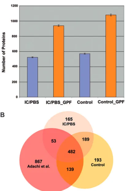
GPF increased protein identification bymore than 60% over the use of one large m/z range
in both sets of urine samples (Figure-1A). A total of
889 IC/PBS and 1003 control proteins with Protein
Probability ≥ 0.8, with error rates ≤ 0.023 and ≤ 0.022
respectively, were identiied. Recently, the normal
urine proteome was extensively analyzed revealing
more than 1,500 proteins (5). A comparative analysis
of our IC/PBS and control urines, and normal urine data by Adachi et al. (5) is shown in a Venn diagram (Figure-1B) created by ProteinCenter (www.proxeon.
com). According to this analysis, 165 proteins ap -peared to be unique to IC/PBS. However, proteins
identiied in only one mutually exclusive subset may
be due to under-sampling in other samples or result
from data iltering (7).
Identiied urine proteins were annotated with Gene Ontology (GO) (9), which assign probable
subcellular compartmentalization and molecular
func-tions. Approximately 50% of the proteins identiied
were annotated as secreted or membrane-associated proteins, which may be a characteristic of the urine proteome (5).
Proteins Associated with IC/PBS
A total of 78 proteins with P-value ≤ 0.1 were considered to be statistically signiicant for dif -ferential expression between IC/PBS and control for this study (Table-1). This P-value was chosen to cast a wider net that includes the most of the differen-tially expressed proteins. By quantitative analysis,
19 were found up-regulated in IC/PBS compared to control, and 59 were down-regulated. Among these, we focused on proteins identiied in all ten IC/PBS subjects and were up-regulated in at least 60% of
this cohort by spectral counts. Similarly, proteins that were found in all ten control subjects, and were
down-regulated in more than 60% of IC/PBS cohort
were also investigated. Using these criteria, two up-regulated and two down-up-regulated proteins were
found. The two up-regulated were α-1B-glycopro -tein (A1BG) and orosomucoid-1 (ORM1) both of which are glycoproteins. This is promising in that
many current biomarkers like prostate speciic anti -gen (PSA) are glycoproteins (7). A1BG is a plasma
glycoprotein of unknown function but over-expres -sion of this protein in pancreatic adenocarcinoma
patients has been reported (10). ORM1 is an acute
phase plasma protein that increases as a result of
acute inlammation (11). The two down-regulated
proteins were transthyretin (TTR) and hemopexin (HPX). TTR is a thyroid hormone-binding protein.
Defects in TTR are the cause of amyloidosis (12).
HPX protects low-density lipoprotein against
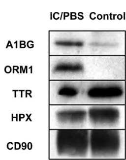
he-moglobin-induced oxidation (13). Differential ex -pression of these proteins was further validated by Western blot analysis of pooled IC/PBS and control samples (Figure-2). Among the other up-regulated proteins, afamin (APF), osteopontin (SPP1),
pancre-atic secretory trypsin inhibitor (SPINK1), proactiva -tor polypeptide (PSAP), and apolipoprotein (LPA) were found from all ten IC/PBS patients.
Enrichment functional analysis, which ranks
the most relevant cellular processes among the differ-entially expressed proteins, was performed by Meta-Core™ pathway analysis tool (www.genego.com).
467
Urinary Proteomics in IC/PBS
were found up-regulated
in IC/PBS, whereas cell
adhesion, extra cellular matrix remodeling, and
stimulus response were found to be
down-regu-lated.
When the 165 IC/PBS proteins
(Figure-1B) were queried for pathways, neurophysiological GABA-A receptor life cycle pathway was mapped
with the most number of proteins. Gamma-amino butyric acid receptors (GABA-A) mediate fast syn-aptic inhibition in the brain and spinal cord (14). Alterations in neuronal surface receptors modulate the synaptic strength, leading to changes in
sensitiv-ity to neurotransmitters (15). When a similar network analysis was performed for the 193 control proteins, Figure 1 - IC/PBS and control urine protein identiications. (A) Average number ± standard deviation (on 4 technical replicates) of identiied proteins with 2 or more peptides (multiple hits) using single m/z or 4 gas-phase m/z fractions for IC/PBS and control urine. (B) Our urinary proteome of IC/PBS and controls were compared to the previously reported normal urine proteome (Adachi et al. 2006). The Venn diagram shows overlapped proteins (621) between our asymptomatic control proteins and previously published healthy urine proteins. This comparative analysis also identiied 165 proteins that may be unique to the IC/PBS proteome. However, proteins identiied in one sample only may be due to under-sampling in other sampleor result from data iltering.
A
468
Urinary Proteomics in IC/PBS
DNA damage regulation pathway was one of the most activated pathways, suggesting a lack of DNA damage
regulation and repair functions in IC/PBS.
In Silico Analysis of Tissue Speciic
Expression
Although the proteins identiied in this study are found in urine, some of the proteins identiied may be more highly expressed in a speciic tissue type, and their tissue speciicity could enhance understanding
of the mechanisms underlying IC/PBS. Among the differentially expressed proteins, cell adhesion mol-ecule with homology to L1CAM (CHL1) is highly expressed in the cortex, brain, and spinal cord based
on UniProt tissue classiications (www.uniprot.org).
CHL1 is a neural recognition molecule involved in signal transduction pathways, and loss of this gene is
responsible for mental defects (16). In our datasets,
CHL1 was down-regulated in IC/PBS.
Protein-protein Interaction Network
Protein-protein interactions are important in signal transduction, which plays a fundamental role in many biological processes and diseases. The dif-ferentially expressed proteins were investigated for novel protein-protein interactions using MetaCore™.
Direct and indirect protein interactions were ranked
and interpreted in terms of GO processes. Two novel
protein network modules with potential importance in IC/PBS were identiied: 1) glucose metabolic process and positive regulation of natural killer cell-mediated
immune response to tumor cells, and 2) response to external stimulus, cell adhesion, wounding, and stress.
COMMENTS
Much effort has been devoted to the search for
useful biomarkers for IC/PBS diagnosis, phenotyping,
and for predicting response to treatment (17). Initial
attempts to develop a urinary biomarker concentrated on mediators of pain such as substance P (18).Other
proposed pain biomarkers have included uroplakin III-δ4 mRNA (19),and heparin-binding epidermal growth
factor-like growth factor (HB-EGF) (20). Antiprolif -erative factor (APF) is another candidate (21).To date,
none of these has been shown to deinitively correlate
with IC/PBS symptoms, clinical course, or response
to treatment. In a recent urine biomarker evaluation study, no robust association among urinary IL-6, cy -clic guanosine monophosphate, HB-EGF, epidermal growth factor, APF, and bladder biopsy was found in IC/PBS (22). Previously, a urinary proteomic method
was applied to identify biomarkers from age-, race-,
and gender-matched IC/PBS and control subjects (23). Three up-regulated proteins (uromodulin and
two kininogens) in the control and one up-regulated
protein (inter-α-trypsin inhibitor heavy chain H4) in the IC/PBS were found. These proteins showed a
cor-relation to IC severity on IC-speciic quality-of-life
scales. All four proteins were also found in our study but their differential expressions were not statistically
signiicant in our datasets.
The goal of this study was to use an in-depth
proteomic approach to identify speciic proteins or Figure 2 – Western blot analysis of differentially expressed
469
Urinary Proteomics in IC/PBS
Table 1 – Differentially expressed proteins identiied from IC/PBS and control urine samples. The protein IPI ID, annotation, ProteinProphet probability score, gene, expression ratio (IC/control, log2), non-adjusted P-value, and GO biological process, GO molecular function, and GO cellular compartment are tabulated. The list is sorted by P-values. ProteinProphet probability 1 represents the highest score of correct identiication. NA = not available.
Up-regulated proteins in IC/PBS
Protein Annotation Probability Gene IC/
Control
p Value
Biological Process
Molecular Function
Cellular compartment
IPI00019943 Afamin 1 AFM 1.69 0.00 transport NA extracellular
IPI00889156 Immunoglobulin
κ variable 3-20 1 IGKV3-20 1.91 0.01 responseimmune
antigen binding extracellular
IPI00022895 α-1B-glycopro -tein
1 A1BG 1.10 0.01 NA NA extracellular
IPI00010402 Uncharacterized protein
1 SH3BGRL3 0.79 0.03 NA NA nucleus
IPI00018136 Vascular cell ad-hesion protein 1
1 VCAM1 3.09 0.03 cell-cell adhesion
protein binding membrane
IPI00784430 Ig κ chain V-III
region VG
1 IGKV3D-11 2.17 0.04 immune
response
antigen binding extracellular
IPI00292150 Latent-trans-forming growth
factor β-binding
protein 2
0.99 LTBP2 3.00 0.04 signaling pathway
calcium ion binding extracellular
IPI00021000 Osteopontin 1 SPP1 1.00 0.05 ossiication cytokine activity extracellular
IPI00022417 Leucine-rich
α-2-glycoprotein
1 LRG1 1.97 0.05 NA protein binding membrane
IPI00884926 Orosomucoid 1 1 ORM1 1.66 0.05 acute-phase
response
protein binding extracellular
IPI00020687 Pancreatic secretory trypsin
inhibitor
1 SPINK1 1.11 0.05 NA serine-type
endo-peptidase inhibitor activity
470
Urinary Proteomics in IC/PBS
IPI00018236 Ganglioside GM2 activator
1 GM2A 1.41 0.06 glycolipid catabolic
process
sphingolipid activa-tor protein activity
lysosome
IPI00007726 Kallikrein-13 0.93 KLK13 1.32 0.07 proteolysis serine-type
endopep-tidase activity
cytoplasm
IPI00012503 Proactivator polypeptide
1 PSAP 0.54 0.07 glycosphin-golipid metabolic
process
α-galactosidase
activity
lysosome
IPI00020091 α-1-acid glyco -protein 2
1 ORM2 1.24 0.08 acute-phase response
binding extracellular
IPI00160384 Protein δ homo -log 1
1 DLK1 0.95 0.08
multi-cellular organismal
develop-ment
calcium ion binding membrane
IPI00217236 Tubulin-speciic
chaperone A
0 TBCA 3.17 0.09 post-chap-eronin tubulin folding pathway
unfolded protein binding
cytoskeleton
IPI00029168 Apolipoprotein (a)
1 LPA 1.15 0.09 proteolysis serine-type endopep-tidase activity
extracellular
IPI00290856 Lymphatic ves-sel endothelial hyaluronic acid
receptor 1
1 LYVE1 1.09 0.09 cell-matrix adhesion
hyaluronic acid binding
membrane Table 1 – continued
IPI00022432 -1.05 0.00
IPI00016334
MUC18
-1.49 0.01
IPI00015199
CD7 CD7 0.01
IPI00153049
ated protein 8
MXRA8 0.01 NA NA
IPI00240345 -3.91 0.01 NA
IPI00019157 -2.09 0.01
IPI00183445 LPHN1 -2.50 0.01
IPI00293057 0.02
IPI00291867 -0.84 0.02
IPI00218834 Low afinity
Ig γ Fc region 0.02
471
Urinary Proteomics in IC/PBS
Down-regulated proteins in IC/PBS
Protein Annotation Probability Gene IC/ Control
p Value Biological Process
Molecular Function Cellular Component
IPI00022432 Transthyretin 1 TTR -1.05 0.00 thyroid
hor-mone genera-tion
thyroid hormone transporter activity
extracellular
IPI00016334 Cell surface glycoprotein
MUC18
1 MCAM -1.49 0.01 cell adhesion protein binding membrane
IPI00015199 T-cell antigen
CD7 1 CD7 -2.32 0.01 calcium ion transport
receptor activity membrane
IPI00153049 Matrix-remod-
eling-associ-ated protein 8
1 MXRA8 -1.55 0.01 NA NA membrane
IPI00240345 C-type lectin domain family
14 member A
1 CLEC14A -3.91 0.01 NA sugar binding membrane
IPI00019157 Chondroitin sulfate
proteo-glycan 4
1 CSPG4 -2.09 0.01 angiogenesis tyrosine phospha-tase signaling
membrane
IPI00183445 Latrophilin-1 1 LPHN1 -2.50 0.01 neuropeptide
signaling pathway
G-protein coupled receptor activity
membrane
IPI00293057 Carboxypepti-dase B2
1 CPB2 -1.14 0.02 proteolysis zinc ion binding extracellular
IPI00291867 Complement factor I
1 CFI -0.84 0.02 proteolysis serine-type
endo-peptidase activity
membrane
IPI00218834 Low afinity Ig γ Fc region
receptor III-A
1 FCGR3A -2.32 0.02 immune response
IgG binding membrane
IPI00298971 Vitronectin 1 VTN -1.08 0.02 immune
response
472
Urinary Proteomics in IC/PBS
IPI00300786 α-amylase 1 1 AMY1A,
AMY1B
-1.30 0.02 carbohydrate metabolic
process
α-amylase activity extracellular
IPI00297124 IL-6 receptor
subunit β 1 IL6ST -1.91 0.02 signal trans-duction
IL-6 receptor activ -ity
membrane
IPI00387119 Ig κ chain V-III
region POM
1 IGKV3 -1.93 0.03 NA NA NA
IPI00026270 Carboxypepti-dase M
1 CPM -1.25 0.03 proteolysis zinc ion binding membrane
IPI00218914 Retinal dehy-drogenase 1
1 ALDH1A1 -3.17 0.03 aldehyde
metabolic process
retinal dehydroge-nase activity
cytosol
IPI00022488 Hemopexin 1 HPX -0.83 0.03 cellular iron
ion homeo-stasis
iron ion binding extracellular
IPI00026944 Nidogen-1 1 NID1 -1.04 0.04
protein-chro-mophore
linkage
calcium ion binding membrane
IPI00299059 Neural cell
adhesion
mol-ecule L1-like
protein
1 CHL1 -1.91 0.04 nervous
sys-tem develop-ment
protein binding membrane
IPI00020996 Insulin-like
growth factor-binding protein
complex acid labile chain
1 IGFALS -2.14 0.04 signal trans-duction
insulin-like growth
factor binding
soluble fraction
IPI00646689 Thioredoxin domain-con-taining protein
17
1 TXNDC17 -3.58 0.04 cell redox
homeostasis
473
Urinary Proteomics in IC/PBS
IPI00025476 Pancreatic
α-amylase
1 AMY1C,
AMY2A
-1.41 0.05 carbohydrate metabolic
process
α-amylase activity extracellular
IPI00169383
Phosphoglycer-ate kinase 1 1 PGK1 -1.22 0.05 glycolysis ATP binding cytoplasm
IPI00032532 Growth
arrest-speciic protein 6
1 GAS6 -1.91 0.05 regulation of
cell growth
calcium ion binding extracellular
IPI00031065 Deoxyribo -nuclease-1
1 DNASE1 -0.80 0.05 DNA catabol
-ic process
Deoxyribonuclease
I activity
nucleus
IPI00027493 4F2 cell-sur-face antigen heavy chain
1 SLC3A2 -1.62 0.05 calcium ion transport
calcium:sodium
antiporter activity
melanosome
IPI00301579 Epididymal se-cretory protein
E1
1 NPC2 -1.26 0.05 cholesterol
homeostasis
cholesterol binding lysosome
IPI00001759 Oxidized low-density
lipopro-tein receptor 1
1 OLR1 -0.68 0.05 proteolysis receptor activity membrane
IPI00219622 Proteasome
subunit α
type-2
1 PSMA2 -2.00 0.05 protein cata-bolic process
threonine endopep-tidase activity
nucleus
IPI00015525 Multimerin-2 1 MMRN2 -1.00 0.05 NA NA extracellular
IPI00073772
Fructose-1,6-bisphosphatase 1
1 FBP1 -1.03 0.06
gluconeogen-esis
phosphatase activity cytosol
IPI00220271 Alcohol dehy-drogenase
1 AKR1A1 -1.74 0.06 glucose
meta-bolic process
aldehyde reductase activity
NA
IPI00032179 Antithrombin III variant
1 SERPINC1 -1.29 0.06 blood
coagu-lation
serine-type endo-peptidase inhibitor
activity
membrane
IPI00816555 IGLV2-14
protein
1 IGLV2-14 -1.58 0.06 NA NA NA
IPI00291136 Collagen
α-1(VI) chain
1 COL6A1 -0.70 0.07 phosphate
transport
474
Urinary Proteomics in IC/PBS
IPI00442294 Neurotrimin
variant 3
1 HNT -3.32 0.07 neuron recog-nition
protein binding membrane
IPI00293088 Lysosomal
α-glucosidase
1 GAA -1.25 0.07 diaphragm
contraction
α-glucosidase activ -ity
lysosome
IPI00029275 Melanotrans-ferrin
1 MFI2 -1.08 0.07 cellular iron
ion homeo-stasis
ferric iron binding membrane
IPI00465248 α-enolase 1 ENO1 -1.12 0.07 glycolysis transcription
core-pressor activity
nucleus
IPI00034319 Protein CutA 1 CUTA -0.91 0.08 response to
metal ion
enzyme binding membrane
IPI00329801 Annexin A5 1 ANXA5 -1.28 0.08 anti-apoptosis phospholipase
inhibitor activity
cytoplasm
IPI00219525 6-phosphoglu -conate
dehy-drogenase
1 PGD 6 -1.05 0.08
pentose-phos-phate shunt, oxidative
branch
phosphogluconate dehydrogenase
NA
IPI00232571 Glypican-4 1 GPC4 -1.74 0.08 cell
prolifera-tion
NA membrane
IPI00032294 Cystatin-S 1 CST4 -3.58 0.08 NA cysteine protease
inhibitor activity
extracellular
IPI00295777 Glycerol-3-phosphate dehydrogenase
1 GPD1 -2.32 0.08 glycerol-3-phosphate catabolic
process
dehydrogenase cytosol
IPI00219425 Poliovirus receptor
1 PVR -0.77 0.08 cell adhesion receptor activity membrane
IPI00000792 Quinone oxido-reductase
1 CRYZ -3.17 0.09 visual percep-tion
quinone reductase activity
cytoplasm
IPI00182728 Vacuolar protein sort-ing-associating
protein 4B
1 VPS4B -2.17 0.09 intracellular cholesterol
transport
475
Urinary Proteomics in IC/PBS
IPI00024284 Basement
membrane-spe-ciic heparan
sulfate proteo-glycan core
protein
1 HSPG2 -0.53 0.09 cell adhesion protein binding membrane
IPI00742696 Vitamin
D-binding protein
1 GC -0.66 0.09 vitamin
trans-port
actin binding extracellular
IPI00007221 Plasma serine protease
inhibi-tor
1 SERPINA5 -0.65 0.09 transport serine-type
endo-peptidase inhibitor activity
membrane
IPI00646304 Peptidyl-prolyl cis-trans
isom-erase B
1 PPIB -1.49 0.09 protein
fold-ing
peptidyl-prolyl cis-trans activity
melanosome
IPI00099670 Carboxyl ester lipase
1 CEL -1.24 0.09 triacylglyc-erol metabolic
process
sterol esterase activity
cytoplasm
IPI00215980 Poliovirus receptor-related
protein 2
1 PVRL2 -0.82 0.09 homophilic cell adhesion
coreceptor activity
membrane
IPI00439446 Mannosidase,
α, class 1A,
member 1
1 MAN1A1 -1.18 0.10 glycosylation mannosidase
activity
Golgi mem-brane
IPI00550640 IGHG4 protein 1 IGHG4 -1.11 0.10 immune
response
copper ion binding membrane
IPI00016786 Cell division control protein
42 homolog
0.94 CDC42 -1.58 0.10 positive
regulation of pseudopodium
formation
GTPase activity cytosol
IPI00437186 Probable G-protein coupled
receptor 116
1 GPR116 -3.17 0.10 neuropeptide
signaling pathway
G-protein coupled receptor activity
membrane
IPI00000073 Pro-epidermal growth factor
1 EGF -0.58 0.11 positive
regulation of
phosphoryla-tion
EGF receptor activating ligand
activity
476
Urinary Proteomics in IC/PBS
protein networks that may be involved in IC/PBS
pathogenesis. A number of urine proteins were found to be differentially expressed between IC/PBS and control. Among the up-regulated proteins in IC/PBS, A1BG and ORM1 were present in all ten IC/PBS patients, with up-regulated expression in ≥ 60% of the cohort. TTR and HPX were found down-regulated in ≥ 60% of the IC/PBS cohort, and were present in
all control urines. None of these four proteins have
been previously implicated in IC/PBS pathogenesis. Two of these, A1BG and ORM1, are glycoproteins.
Currently, many clinical biomarkers and therapeutic
targets are glycoproteins, e.g. Her2/neu, PSA, and CA125.
165 and 193 proteins were found either in the
IC/PBS or control urines, respectively. One
interest-ing indinterest-ing from these datasets was that pathway and network analyses identiied possible activation of
neurophysiological processes involved in synaptic
inhibition, and lack of DNA damage repair in IC/PBS. One of the most important indings in pain research has been the identiication of changes in the central nervous system (CNS), which may explain the per -petuation of pain in chronic pain syndromes (24). For example, in male chronic pelvic pain syndrome, responses to painful stimuli are changed, and evidence of nervous system alterations is present (25). CHL1 is
known to be abundantly expressed in the CNS (e.g.,
brain, and spinal cord). Overall expression level of CHL1 was down-regulated in IC/PBS. Although no conclusions are being made as a result of these
data, altered response to stimuli taken together with down-regulation of CNS proteins such as CHL1 may
represent neurophysiological changes that contribute to IC/PBS pathogenesis.
Our strategy identiied many differentially
expressed proteins not previously associated with IC/PBS and this led to hypotheses around several
novel network modules. These included glucose me
-tabolism, alteration of which has been linked to human diseases (26). Natural killer cell mediated immune
response has been well documented in various hu-man diseases including prostate cancer (27). Little
is known about involvement of glucose metabolism and natural killer cell immune response in IC/PBS.
However, an association of glucose metabolism in IC/PBS has been recently detected by gene array
analysis of experimentally induced IC in mice (28). Although the implication of these networks needs
to be further investigated, any alteration to protein-protein interactions could impact the natural cascade signaling process.
Although there is clear correspondence be-tween pathological events and changes in protein
ex-pression in relevant networks and modules, whether
any of the differentially expressed proteins are true
markers for IC/PBS will require further investiga -tion. In urine analysis, an individual’s lifestyle, diet, medication history, and time of urine collection can
inluence the proteome proile; none of these fac -tors were considered in urine collection in this or other studies. Another limitation of this study is the
lack of detailed phenotyping of the subjects, which
might aid data interpretation. However, this is a pilot study, and we were attempting to determine if our methods held merit for identifying selected
proteins; furthermore, this small cohort would likely
preclude any conclusions based on demographic or clinical variables.
CONCLUSION
Our preliminary data indicate that there are qualitative and quantitative differences between the urinary proteomes of women with and without
IC/PBS. We identiied a number of proteins as well as pathways/networks that might contribute to the
pathology of IC/PBS or result from perturbations induced by this condition.
ACKNOWLEDGMENT
This work was supported by: National Institute of Diabetes and Digestive and Kidney Diseases U01 DK065202; National Institute of Environmental Health Sciences 5P30ES007033-12; National Center For Research Resources 1S10RR023044-01, and Robert Wood Johnson Foundation 64189.
The authors thank Dr. Priska von Haller at the University of Washington South Lake Union
477
Urinary Proteomics in IC/PBS
CONFLICT OF INTEREST
None declared.REFERENCES
1. Hanley RS, Stoffel JT, Zagha RM, Mourtzinos A, Bresette JF: Multimodal therapy for painful bladder syndrome / interstitial cystitis: pilot study combining
behavioral, pharmacologic, and endoscopic therapies.
Int Braz J Urol. 2009; 35: 467-74.
2. Hohlbrugger G, Lentsch P: Intravesical ions, osmolal
-ity and pH inluence the volume pressure response in
the normal rat bladder, and this is more pronounced
after DMSO exposure. Eur Urol. 1985; 11: 127-30. 3. Wesselmann U: Neurogenic inlammation and chronic
pelvic pain. World J Urol. 2001; 19: 180-5.
4. Anderson NL, Anderson NG: The human plasma proteome: history, character, and diagnostic prospects. Mol Cell Proteomics. 2002; 1(11): 845-67. Erratum in: Mol Cell Proteomics. 2003; 2: 50.
5. Adachi J, Kumar C, Zhang Y, Olsen JV, Mann M: The human urinary proteome contains more than 1500
proteins, including a large proportion of membrane
proteins. Genome Biol. 2006; 7: R80.
6. Scherl A, Shaffer SA, Taylor GK, Kulasekara HD, Miller SI, Goodlett DR: Genome-speciic gas-phase
fractionation strategy for improved shotgun proteomic
proiling of proteotypic peptides. Anal Chem. 2008; 80: 1182-91.
7. Goo YA, Liu AY, Ryu S, Shaffer SA, Malmström L,
Page L, et al.: Identiication of secreted glycoproteins
of human prostate and bladder stromal cells by
com-parative quantitative proteomics. Prostate. 2009; 69: 49-61.
8. Liu H, Sadygov RG, Yates JR 3rd: A model for random
sampling and estimation of relative protein abundance in
shotgun proteomics. Anal Chem. 2004; 76: 4193-201. 9. Ashburner M, Ball CA, Blake JA, Botstein D, Butler
H, Cherry JM, et al.: Gene ontology: tool for the uni
-ication of biology. The Gene Ontology Consortium. Nat Genet. 2000; 25: 25-9.
10. Tian M, Cui YZ, Song GH, Zong MJ, Zhou XY, Chen Y, et al.: Proteomic analysis identiies MMP-9, DJ-1
and A1BG as overexpressed proteins in pancreatic juice from pancreatic ductal adenocarcinoma patients.
BMC Cancer. 2008; 8: 241.
11. Narita T, Sasaki H, Hosoba M, Miura T, Yoshioka N, Morii T, et al.: Parallel increase in urinary excretion
rates of immunoglobulin G, ceruloplasmin, transferrin, and orosomucoid in normoalbuminuric type 2 diabetic
patients. Diabetes Care. 2004; 27: 1176-81.
12. Altland K, Benson MD, Costello CE, Ferlini A, Hazen
-berg BP, Hund E, et al.: Genetic microheterogeneity of
human transthyretin detected by IEF. Electrophoresis.
2007; 28: 2053-64.
13. Miller YI, Smith A, Morgan WT, Shaklai N: Role of
hemopexin in protection of low-density lipoprotein against hemoglobin-induced oxidation. Biochemistry.
1996; 35: 13112-7.
14. Kneussel M: Dynamic regulation of GABA(A) recep
-tors at synaptic sites. Brain Res Brain Res Rev. 2002; 39: 74-83.
15. Kanematsu T, Mizokami A, Watanabe K, Hirata M:
Regulation of GABA(A)-receptor surface expression with special reference to the involvement of GAB-ARAP (GABA(A) receptor-associated protein) and PRIP (phospholipase C-related, but catalytically
inac-tive protein). J Pharmacol Sci. 2007; 104: 285-92. 16. Montag-Sallaz M, Baarke A, Montag D: Aberrant neu
-ronal connectivity in CHL1-deicient mice is associated
with altered information processing-related immediate
early gene expression. J Neurobiol. 2003; 57: 67-80. 17. Dimitrakov J: A road map to biomarker discovery and
validation in urological chronic pelvic pain syndrome.
J Urol. 2008; 179: 1660-1.
18. Vera PL, Meyer-Siegler KL: Substance P induces
localization of MIF/alpha1-inhibitor-3 complexes to umbrella cells via paracellular transit through the
uro-thelium in the rat bladder. BMC Urol. 2006; 6: 24. 19. Zeng Y, Wu XX, Homma Y, Yoshimura N, Iwaki H,
Kageyama S, et al.: Uroplakin III-delta4 messenger RNA as a promising marker to identify nonulcerative interstitial cystitis. J Urol. 2007; 178: 1322-7; discus -sion 1327.
20. Kim J, Keay SK, Freeman MR: Heparin-binding epi
-dermal growth factor-like growth factor functionally
antagonizes interstitial cystitis antiproliferative factor
via mitogen-activated protein kinase pathway activa
-tion. BJU Int. 2009; 103: 541-6.
21. Keay SK, Zhang CO, Shoenfelt J, Erickson DR, Whit
-more K, Warren JW, et al.: Sensitivity and speciicity
of antiproliferative factor, heparin-binding epidermal
growth factor-like growth factor, and epidermal growth factor as urine markers for interstitial cystitis. Urology. 2001; 57(6 Suppl 1): 9-14.
22. Erickson DR, Tomaszewski JE, Kunselman AR, Stet
478
Urinary Proteomics in IC/PBS
cystitis/painful bladder syndrome. J Urol. 2008; 179: 1850-6.
23. Canter MP, Graham CA, Heit MH, Blackwell LS, Wilkey DW, Klein JB, et al.: Proteomic techniques
identify urine proteins that differentiate patients with interstitial cystitis from asymptomatic control subjects.
Am J Obstet Gynecol. 2008; 198: 553. e1-6.
24. Coderre TJ, Katz J, Vaccarino AL, Melzack R: Contri
-bution of central neuroplasticity to pathological pain:
review of clinical and experimental evidence. Pain.
1993; 52: 259-85.
25. Yang CC, Lee JC, Kromm BG, Ciol MA, Berger RE: Pain sensitization in male chronic pelvic pain syndrome: why are symptoms so dificult to treat? J Urol. 2003; 170: 823-6; discussion 826-7.
26. Holroyde CP, Gabuzda TG, Putnam RC, Paul P, Reich
-ard GA: Altered glucose metabolism in metastatic carcinoma. Cancer Res. 1975; 35: 3710-4.
27. Suzuki K, Nakazato H, Matsui H, Hasumi M, Shibata Y, Ito K, et al.: NK cell-mediated anti-tumor immune response to human prostate cancer cell, PC-3: im -munogene therapy using a highly secretable form of
interleukin-15 gene transfer. J Leukoc Biol. 2001; 69:
531-7.
28. Tseng LH, Chen I, Chen MY, Lee CL, Lo TS, Lloyd LK: Genome-based expression proiles as a single
standardized microarray platform for the diagnosis of
experimental interstitial cystitis: an array of 75 genes model. Int Urogynecol J Pelvic Floor Dysfunct. 2009; 20. [Epub ahead of print]
Accepted after revision:
March 15, 2010
Correspondence address:
Dr. Claire C. Yang
University of Washington, Department of Urology Box 356510
Seattle, WA, 98195-6510, USA Fax: + 1 206 543-3272
E-mail:[email protected]
EDITORIAL COMMENT
I will say in order that biological meaning may be derived and testable hypotheses may be built from proteomic experiments in relation to IC/PBS, assignments of proteins detected by mass spectrom-etry must be supplemented with additional notation,
such as information on known protein functions,
protein-protein interactions, or biological pathway associations.
Visualizing this bulk of proteomic informa -tion and summariza-tion the resulting significant differential expressed proteins underlying IC/PBC
in an easy to navigate tabular formats, including meta-information on those proteins in addition to complementary gene ontology (GO) terminology, is also important so that in-house expertise on particular proteins may be integrated into the larger datasets.
479
Urinary Proteomics in IC/PBS
articles can make use of certain proteomic mapping
and comparison tools (1-4) further investigating mass spectrometry and proteomic outputs in order to derive insight into the signaling pathway underlying IC/
PBS.
REFERENCES
1. Schmidt T, Frishman D: PROMPT: a protein map -ping and comparison tool. BMC Bioinformatics.
2006; 7: 331.
2. Gehlenborg N, O’Donoghue SI, Baliga NS, Goes
-mann A, Hibbs MA, Kitano H, et al.: Visualization of omics data for systems biology. Nat Methods. 2010; 7(3 Suppl): S56-68.
3. Yu K, Sabelli A, DeKeukelaere L, Park R, Sindi S, Gatsonis CA, et al.: Integrated platform for manual
and high-throughput statistical validation of tandem
mass spectra. Proteomics. 2009; 9: 3115-25.
4. Pruess M, Apweiler R: Bioinformatics Resources for In Silico Proteome Analysis. J Biomed Biotechnol. 2003; 2003: 231-236.
Dr. Ling-Hong Tseng
Department of Obstetrics and Gynecology Chang Gung Memorial Hospital Chang-Gung University College of Medicine
Tao-Yuan, Taiwan E-mail: [email protected]
EDITORIAL COMMENT
Interstitial cystitis (IC) is a debilitating chronic disease caused by undetermined and
un-known factors, which impedes the development of
accurate diagnostic methods, therefore delaying the treatment of patients that otherwise would promptly be cared for. Currently, the diagnosis of IC in most cases is given by exclusion mainly because this
pathology lacks adequate biologic markers in blood
and urine.
Exact etiology of interstitial cystitis (IC) is
unknown, however the impermeability of the uro -thelial barrier of the bladder just as an alteration in the production of urine proteins could play important physiological roles in lower urinary tract dysfunction (1).
Recently, the study of proteome has been
introduced as a diagnostic tool for inlammatory dis
-eases of dificult diagnoses. In diabetic nephropathy the proteomic marker can be a prognostic and/or a
therapeutic factor (2).
In a preliminary study, the authors showed that there were qualitative and quantitative differ-ences between the urinary proteomes of women with and without IC/PBS. Furthermore, future studies
researching the proteome characteristics associated with IC could provide not only a better comprehension of physiopathology but also lead to further develop-ment of new drugs or therapies for treatdevelop-ment and/or prevention of IC and related disorders.
REFERENCES
1. Deng FM, Ding M, Lavker RM, Sun TT: Urothelial function reconsidered: a role in urinary protein se
-cretion. Proc Natl Acad Sci U S A. 2001; 98: 154-9. 2. Thongboonkerd V: Current status of renal and uri
-nary proteomics: ready for routine clinical applica
-tion? Nephrol Dial Transplant. 2010; 25: 11-6.
Dr. João Luiz Amaro
Departamento de Urologia Faculdade de Medicina de Botucatu