Prostatic Atrophy. Clinicopathological Significance
Athanase Billis
Department of Anatomic Pathology, School of Medicine, University of Campinas (Unicamp), Campinas, Sao Paulo, Brazil
ABSTRACT
Prostatic atrophy is a benign lesion that may mimic adenocarcinoma histologically and on imaging. It is more frequent in the peripheral zone and has gained importance with the increasing use of needle biopsies. Diffuse atrophy occurs sec-ondarily to radiotherapy and/or endocrine therapy. Inflammation and/or chronic local ischemia may cause focal atrophy with an increasing frequency in age. Atrophy may be classified morphologically into diffuse and focal. The latter may be partial, complete or combined. Partial focal atrophy is the most frequent mimicker of adenocarcinoma on needle biopsies. Complete focal atrophy may be subtyped into simple, sclerotic and hyperplastic (or postatrophic hyperplasia). Combined lesions are frequent and partial atrophy may precede complete atrophy. The several morphologic types of focal atrophy may represent a morphologic continuum and the hyperplastic (or postatrophic hyperplasia) subtype seems to be at the extreme end of this continuum. Chronic inflammation associated to focal atrophy (proliferative inflammatory atrophy) has been linked to high-grade prostatic intraepithelial neoplasia and/or carcinoma. This link, however, remains controversial in the literature. The question whether inflammation directly produces tissue damage and atrophy or some other insult induces atrophy directly, with inflammation occurring secondarily, is still unresolved. An intriguing finding that needs further studies is a possible association of extent of atrophy to serum PSA elevation.
Key words: prostate; atrophy; prostatic neoplasms; carcinoma; biopsy; needle Int Braz J Urol. 2010; 36: 401-9
INTRODUCTION
Prostatic atrophy (PA) is one of the most fre-quent histologic mimics of prostatic adenocarcinoma (1). On conventional and color Doppler transrectal ultrasound and on magnetic resonance spectroscopic imaging studies, PA may also simulate prostate cancer (2,3). Thus, PA should be considered together with prostatitis as an important cause of false-positive results on imaging of the prostate (2). Of further interest as regards this lesion relates to a possible link to high-grade intraepithelial neoplasia (HGPIN) and/or carcinoma (4). PA occurs most frequently in
doi: 10.1590/S1677-55382010000400003
the peripheral zone (5-7) and gained importance with the increasing use of needle biopsies for the detection of prostatic carcinoma (8). It is a very frequent lesion: 83.7% on needle biopsies (9) and 85% in autopsies (10).
HISTORICAL BACKGROUND AND NOMENCLATURE
autopsy study. He found that there was a strong cor-relation with age and, according to his study, prostatic atrophy is initiated during the 5th decade and con-tinues as a progressive process into the 8th decade. Moore classified PA into simple acinar atrophy and sclerotic atrophy.
In 1954, Franks (6) added to simple acinar atrophy and sclerotic atrophy a lesion that he called postatrophic hyperplasia. When hyperplasia followed simple acinar atrophy, it was called lobular hyper-plasia. Sclerotic atrophy with hyperplasia was called postsclerotic hyperplasia. According to the author, lobular hyperplasia and postsclerotic hyperplasia are closely related existing conditions in which it is impossible to decide to which group a particular area of hyperplasia belongs.
Since these first studies, authors have de-scribed similar morphologic features with different names. For example, despite not using the term hyperplasia, Moore’s Figure-14 called by the author as simple acinar atrophy (5) is identical to the lesion shown in Figure-8 and called lobular hyperplasia in Franks’ article (6).
PATHOGENESIS
Radiotherapy and/or endocrine therapy are established causes of diffuse atrophy. Inactive or ac-tive inflammation is a frequent cause of focal atrophy. Based on a study of autopsies there is evidence that chronic local ischemia may be a cause of focal atro-phy, which was clearly more frequent in advanced age (10). A further evidence for a possible role of chronic local ischemia in the pathogenesis of prostatic atrophy was the finding that blood flow, assessed by color Doppler transrectal ultrasound, was absent in 60% of hypoechoic nodules due to prostatic atrophy (3). However, many examples of atrophy are still considered idiopathic in nature (11).
MORPHOLOGIC CLASSIFICATION
A morphologic classification of prostatic at-rophy is shown in Table-1. In diffuse atat-rophy, all acini of the gland are atrophic. This occurs secondarily to
radiotherapy and/or hormonal therapy. Focal atrophy is the usual lesion seen by the pathologist on routine practice. It occurs in patches and preserves the lobular architecture of the gland. The latter feature helps in the histological differential diagnosis of adenocarci-noma.
Partial atrophy. This is the most common benign variant of focal atrophy that causes difficulty in the differential diagnosis of adenocarcinoma (1,12). The lesion was reported in the literature in 1998 (13). Architecturally, partial atrophy consists of crowded glands often with a disorganized growth pattern. In contrast to complete atrophy, which can typically be diagnosed at scanning magnification owing to the presence of well-formed glands with a very basophilic appearance, partial atrophy has a pale cytoplasm later-al to the nuclei giving rise to plater-ale staining glands that more closely mimic cancer (Figure-1). An additional difficulty in distinguishing cancer from partial atrophy is the positivity for alpha-methylacyl coenzyme A racemase (AMACR) in some acini (1,12,14).
Complete atrophy. The glands show a ba-sophilic appearance due to the scant cytoplasm and crowding of nuclei. The hyperplastic (or postatrophic hyperplasia) subtype most frequently mimics adeno-carcinoma. In a working group classification (15), complete focal atrophy was subtyped into simple atrophy, simple atrophy with cyst formation, and postatrophic hyperplasia. Based on an autopsy study, complete focal atrophy was subtyped into simple atrophy, sclerotic atrophy, and hyperplastic atrophy (or postatrophic hyperplasia) (10). Sclerotic atrophy in the morphologic classification shown in Table-1 corresponds to simple atrophy with cyst formation in the working group classification.
Simple atrophy. Usually involves an entire lobule, although isolated acini may be affected. The acini are small showing scant cytoplasm and decrease in the height of the epithelial cells (Figure-2A). They may be cystic with flattened epithelium (Figure-2B). The surrounding stroma may or may not show any fibrosis.
Figure 1 – Partial atrophy. A) Crowded acini with pale cytoplasm lateral to the nuclei giving rise to pale staining glands that more closely mimic cancer. There is scant apical cytoplasm with nuclei extending to the full cell height (HE, X165); B) Higher power view
Table 1 – Morphologic classification of prostatic atro-phy.
A - Diffuse B - Focal
1. Partial 2. Complete
a) Simple b) Sclerotic
c) Hyperplastic (or postatrophic hyperplasia) 3. Combined
Figure 2 – Simple atrophy. A) Isolated small acini showing crowded nuclei, scant cytoplasm and decrease in the height of the epithelial cells (HE, X220); B) Simple atrophy with cystic dilatation (HE, X165).
collar frequently associated to elastosis that involves the acinus. With higher degrees of hyalinization the acini dilate, sometimes prominently, and the epithe-lium becomes extremely flattened and eventually no longer can be identified (Figure-3A). In advanced stages, the lumen is replaced by loose mesenchyme-like tissue with scattered inflammatory round-cells (Figure-3B).
Hyperplastic atrophy (or postatrophic hyper-plasia). Shows small acini closely packed together and lined by atrophic epithelium (Figure-4A). The
A
A
B
acini may also show cystic dilatation (Figure-4B). In contrast to partial atrophy, the cytoplasm is very scant and the nuclei are crowded conferring a baso-philic tinge to the lesion. When fibrosis is present in the stroma, the proliferation is irregular and can result in distortion of the acini mimicking infiltrative adenocarcinoma. Elastosis of the stroma (Figure-4A) may be seen in all subtypes of complete atrophy and is a microscopic feature useful for the differential di-agnosis of adenocarcinoma (16). The cysts of simple or hyperplastic atrophy must be differentiated from
Figure 3 – Sclerotic atrophy. A) Dilated acini, atrophy of the epithelium and a thick fibrohyaline collar involving the acinus (HE, X165); B) Advanced stage of sclerotic atrophy. The epithelium disappeared and the lumen is replaced by loose mesenchyme-like tissue with scattered inflammatory round-cells (HE, X165).
other simple or multiple parenchymal cysts due to retention of prostatic secretions, difficult drainage of secretions due to benign prostatic hyperplasia (BPH) nodule, inflammation of ducts that leads to obstruc-tion, and cystic degeneration of BPH (17).
Combined. Subtypes of focal complete atro-phy are frequently combined (10). They may occur in adjacent but separate foci or merging in the same focus. Hyperplastic atrophy with a central duct or aci-nus showing sclerotic atrophy is a frequent combined lesion in the same focus and very characteristic of
Figure 4 – Hyperplastic atrophy (or postatrophic hyperplasia). A) Small acini closely packed together and lined by atrophic epithelium. In contrast to partial atrophy, the cytoplasm is very scant and the nuclei are crowded. The stroma shows fibrosis and elastosis (HE, X220); B) Hyperplastic atrophy with cystic dilatation (HE, X165).
A
A
B
prostatic atrophy being a feature useful for the differ-ential diagnosis with adenocarcinoma (Figure-5). The existence of combined subtypes supports the hypoth-esis that complete prostatic atrophy is a morphologic continuum and that hyperplastic type (or postatrophic hyperplasia) seems to be at the extreme end of this morphologic continuum (8). The hyperplastic small acini seem to represent a regenerative process. A high cell proliferation using immunohistochemistry seems to support this hypothesis (18).
Partial and complete atrophy may be also combined. Studying needle prostatic biopsies we found that partial atrophy and complete atrophy were present concomitantly in 47/75 (63%) biopsies. In 20/75 (27%) biopsies, we found topographical mer-gence of partial and complete atrophy. In these areas, transitions between partial and complete atrophy could be appreciated in the same gland (Figure-6). Based on the aberrant phenotypic expression of the secretory compartment in complete atrophy (19) but not in partial atrophy, immunohistochemistry may highlight these transitions in the same gland (Figure-7).
In a study by Oppenheimer et al. (13) partial atrophy was present simultaneously with more fully developed atrophy (complete atrophy) in 35.35% of biopsies. In the study by Wang et al. (12), 48 of 278 (17.3%) partial atrophy cases were mixed with postatrophic hyperplasia. Przybycin et al. (20)found a much higher frequency. The authors described that
Figure 5 – Combined atrophy. Hyperplastic atrophy with a dilated central duct or acinus with sclerotic atrophy (HE, X165).
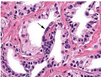
Figure 7 – Immunohistochemistry for 34βE12. Negative in cells of the secretory compartment in segment of partial atrophy (short arrow) and aberrant positivity in the segment of complete atrophy (long arrow) (HE, X220).
Figure 6 – Morphologic transition between partial and complete atrophy in the same gland (arrow shows segment of complete atrophy) (HE, X220).
complete atrophy was present but distinct from partial atrophy foci in 41/45 (91%) needle biopsies.
An intriguing finding in partial atrophy foci is the very rare presence of chronic unspecific inflam-mation. Przybycin et al. (20), found inflammation in an insignificant 1% of partial atrophy foci. In a study on 75 biopsies, we did not find chronic unspecific inflammation in partial atrophy foci as well as in areas of topographic mergence between these lesions. On the other hand, inflammation was frequently seen in complete atrophy foci: 56.2%, 48%, and 54.3%, in simple, sclerotic, and hyperplastic atrophy, respec-tively.
PRECANCEROUS LESION?
The term “proliferative inflammatory atro-phy” (PIA) was proposed by De Marzo et al. (4) to designate focal simple or postatrophic hyperplasia occurring in association with inflammation. Several studies have postulated that PIA may represent a precursor lesion to high-grade prostatic intraepi-thelial neoplasia (HGPIN) and, therefore, prostatic carcinoma (4,19,21-23). Chronic inflammation of longstanding duration has been linked to the devel-opment of carcinoma in several organ systems and HGPIN is considered the most likely precursor of prostate carcinoma (4,24).
Several separate findings provide supportive evidence for this novel hypothesis:
1) There is a shift in the topographic fidelity of proliferation in PIA similar to HGPIN and carcinoma (4). Most cell division in the normal human prostate epithelium occurs in the basal cell compartment, yet HGPIN and adenocarcinoma cells possess phenotypic and morphologic features of secretory cells. Thus, cell proliferation has been shifted up from the basal into the secretory compartment in HGPIN and carci-noma;
2) The phenotype of many of the cells in PIA is most consistent with that of an immature secretory-type cell similar to that for the cells of HGPIN and carcinoma (4,19,22). Atrophic luminal cells show an intermediate phenotype in that many cells express bcl-2 (normally a basal cell marker in the prostate), and virtually all of the cells express high levels of keratins 8/18. Intermediate cell population has been suggested to represent amplifying cells modulating
the expansion and development of the prostate epi-thelium. Increased proliferation has been observed in atrophic glandular epithelium (18);
3) PIA, HGPIN, and carcinoma all occur with high prevalence in the peripheral zone and low preva-lence in the central zone of the human prostate (21);
4) Topographic relation of PIA to HGPIN, i.e. areas of atrophy merging directly with areas of HGPIN within the same glands. In radical prostatec-tomy specimens, Putzi and De Marzo (21) identified morphologic merging between PIA and HGPIN in 34% of the PIA lesions. They also found frequent oc-currences of small carcinoma lesions in the vicinity of focal atrophy. In a study by Wang et al. (22), a total of 1,188 HGPIN lesions were identified, of which 17% (198) were in the morphological process of merging with PIA. Thirty-six PIA-merging prostatic carcinoma lesions were also detected. The atrophic epithelial cells in such merging lesions had increased Ki-67 proliferative index and an intermediate phenotype: increased expression for cytokeratin 5, GSTP1, c-MET, and C/EBPβ.
The link of PIA to prostatic carcinogenesis and the morphological transition of PIA, HGPI, and invasive carcinoma, however, are not favored in other studies. In autopsies, prostates with atrophy showed no association with histologic carcinoma and/or HG-PIN (10). The only significant association found was with arteriosclerosis. In this study ischemia caused by local intense arteriosclerosis seemed to be a potential factor for the pathogenesis of atrophy. In a subsequent study, the same lack of association was found to HG-PIN and/or histologic carcinoma comparing atrophy with and without inflammation (9).
Postma et al. (27) evaluated whether the incidence of atrophy reported on sextant biopsies was associated with subsequent prostate cancer detection. The au-thors concluded that atrophy, especially in its simple form, is a very common lesion in prostate biopsy cores (94%). Atrophy in an asymptomatic popula-tion undergoing screening was not associated with a greater prostate cancer or HGPIN incidence during subsequent screening rounds.
A question to be raised is whether atrophy by itself is implicated in carcinogenesis or the key event is chronic inflammation leading to atrophy, HGPIN and cancer. This question is still unresolved (28,29). Inflammation directly may produce tissue damage and atrophy or, alternatively, some other insult like ischemia induces the atrophy directly, with inflammation occurring secondarily. Hypothesizing that partial atrophy precedes complete atrophy, the absence of inflammation in the former as well as in areas of mergence between these two lesions seems to favor that chronic inflammation in complete focal atrophy may be a secondary phenomenon.
PROSTATIC ATROPHY AND PSA
An intriguing finding was a positive and significant association between extent of atrophy and total or free serum prostate-specific antigen (PSA) elevation (30). The study was based on 131 needle prostatic biopsies corresponding to 107 patients. The only diagnosis in all biopsies was focal prostatic atrophy without presence of cancer, HGPIN or sus-picious for cancer (ASAP). In a subsequent study it was shown that this association is not related to the type of atrophy (31).
What would be a possible pathogenesis for the serum PSA elevation associated with focal pros-tatic atrophy? It is intriguing that cells of the secretory compartment of atrophic acini may produce higher levels of PSA. The authors hypothesize that injurious stimuli causing focal prostatic atrophy may interfere in the physiologic barrier that prevents the escape of any significant amounts of PSA to the general circula-tion.
PSA is a single chain glycoprotein with pro-teolytic enzyme activity mainly directed against the
major gel-forming protein of the ejaculate (semenoge-lin). PSA induces liquefaction of semen with release of progressively motile spermatozoa (32). There are several efficient physiologic barriers to prevent the escape of any significant amounts of PSA from the prostatic ductal system: basement membrane of the acini, basal cells lining the acini, prostatic stroma, basement membrane of capillary endothelial cells, and endothelial cells. These barriers normally prevent PSA from entering the general circulation at concentrations of more than 3 ng/mL (32).
Focal prostatic atrophy represents a form of adaptive response to injury most commonly to inflam-mation and/or local ischemia. Inflaminflam-mation and/or ischemia are injurious stimuli resulting in diminished oxidative phosphorilation, membrane damage, influx of intracellular calcium, and accumulation of oxygen-derived free radicals (oxidative stress) (4). Studies showing elevated levels of glutathione S-transferase P1, glutathione S-transferase A1, and Cox-2 in pros-tatic atrophic epithelial cells suggest a stress-induced response (33-35). We do not know which mechanisms are involved in the physiologic barrier that prevents the escape of any significant amounts of PSA to the general circulation, however, all these stress-induced responses may affect this barrier. Inflammation and particularly ischemia may have also a field effect af-fecting the physiologic barrier of normal acini close to atrophic acini.
CONCLUSION
seem to be at the extreme end of this continuum. A possible link of prostatic atrophy to HGPIN and/or carcinoma remains controversial in the literature. The possible association of extent of prostatic atrophy to serum PSA elevation needs further studies.
CONFLICT OF INTEREST
None declared.
REFERENCES
1. Herawi M, Parwani AV, Irie J, Epstein JI: Small glan-dular proliferations on needle biopsies: most common benign mimickers of prostatic adenocarcinoma sent in for expert second opinion. Am J Surg Pathol. 2005; 29: 874-80.
2. Prando A, Billis A: Focal prostatic atrophy: mimicry of prostatic cancer on TRUS and 3D-MRSI studies. Abdom Imaging. 2009; 34: 271-5.
3. Meirelles LR, Billis A, Cotta AC, Nakamura RT, Caserta NM, Prando A: Prostatic atrophy: evidence for a possible role of local ischemia in its pathogenesis. Int Urol Nephrol. 2002; 34: 345-50.
4. De Marzo AM, Marchi VL, Epstein JI, Nelson WG: Proliferative inflammatory atrophy of the prostate: implications for prostatic carcinogenesis. Am J Pathol. 1999; 155: 1985-92.
5. Moore RA: The Evolution and Involution of the Pros-tate Gland. Am J Pathol. 1936; 12: 599-624.
6. Franks LM: Atrophy and hyperplasia in the prostate proper. J Pathol Bacteriol. 1954; 68: 617-21.
7. Liavåg I: Atrophy and regeneration in the pathogenesis of prostatic carcinoma. Acta Pathol Microbiol Scand. 1968; 73: 338-50.
8. Cheville JC, Bostwick DG: Postatrophic hyperplasia of the prostate. A histologic mimic of prostatic adeno-carcinoma. Am J Surg Pathol. 1995; 19: 1068-76. 9. Billis A, Magna LA: Inflammatory atrophy of the
prostate. Prevalence and significance. Arch Pathol Lab Med. 2003; 127: 840-4.
10. Billis A: Prostatic atrophy: an autopsy study of a his-tologic mimic of adenocarcinoma. Mod Pathol. 1998; 11: 47-54.
11. Srigley JR: Benign mimickers of prostatic adenocar-cinoma. Mod Pathol. 2004; 17: 328-48.
12. Wang W, Sun X, Epstein JI: Partial atrophy on prostate needle biopsy cores: a morphologic and immunohisto-chemical study. Am J Surg Pathol. 2008; 32: 851-7.
13. Oppenheimer JR, Wills ML, Epstein JI: Partial atrophy in prostate needle cores: another diagnostic pitfall for the surgical pathologist. Am J Surg Pathol. 1998; 22: 440-5.
14. Worschech A, Meirelles L, Billis A: Expression of AMACR (alpha-methylacyl coenzyme A racemase) in partial and complete focal atrophy on prostate needle biopsies. Anal Quant Cytol Histol. 2010; (in press). 15. De Marzo AM, Platz EA, Epstein JI, Ali T, Billis A,
Chan TY, et al.: A working group classification of fo-cal prostate atrophy lesions. Am J Surg Pathol. 2006; 30: 1281-91. Erratum in: Am J Surg Pathol. 2006; 30: 1489.
16. Billis A, Magna LA: Prostate elastosis: a microscopic feature useful for the diagnosis of postatrophic hyper-plasia. Arch Pathol Lab Med. 2000; 124: 1306-9. 17. Galosi AB, Montironi R, Fabiani A, Lacetera V, Gallé
G, Muzzonigro G: Cystic lesions of the prostate gland: an ultrasound classification with pathological correla-tion. J Urol. 2009; 181: 647-57.
18. Ruska KM, Sauvageot J, Epstein JI: Histology and cellular kinetics of prostatic atrophy. Am J Surg Pathol. 1998; 22: 1073-7.
19. van Leenders GJ, Gage WR, Hicks JL, van Balken B, Aalders TW, Schalken JA, et al.: Intermediate cells in human prostate epithelium are enriched in prolifera-tive inflammatory atrophy. Am J Pathol. 2003; 162: 1529-37.
20. Przybycin CG, Kunju LP, Wu AJ, Shah RB: Partial atrophy in prostate needle biopsies: a detailed analysis of its morphology, immunophenotype, and cellular kinetics. Am J Surg Pathol. 2008; 32: 58-64.
21. Putzi MJ, De Marzo AM: Morphologic transitions between proliferative inflammatory atrophy and high-grade prostatic intraepithelial neoplasia. Urology. 2000; 56: 828-32.
22. Wang W, Bergh A, Damber JE: Morphological tran-sition of proliferative inflammatory atrophy to high-grade intraepithelial neoplasia and cancer in human prostate. Prostate. 2009; 69: 1378-86.
23. De Marzo AM, Meeker AK, Zha S, Luo J, Nakayama M, Platz EA, et al.: Human prostate cancer precursors and pathobiology. Urology. 2003; 62(5 Suppl 1): 55-62.
24. Montironi R, Mazzucchelli R, Scarpelli M: Precan-cerous lesions and conditions of the prostate: from morphological and biological characterization to chemoprevention. Ann N Y Acad Sci. 2002; 963: 169-84.
asso-ciation with prostate cancer. Am J Surg Pathol. 1999; 23: 932-6.
26. Billis A, Freitas LL, Magna LA, Ferreira U: Inflam-matory atrophy on prostate needle biopsies: is there topographic relationship to cancer? Int Braz J Urol. 2007; 33: 355-60; discussion 361-3.
27. Postma R, Schröder FH, van der Kwast TH: Atrophy in prostate needle biopsy cores and its relationship to prostate cancer incidence in screened men. Urology. 2005; 65: 745-9.
28. Mikuz G, Algaba F, Beltran AL, Montironi R: Prostate carcinoma: atrophy or not atrophy that is the question. Eur Urol. 2007; 52: 1293-6.
29. Tomas D, Kruslin B, Rogatsch H, Schäfer G, Belicza M, Mikuz G: Different types of atrophy in the prostate with and without adenocarcinoma. Eur Urol. 2007; 51: 98-103; discussion 103-4.
30. Billis A, Meirelles LR, Magna LA, Baracat J, Prando A, Ferreira U: Extent of prostatic atrophy in needle biopsies and serum PSA levels: is there an association? Urology. 2007; 69: 927-30.
31. Billis A, Meirelles L, Freitas LL, Magna LA, Ferreira U: Does the type of prostatic atrophy influence the
association of extent of atrophy in needle biopsies and serum prostate-specific antigen levels? Urology. 2009; 74: 1111-5.
32. Oesterling JE, Lilja H: Prostate-specific antigen. The value of molecular forms and age-specific reference ranges. In: Vogelzang NJ, Scardino PT, Shipley WU et al. (ed.), Comprehensive Textbook of Genitourinary Oncology. Baltimore, Williams & Wilkins. 1996; pp. 668-80.
33. Kumar V, Abbas AK, Fausto N: Robbins and Cotran Pathologic Basis of Disease, 7th ed. Philadelphia, Elsevier Sanders. 2005; pp. 3-46.
34. Parsons JK, Nelson CP, Gage WR, Nelson WG, Kensler TW, De Marzo AM: GSTA1 expression in normal, preneoplastic, and neoplastic human prostate tissue. Prostate. 2001; 49: 30-7.
35. Zha S, Gage WR, Sauvageot J, Saria EA, Putzi MJ, Ewing CM, et al.: Cyclooxygenase-2 is up-regulated in proliferative inflammatory atrophy of the prostate, but not in prostate carcinoma. Cancer Res. 2001; 61: 8617-23.
Accepted: January 21, 2010
Correspondence address:
Dr. Athanase Billis
Anatomia Patológica, FCM, Unicamp Caixa Postal 6111
Campinas, SP, 13084-971, Brazil Fax: + 55 19 3289-3897