Retrospective Study of a Series of Choanal
Atresia Patients
Denise Manica
1,2Cláudia Schweiger
1,2Cátia C Saleh Netto
1,2Gabriel Kuhl
1,21Hospital de Clínicas de Porto Alegre, Universidade Federal do Rio
Grande do Sul, Porto Alegre, RS, Brazil
2Serviço de Otorrinolaringologia, Hospital de Clínicas de Porto Alegre,
Porto Alegre, RS, Brazil
Int Arch Otorhinolaryngol 2014;18:2–5.
Address for correspondence Denise Manica, MD, MSc, Avenida João Pessoa, 1051/408, Bairro Cidade Baixa, Porto Alegre/RS, CEP 90040-000, Brazil (e-mail: [email protected]).
Introduction
Choanal atresia is defined as a malformation of the posterior nasal aperture that interferes with airflow from the nose to the rhinopharynx. It was first described by Roederer in 1755.1
Since then, several studies have been published on the subject, mainly case series without adequate standardization that compromised comparison between data. Therefore, there are many doubts about this congenital abnormality, including timing of surgery, access to the surgical field, procedure Keywords
►
CHARGE syndrome
►
choanal atresia
►
mouth breathing
►
nasopharynx
►
nose diseases
Abstract
Introduction
Although it has been more than 250 years since the
fi
rst description of
choanal atresia (CA), there are still doubts about this abnormality. The differences
between unilateral and bilateral forms are seldom discussed.
Objectives
Aggregate data from patients diagnosed with CA, grouping patients with
unilateral and bilateral forms.
Methods
Retrospective study.
Results
Eighteen patients were included: 12 (66.6%) presented bilateral atresia, of
which 77.8% were mixed bony-membranous type and 22.2% were pure bony type. From
the 12 patients with bilateral atresia, 10 presented related malformations, 3 of whom
had CHARGE syndrome (coloboma, heart defects, choanal atresia, retardation of
growth and development, genitourinary problems, ear abnormalities). From the
remaining 6 patients with unilateral atresia, only 2 showed malformations, 1 renal
and 1 cardiac. All patients with unilateral atresia needed only 1 surgical procedure, and
patients with the bilateral form needed a median of 2.85 interventions (
p
¼
0.003). The
median age of surgical procedure in the unilateral group was 6 years, ranging from
6 months to 18 years, and in the bilateral group was 25 days, ranging from 6 days to
6 years (
p
¼
0.003). The median interval between diagnosis and surgery was 9 months
in the unilateral group, ranging from 1 month to 18 years, and in the bilateral group was
1 day, ranging from 1 day to 2 months (
p
¼
0.001).
Discussion and Conclusions
Success rates with the endoscopic approach vary from 62
to 100%. Nonetheless, most of these reports present results without considering the
number of compromised sides. In our opinion, unilateral and bilateral cases involve
distinct patients (taking into account the related malformations), have diverging clinical
presentations, and show discrepant restenosis rates and therefore could be considered
in different groups of analysis.
received
May 26, 2013
accepted
July 23, 2013
DOI http://dx.doi.org/ 10.1055/s-0033-1358581.
ISSN 1809-9777.
Copyright © 2014 by Thieme Publicações Ltda, Rio de Janeiro, Brazil
Original Article
technique, pharmacologic approach before and during surgery, and stent application and maintenance. Cedin et al showed in a systematic review that there is no definitive evidence to demonstrate the potential advantages and disadvantages of any specific surgical technique and recommend multicenter controlled trials to better study these patients.1An important aspect neglected on most reports is differentiation between patients presenting unilateral and bilateral choanal atresia. Few case series separate patients under this parameter.2–4
The aim of this study was to add data to the current knowledge on choanal atresia through evaluation of clinical and surgical characteristics from patients with choanal atresia, separated by its unilateral or bilateral form of presentation.
Materials and Methods
A retrospective cohort study was undertaken through review of medicalfiles from patients submitted to choanal atresia management in the Otolaryngology Department of Hospital de Clínicas de Porto Alegre (HCPA) between 2000 and 2012. Choanal atresia composition classification was performed through tomographic analysis, and its opening was per-formed with transnasal endoscopy with removal of the posterior septum in all cases. Parallel procedures as stenting, mitomycin, and dilatation were performed sporadically, mainly in the second intervention.
Surgical procedures were undertaken by trained yngologists under the supervision of HCPA consultant otolar-yngologists or professors.
Statistical analysis was performed with PASW version 17.0.2 (2009; Somers, NY, United States). Quantitative param-eters were reported as median, 25th and 75th percentiles, and minimum and maximum values. Differences between strata were analyzed with Mann-WhitneyUtest. Categorical variables were described by the total number of patients (n) and percentage (%). Differences between proportions were calculated through chi-square distribution with the Fischer exact test.
This study was approved by the HCPA Research Ethics Committee under the identification number 110256, and the authors have signed a Data Utilization Responsibility Form.
Results
Eighteen patients were enrolled from 2000 to 2012. Among them, 9 (50%) were male and 12 (66.6%) showed bilateral choanal atresia, 77.8% were mixed forms and 22.2% osseous presentations, as shown in ►Table 1. Pure membranous choanal atresia was not found in this cohort. From the six cases of unilateral choanal atresia,five were right-sided.
As presented in ►Table 2, of 12 patients with bilateral choanal atresia, 10 patients had concomitant malformations (83.3%), 3 of them with CHARGE syndrome (coloboma, heart defects, atresia choanae, retardation of growth and develop-ment, genitourinary problems, ear abnormalities), 1 with Treacher-Collins syndrome, 3 with encephalocele, 1 with neuropsychomotor retardation, 1 with corpus callosum hy-poplasia, and 1 with micropenis and accessory nipple. Table 1 Group characteristics
Sex Side Composition
Male Female Right Left Osseous Mixed
Bilateral (n¼12) 7 5 3 9
Unilateral (n¼6) 2 4 5 1 1 5
Total (n¼18) 9 9 5 1 4 14
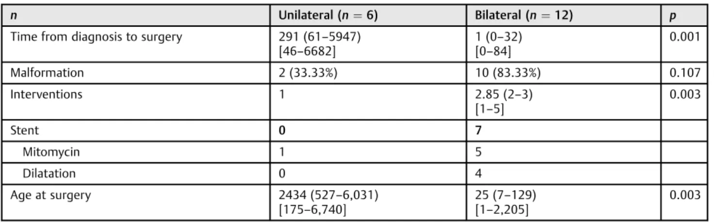
Table 2 Group characteristics and analysis
n Unilateral (n¼6) Bilateral (n¼12) p
Time from diagnosis to surgery 291 (61–5947) [46–6682]
1 (0–32) [0–84]
0.001
Malformation 2 (33.33%) 10 (83.33%) 0.107
Interventions 1 2.85 (2–3)
[1–5]
0.003
Stent 0 7
Mitomycin 1 5
Dilatation 0 4
Age at surgery 2434 (527–6,031)
[175–6,740]
25 (7–129) [1–2,205]
0.003
Quantitative variables are described by median (25th and 75th percentiles) [minimum and maximum]. Categorical variables are described by number of patients and percentage (%).
Of 6 patients with unilateral choanal atresia, only 2 presented associated malformations (33.3%), namely 1 cardiac and 1 renal malformation (p¼0.107).
All patients with unilateral choanal atresia demanded only 1 surgical intervention, and patients with bilateral choanal atresia had a median of 2.85 surgical procedures, ranging from 1 to 5 interventions to attain airway patency (p¼0.003). As parallel procedures, only 1 patient with unilateral presentation used mitomycin, and in the bilateral group, 7 needed stenting; 5, mitomycin; and 4, dilatation. In those patients with stents, the material used was thoracos-tomy tubes, nelaton catheters, or orotracheal tubes and was maintained for a mean period of 6 weeks, ranging from 2 to 12 weeks.
Median age at surgery for patients with unilateral presen-tation was 6 years, ranging from 6 months to 18 years. From patients with bilateral choanal atresia, there was a median of 25 days of life, ranging from 1 day to 6 years (p¼0.003).
Median time from diagnosis to surgery was 9 months in the unilateral group, ranging from 1 month to 18 years, and 1 day in the bilateral group, ranging from 1 day to 2 months (p¼0.001).
Follow-up of patients ranged from 12 years to 6 months, with a median of 6 years.
Discussion
Choanal atresia is the most prevalent major nasal congenital abnormality, presenting an incidence of 1:4,000 to 1:10,000 live births. It is most common in the female sex in some series,5,6but an analysis of 444 cases did not show differences between sex incidences.7The present study also did notfind gender differences in frequency.
It is unilateral in 50 to 60% of cases, the remaining being bilateral presentations.8 In our cohort, there was a higher number of bilateral cases, probably a consequence of referral bias to a high complexity hospital.
There seem to be no difference between right- or left-side presentation in unilateral choanal atresia.6,7 Teissier et al,
however, described a higher prevalence on right-sided atre-sia, as our research showed.9
Composition can be classified into osseous-membra-nous (two-thirds of cases) or purely osseous.8In our cohort, 77.8% of the cases were mixed type. No membranous case was identified, corroborating the idea that choanal atresia is not only a membrane in the posterior nasal aperture but actually a medialization of the pterygoid processes and the whole lateral nasal wall.10 Computed tomography (CT) scanning is the preferred method for the radiologic exami-nation in children with suspected choanal atresia. Careful suctioning and application of a topical decongestant can improve resolution. This is especially important prior to CT to eliminate viscous secretions thatfill the occluded nasal cavity that can obscure the true thickness of membranous occlusion and even lead to misinterpretation of associated cranial base anomalies11 as we can see in this series of patients (►Fig. 1).
Choanal atresia can present as an isolated abnormality or be part of a syndrome, and it was already described with more than 20 different congenital syndromes. The most frequently associated syndrome is known by the acronym CHARGE (coloboma, heart defects, choanal atresia, retardation of growth and development, genitourinary problems, ear ab-normalities), where choanal atresia is seen in 60% of cases.12 Burrow et al described association of choanal atresia with other malformations in 73.6% of cases.6In the present study, an association was found in 33.3% of unilateral cases and 83.3% of bilateral cases. This difference was not statistically significant due probably to the limited number of cases, but it is emphasized that severity of malformations seemed dis-crepant as bilateral cases showed more neurologic abnormal-ities. Bearing this in mind, it is recommended that a geneticist evaluate patients diagnosed with choanal atresia, especially when it is bilateral.
Early diagnosis is easier in bilateral cases as newborn nasal obstructions tend to show severe clinical manifestations.13 However, unilateral cases are often diagnosed later in life, when patients seek medical attention because of
long-Fig. 1 View of choanal atresia after suctioning of viscous secretions.
International Archives of Otorhinolaryngology Vol. 18 No. 1/2014
standing unilateral nasal obstruction, anosmia, and rhinor-rhea. This difference in clinical manifestations can be inferred from the statistically significant difference between the me-dian age at surgery and between the interval from diagnosis to surgery, which is shorter in the bilateral group.
Surgical success rates are difficult to compare between a series of patients, as results are not standardized, due to a lack of clear definition in some series of thefirst intervention and of review procedures, inadequate age reporting, and espe-cially no distinction in reporting of unilateral or bilateral presentation. First intervention success, however, seems to vary between 62 and 100%.10,14 This fact is evidence that choanal atresia management is still a major challenge. Devel-opment offlap technique and surgical equipment designed for the newborn nasal structure bring great expectations to minimize restenosis rates.
This study has unequivocal limitations that imply cautious interpretation of presented results, mainly related to its retrospective design and exiguous sample. However, it sug-gests that uni- and bilateral choanal atresia behave as distinct spectra of the disease, based on discrepancies found on clinical manifestation severity, characteristics inherent to malformation type, and restenosis rate. It is imperative that multicentric studies, allowing for a higher number of patient accrual, are developed to ascertain whether it would effec-tively be more reasonable to report surgical success on this disease by laterality, possibly augmenting comparability of results.
Conclusions
It is the authors’opinion that case series of choanal atresia reported in the literature should describe separately bilateral and unilateral cases, as they seem to concern different clinical presentations, have different patient inherent characteristics (based on associated malformations), and show discrepant restenosis rates.
References
1 Cedin AC, Atallah AN, Andriolo RB, Cruz OL, Pignatari SN. Surgery
for congenital choanal atresia. Cochrane Database Syst Rev 2012;2: CD008993 10.1002/14651858.CD008993.pub2
2 Gujrathi CS, Daniel SJ, James AL, Forte V. Management of bilateral
choanal atresia in the neonate: an institutional review. Int J Pediatr
Otorhinolaryngol 2004;68(4):399–407
3 Wiatrak BJ. Unilateral choanal atresia: initial presentation and
endoscopic repair. Int J Pediatr Otorhinolaryngol 1998;46(1–2):
27–35
4 Liktor B, Csokonai LV, Gerlinger I. A new endoscopic surgical
method for unilateral choanal atresia. Laryngoscope 2001;
111(2):364–366
5 Hengerer AS, Brickman TM, Jeyakumar A. Choanal atresia:
em-bryologic analysis and evolution of treatment, a 30-year
experi-ence. Laryngoscope 2008;118(5):862–866
6 Burrow TA, Saal HM, de Alarcon A, Martin LJ, Cotton RT, Hopkin RJ.
Characterization of congenital anomalies in individuals with choanal atresia. Arch Otolaryngol Head Neck Surg 2009;135(6):
543–547
7 Harris J, Robert E, Källén B. Epidemiology of choanal atresia with
special reference to the CHARGE association. Pediatrics 1997;
99(3):363–367
8 Brown OE, Pownell P, Manning SC. Choanal atresia: a new
ana-tomic classification and clinical management applications.
Laryn-goscope 1996;106(1 Pt 1):97–101
9 Teissier N, Kaguelidou F, Couloigner V, François M, Van Den
Abbeele T. Predictive factors for success after transnasal endo-scopic treatment of choanal atresia. Arch Otolaryngol Head Neck
Surg 2008;134(1):57–61
10 Ramsden JD, Campisi P, Forte V. Choanal atresia and choanal
stenosis. Otolaryngol Clin North Am 2009;42(2):339–352, x
11 Kearns DB, Wickstead M, Choa DI, Leitch RN, Bailey CM, Evans JN.
Computed tomography in choanal atresia. J Laryngol Otol 1988;
102(5):414–418
12 Vanzieleghem BD, Lemmerling MM, Vermeersch HF, et al. Imaging
studies in the diagnostic workup of neonatal nasal obstruction. J
Comput Assist Tomogr 2001;25(4):540–549
13 Manica D, Smith MM, Schweiger C, Bruneli DS, Kuhl G. Nasal
obstruction of the newborn: a differential diagnosis. Int Arch
Otorhinolaryngol 2009;13(3):340–345
14 De Freitas RP, Berkowitz RG. Bilateral choanal atresia repair in
neonates—a single surgeon experience. Int J Pediatr
Otorhinolar-yngol 2012;76(6):873–878