Instituto do Coração do Hospital das Clínicas - FMUSP
Mailing address: Marcelo Biscegli Jatene Rua João Moura, 1535 05412003 -São Paulo, SP - Brazil
Purpose – To assess anatomical characteristics of the aortic valve, so that they may be useful in diagnostic situations and surgical treatment.
Methods – The study analyzed 100 healthy fixed human hearts; 84% of them obtained from males, 61% of them from Caucasian individuals. The ages of the indivi-duals ranged from 9 to 86 years (mean 30±15.5 years). The characteristics assessed related to age, sex, and race were the following: number and height of the cusps, size of the lunulae, internal and external intercommissural distance, position of the coronary ostium in relation to the aortic valve, position of the ventricular septum in relation to the aortic valve, thickness of the cusps.
Results – All hearts assessed had a tricuspidal aortic valve. In regard to the height of the cusps and size of the lunula, the left coronary cusp was larger, followed by the right coronary cusp and the noncoronary cusp. The internal and external intercommissural distances had mean values of 24.6±5.7mm and 19.7±7mm, respectively. In regard to the position of the coronary ostia, in one heart two ostia emerged from the left coronary sinus, and in another, the ostium was supracommissural. The mean diameter of the aorta was 21.8±3.6mm, and there were no significant sexual or racial differences, but the diameter increased progressively with the increase in age. The thickness of the cusps did not show any significant difference in the 3 points assessed.
Conclusion – The aortic valve annulus did not show a perfect circumference, with some variations in the mea-surements of the annulus, in the cusps and in the relation with the ventricular septum.
Key words : heart, anatomy, aortic valve
Arq Bras Cardiol, volume 73 (nº 1), 81-86, 1999
Marcelo Biscegli Jatene, Rosangela Monteiro, Maria Helena Guimarães, Siomara Christina Veronezi, Márcia Kyiomi Koike, Fabio Biscegli Jatene, Adib Domingos Jatene
São Paulo, SP - Brazil
Aortic Valve Assessment. Anatomical Study of
100 Healthy Human Hearts
In recent decades, some events have increased the interest in the anatomy of the heart, one such event being the increasingly frequent use of conservative surgical techniques for repairing or replacing cardiac valves. Thromboembolic complications in the mechanical pros-theses, as well as ruptures, restenoses and calcifications in the bioprostheses, are the major causes of morbidity and mortality in patients undergoing valvar replacement. This shows that, despite the great technological evolution and constant studies, an ideal substitute for the natural valve, either mechanical or biological, providing longevity and low thrombogenicity, is yet to be found 1-5. Because of that,
conservative techniques have been progressively used in patients with valvopathies in a number of centers 2-6. Thus,
the interest in the morphology of cardiac valves, which during the 60’s and 70’s was limited to the pathological features due to the large number of valvar replacements, was extended. The more accurate knowledge of normal anatomy began to play an important role in the success of valvoplasties, as many decisions during surgery are based on the visual assessment of the changes. Detailed knowled-ge of the anatomical characteristics of the aortic valve should improve the understanding of its anatomy and help obtain better results in conservative procedures, promoting return to anatomical and functional normality. This precise knowledge also defines some details of the architecture of the aortic valve that are necessary for the development and manufacture of the prostheses. In addition, the knowledge and understanding of the structure and function of the nor-mal and pathological cardiac valves are crucial for the selec-tion of the patients for surgery and for planning the best treatment to be adopted.
Technical difficulties found in the surgical repair of the aortic valve result from its anatomical characteristics, as well as from the condition of its cusps 2,7. The closure
Specific studies of the anatomy of the aortic valve were carried out by Silver and Roberts 12, Swanson and
Clark 13, Thubrikar et al 14, and Angelini et al 15, aiming to
better define peculiarities of this valve. In addition to perfor-ming an anatomical assessment, they analyzed some linear variables in each cusp and documented the presence of some fenestrations close to the commissural attachments.
Despite the above studies, there is a need for further investigation of this subject and this study aims to assess the anatomy of the aortic valve. Data about the cusps, the position of the ostia, and anatomical relations facilitate the conservative surgeries as well as those involving valve replacement.
Methods
This study analyzed 100 healthy hearts of individuals without previous heart disease. The hearts were fixed according to the technique of molding of cavities and structures with cotton embedded in 10% formalin 16. 84% of
the individuals were males, 61% Caucasians, and the ages varied from 9 to 86 years (30±15.45 years). The cardiac mass varied from 107 to 463 g (259.47±59.3g). According to information provided by the Institute of Forensic Medicine of the State of São Paulo and by the analysis of patho-logists, all hearts belonged to individuals without previous heart disease. In 90% of the cases, the cause of death related to trauma and, in the remaining 10%, death resulted from diseases not related to the cardiovascular system.
Using a pachymeter and a # 0 cotton thread, the Valsalva’s sinuses were assessed as well as the 3 cusps of the aortic valve: left coronary cusp (LC), right coronary cusp (RC), and noncoronary cusp (NC), according to the following parameters:
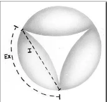
Number and height of the cusps (fig. 1): height obtai-ned by stretching the cotton thread from the bottom of the Valsalva’s sinus until the free margins of the cusps in the middle point between the commissures, respecting its curve. This measure was then transferred to the pachymeter;
Size of the lunula (fig. 1) according to two parameters: width – assessed at the commissural level; and length –
measured by stretching the cotton thread at the surface of each cusp at the free margins, following its curve;
Intercommissural distances (fig. 2): external – measu-red by stretching the cotton thread along the aortic wall uniting one commissure with the other, so that the addition of the 3 measures represents the aortic circumference; internal – obtained by uniting the commissures with the cotton thread at the smaller distance between them;
Position of the ostium and its relation with the corres-pondent Valsalva’s sinus (fig. 3): using the cotton thread, the distance between the ostium and the commissures and the bottom of the Valsalva’s sinus was measured;
Position of the ventricular septum in relation to the aortic valve (fig. 4), assessed in the following parameters: distance between the septal extremity and the RC-LC com-missure (measure A); distance between the septal extremity and the RC-NC commissure (measure B); distance between the septum and the NC-LC commissure (measure C); aortic diameter (measure D).
The ratio C/D x 100 was also established to determine the percentage of free area of the aortic valve annulus and, consequently, to evaluate the position and percentage of the area occupied by the septum in relation to the left ventricle outflow tract (LVOT).
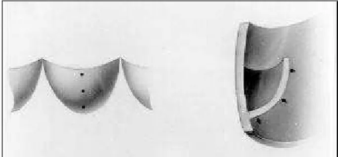
Thickness of the cusps (fig. 5): grossly assessed at 3 different points of the surface of each cusp: free margins, bottom of the Valsalva’s sinus, and the intermediate point between the other 2. The assessment of thickness was per-formed by visual inspection without using specific instru-ments because the tissue of the cusps is very delicate. Even though a histological study of the cusps would provide more accurate information, it was not carried out because the specimen had to be kept intact for further studies.
All parameters analyzed were related to sex, age and race.
Fig. 2 – Sketch demonstrating the external (Ex) and internal (I) intercommissural distances.
Fig. 1 – Sketch demonstrating the site of measurement in one of the 3 cusps. H- height of the cusp; W – width of the lunula; L – length of the lunula.
L
W
Results
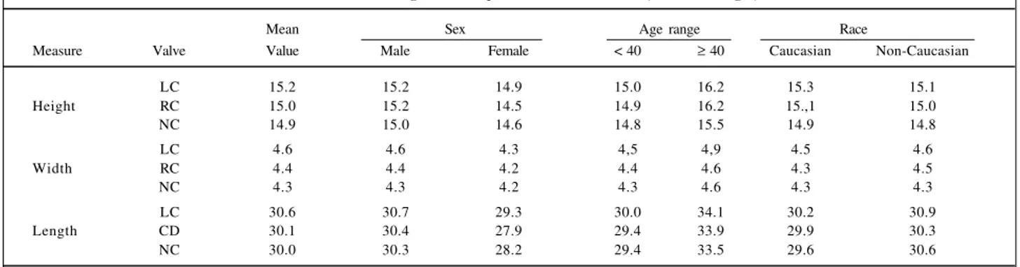
All hearts analyzed had tricuspide aortic valves. The mean height of the cusps was 15.0±1.98mm and the LC cusp had the largest dimensions, followed by the RC and the NC cusps, whose dimensions were similar. The same pattern was observed for the width and length of the lunula, whose mean values were 4.4±0.96mm and 30.2 ± 7.1mm, respectively (table I).
In regard to the intercommissural distances, the external intercommissural distance was larger in the LC Valsalva’s sinus, followed by that of the RC and NC. The internal intercommissural distance, however, was larger in the NC sinus, followed by that of the LC and RC. The mean external intercommissural distance was 24.6±5.7mm and the internal was 19.7±7.0mm; the mean individual values of each valve were grouped according to sex, age and race (table II). In regard to the position of the coronary ostia, in one
case, both ostia were in the LC sinus and, in another case, the ostium was supracommissural. In the remaining cases, there were small variations regarding the position of the respective ostium in relation to the correspondent Val-salva’s sinus (table III).
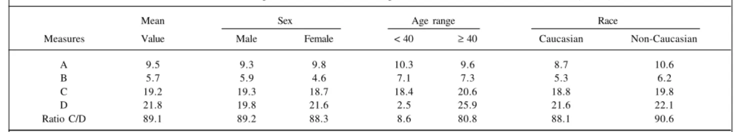
Studying the position of the ventricular septum (IVS) in regard to the aortic valve annulus and its structures, the mean values of the A, B and C measurements were, respec-tively, 9.5±5.3mm; 5.7±4.2mm; and 19.2±2.9mm. There were no statistically significant differences in regard to sex, race and age. The mean aortic diameter (measure D) was 21± 3.6mm, and there were no significant sexual and racial differences. The measure D, however, was smaller in hearts of individuals under the age of 40 years, and it showed a progressive increase with age. Therefore, the ratio C/D x 100 showed mean values of 89.1±14.6mm, and it was larger in the hearts of individuals under the age of 40 years (table IV).
The gross assessment of the thickness of the cusps, evaluated in 3 points of their surface showed no differences and was practically constant.
Discussion
Considering that the present study aimed to analyze only normal hearts, the selection of the material played an extremely important role. Before being included in the sample, all hearts were carefully analyzed and had the hypothesis of previous heart disease excluded; therefore the possibility of obtaining of anatomical pieces with morphological alterations due to cardiac disease or to repercussion of functional disorders in other organs was very remote.
A point that deserves to be discussed is heart weight. According to Gardner et al17, the mean heart weight in males
is around 328g, varying from 256g to 390g and, in females, it is around 244g, ranging from 198g to 270g. Linzbach 18,
however, states that the weight of the human heart without pathological alterations can reach 500g. In situations where the work of the heart is chronically increased, as in athletes or workers who exercise intensely, the increase in heart weight is not caused by an increase in the number of mus-cle fibers but by an increase in the thickness and length of these fibers. In addition, as already demonstrated by se-veral authors, the heart weight is directly related to the indi-vidual body surface 19,20.
Fig. 5 – Sketch (front and side view) demonstrating the 3 sites of evaluation of the thickness of the cusps.
Fig. 3 – Sketch of the position of the ostium in relation to the left commissure (L); right commissure (R) and bottom of the Valsalva’s sinus (V).
L
R
V
Fig. 4 – Sketch of the position of the ventricular septum (IVS) in relation to the aortic valve. LC – left coronary; RC – right coronary; NC - noncoronary; A- distance between the septal extremity and the RC-LC commissure; B- distance between the septal extremity and the RC-NC commissure; C- distance between the septum and the NC-LC commissure; D- aortic diameter.
LC
RC
Among the data obtained in our study, some proved to be more interesting than others for the study of the aortic valve and its relations.
In the present analysis, the external intercommissural distance proved to be the largest in the LC Valsalva’s sinus, followed by the RC and the NC. Analyzing the internal intercommissural distance, however, we observed some changes, with larger values in the NC sinus, followed by the LC and the RC in most of the studied groups, emphasizing the observation that the aortic valve annulus has variable dimensions and that it is not a perfect circumference at the level of the commissures. Vollebergh and Becker 21,
stu-dying the intercommissural distances of 200 human hearts, also demonstrated variations; the RC sinus, however, was the largest one followed by the NC and LC sinuses, just the
opposite of our observations. In addition, Thubrikar et al 22
showed that, in in vivo investigations of canine hearts, the
aortic valve is a dynamic structure and that the geometrical parameters change at each cardiac cycle. An exclusively anatomical investigation of the specimen would be mainly useful to provide information that the surgeon would need at the moment of approaching the aortic valve. Studies of specimens show some aspects such as rigor mortis, that
differ from the heart at the time of a surgery 23 and the
retraction after fixation. The influence of these factors is more significant in the ventricular chambers. Tei et al 24, in a
control group of a study of the right atrioventricular valve and in a comparative evaluation between 3 groups, obser-ved similar values for nonfixed and fixed hearts, as well as for those evaluated by echocardiograms. Scholz et al 25 and
Table II - Mean values of the external and internal intercommissural distances (in mm)
Intercommissural Mean Sex Age range Race
Distance Valve Value Male Female < 40 ≥ 40 Caucasian Non-Caucasian
LC 25.2 25.4 23.5 24.4 29.3 24.0 25.5
External RC 24.5 24.5 23.8 23.6 29.4 23.1 25.2
NC 24.1 24.4 22.1 23.6 27.0 23.3 23.8
LC 19.8 20.0 18.5 19.3 22.7 18.8 21.4
Internal RC 19.2 19.2 18.7 18.7 22.2 17.9 20.9
NC 20.0 20.1 19.1 19.6 22.3 18.5 21.8
LC - left coronary; RC - right coronary; NC - noncoronary.
Table I - Mean values of the height of the cups and size of the lunulae (width and length) in mm
Mean Sex Age range Race
Measure Valve Value Male Female < 40 ≥ 40 Caucasian Non-Caucasian
LC 15.2 15.2 14.9 15.0 16.2 15.3 15.1
Height RC 15.0 15.2 14.5 14.9 16.2 15.,1 15.0
NC 14.9 15.0 14.6 14.8 15.5 14.9 14.8
LC 4.6 4.6 4.3 4,5 4,9 4.5 4.6
Width RC 4.4 4.4 4.2 4.4 4.6 4.3 4.5
NC 4.3 4.3 4.2 4.3 4.6 4.3 4.3
LC 30.6 30.7 29.3 30.0 34.1 30.2 30.9
Length CD 30.1 30.4 27.9 29.4 33.9 29.9 30.3
NC 30.0 30.3 28.2 29.4 33.5 29.6 30.6
LC - left coronary; RC - right coronary; NC - noncoronary.
Table III - Mean values of the distances of the ostium and its relation to the corresponding Valsalva's sinus (in mm)
Distance between Valsalva's General Sex Age range Race
the ostium and sinus mean Male Female < 40 ≥ 40 Caucasian Non-Caucasian
Left LC 9.6 9.7 9.3 9.3 12.0 9.5 9.8
commissure RC 11.1 11.2 10.7 10.7 13.9 11.2 11.1
Right LC 110 10.9 10.8 10.5 13.4 10.5 11.6
commissure RC 11.1 11.3 9.9 10.8 13.0 10.7 11.7
Bottom of the LC 13.3 13.4 13.0 13.0 14.9 13.3 13.4
Valsalva's sinus RC 14.8 15.0 13.8 14.6 16.1 14.9 14.7
Table IV - Mean values of the position of the ventricular septum and its relation with the aortic valve (in mm)
Mean Sex Age range Race
Measures Value Male Female < 40 ≥ 40 Caucasian Non-Caucasian
A 9.5 9.3 9.8 10.3 9.6 8.7 10.6
B 5.7 5.9 4.6 7.1 7.3 5.3 6.2
C 19.2 19.3 18.7 18.4 20.6 18.8 19.8
D 21.8 19.8 21.6 2.5 25.9 21.6 22.1
Ratio C/D 89.1 89.2 88.3 8.6 80.8 88.1 90.6
A- distance between the septal extremity and the RC-LC commissure; B- distance between the septal extremity and the RC-NC commissure; C- distance between the septum and the NC-LC commissure; D - aortic diameter.
Maron et al 26, in a study of cardiac mass, valvar
circum-ferences and ventricular thickness, found similar results. Knowledge of the design of the normal aortic valve, responsible for its efficiency and longevity, as well as the knowledge of variations of its geometry is important for the conservative surgical techniques and for the develop-ment and manufacture of prostheses similar to the natural valve 9,13,22,27. In this regard, Fontes et al 28 carried out an
interesting study in which the aortic root proved to have a structure similar to a sphere, encompassing the aortic valve and its annulus. This information has important implications in the manufacture of prostheses that must conform to this anatomical configuration.
When we analyze the size of the lunula, the mean of the intercommissural distances and the mean height of the cusps in relation to sex and age, the values are greater in the hearts of males and these values increase with age. Krovetz et al 29, in a clinical study relating age and the size
of the aortic valve, observed that there is an increase in the volume of the aorta with the increase in age and also that, after the age of 40 years, the aortic valve and its annulus become progressively dilated. This supports the findings of our study.
Our gross evaluation of the thickness of the cusps performed at 3 different points did not show any significant change. We know there are structural alterations and differences in thickness of the cusps, but these can only be demonstrated through specific microscopic studies. We chose the gross examination, however, because in surge-ries, some decisions are made during the procedure based only on the surgeon’s observations. Sahasakul et al 30, with
the aid of a highly accurate micrometer, measured different points of the aortic valve in 200 human hearts. They observed that in hearts of women <20 years, the thickness
of the middle part of the LC and NC cusps was greater than in the other points. They also noticed that the thickness of each cusp increased significantly with age, not only during childhood and adolescence but also in adulthood. These variations, however, were not statistically significant when compared with the other parameters.
Another point to be considered is the relation between the ventricular septum (VS) and the aortic valve and the LVOT. The ratio C/D was higher in hearts of individuals <40 years of age and progressively decreased with the increase in age. This may suggest that perhaps LVOT shows a trend to proportionally decrease its area in relation to the aortic diameter with increasing age. This may be related to some clinical process leading to hypertrophy of the muscle structures – VS and the muscle of the free wall of the LV – combined with an increase in the aortic diameter. Conside-ring still the relation between VS and the aortic valve, a practical consideration plays an important role in surgical cases, where it is necessary to widen the aortic valve annu-lus to place the valvar prosthesis. According to Stolf et al 31,
who widened the aortic valve annulus in 25 patients with the diagnosis of stenosis or dysfunction of the aortic prosthesis with satisfactory results, the perfect knowledge of the position of the VS help the access and the surgical procedure without major risks of damaging the VS and severe consequences, such as infarctions of the VS.
1. Braunwald E. Heart disease: A textbook of cardiovascular medicine. 4ª ed. Philadelphia: WB Saunders Co, 1992: 1043-53.
2. Lavítola PL, Dallan LA, Tarasoutchi F, et al. Procedimento conservador na correção de defeito valvar aórtico em associações com outras cardiopatias. Arq Bras Cardiol 1987; 48: 351-3.
3. McClung JA, Stein JH, Ambrose JA, Herman MV, Reed GE. Prosthetic heart valves - A review. Prog Cardiovasc Dis 1983; 26: 237-71.
4. Pomerantzeff PMA, Azevedo JG, Ratti M, et al. Plástica da valva mitral em pacientes consecutivos: como é a evolução tardia? Avaliação clínica e ecocardiográficas. Rev Bras Cir Cardiovasc 1991; 6: 63-79.
5. Pomerantzeff PMA, Yochtomi Y, Fabri HA, et al. Reoperações valvares: experiência do InCor-FMUSP. Rev Bras Cir Cardiovasc 1991; 6: 182-9. 6. Jatene FB, Monteiro R, Jatene MB, Magalhães MHG, Fukushima JT, Jatene AD.
Estudo do anel mitral e trígonos fibrosos com diferentes variáveis. Rev Bras Cir Cardiovasc 1991; 6: 190-4.
7. Trusler GA, Williams WG, Smallhorn JF, Freedom RM. Late results after repair of aortic insufficiency associated with ventricular septal defect. J Thorac Cardiovasc Surg 1992; 103: 276-81.
8. Bongiovani HL, Ribeiro PJF, Évora PRB, Brasil JCF, Reis CL, Sgarbieri RN -Plastia valvar aórtica por ampliação de válvula com pericárdio bovino: nota prévia. Rev Bras Cir Cardiovasc 1988; 3: 130-3.
9. Mercer JL, Benedicty M, Bahnson HT. The geometry and construction of the aortic leaflet. J Thorac Cardiovasc Surg 1973; 65: 511-8.
10. Ribeiro PJF, Bongiovani HL, Évora PRB, et al. Plastia valvar aórtica por ampliação de válvulas com pericárdio bovino. Rev Bras Cir Cardiovasc 1990; 5: 99-105. 11. Carpentier A. Cardiac valve surgery: The “French correction”. J Thorac
Cardiovasc Surg 1983; 86: 323-37.
12. Silver MA, Roberts WC. Detailed anatomy of the normally functioning aortic valve in hearts of normal and increased weight. Am J Cardiol 1985; 55: 454-61. 13. Swanson WM, Clark RE. Dimensions and geometric relationships of the human
aortic valve as a function of pressure. Cir Res 1974; 35: 871.
14. Thubrikar M, Nolan SP, Boscher LP. The ciclic changes and structure of the base of the aortic valve. Am Heart J 1980; 99: 217-24.
15. Angelini A, Ho SY, Anderson RH, et al. The morphology of the normal aortic valve as compared with the aortic having two leaflets. J Thorac Cardiovasc Surg 1989; 98: 362-7.
16. Jatene FB, Magalhães MHG, Oliveira PR, Jatene AD. Técnica de preservação de corações através de perfusão sob pressão. Arq Bras Cardiol 1991; 57: 93-6.
References
17. Gardner E, Gray DJ, O’Rahilly R. Anatomia. Estudo Regional do Corpo Humano. 3ª ed. Rio de Janeiro: Guanabara Koogan, 1971: 325-46.
18. Linzbach AJ. Heart failure from the point of view of quantitative anatomy. Am J Cardiol 1960; 5: 370-82.
19. Gutgesell HP, Rembold CM. Growth of the human heart relative to body surface area. Am J Cardiol 1990; 65: 662-8.
20. Eckner FAO, Brown BW, Davidson DL, Glagov S. Dimensions of normal hearts: after standard fixation by controlled pressure coronary perfusion. Arch Pathol 1969; 88: 497-507.
21. Vollebergh FEMG, Becker AE. Minor congenital variations of cusp size in tricuspid aortic valves. Possible link with isolated aortic stenosis. Br Heart J 1977; 39: 1006-11.
22. Thubrikar M, Piepgrass WC, Shaner TW, Nolan SP. The design of the normal aortic valve. Am J Physiol 1981: H795-801.
23. Curti HJV, Sanches PCR, Carvalhal SS. Rigor mortis cardíaco. Arq Bras Cardiol 1985; 45: 439-46.
24. Tei C, Pilgrim JP, Shah PM, Ormiston JA, Wong M. The tricuspid valve annulus: study of size and motion in normal subjects and in patients with tricuspid regurgitation. Circulation 1982; 66: 665-71.
25. Scholz DG, Kitzman DW, Hagen PT, Ilstrup DM, Edwards WD. Age-related changes in normal human hearts during the first 10 decades of life. Part I (growth): a quantitative anatomic study of 200 specimens from subjects from birth to 19 years old. Mayo Clin Proc 1988; 63: 126-36.
26. Maron BJ, Henry WL, Roberts WC, Epstein SE. Comparison of echocardiographic and necropsy measurements of ventricular wall thicknesses in patients with and without disproportionate septal thickening. Circulation 1977; 55: 341- 6. 27. Davila JC. The mechanics of cardiac valves. In: Merendino KA (editor).
Prosthetic Valves for Cardiac Surgery. Springfield: Charles C. Thomas Publishers, 1960: 3-21. 28. Fontes RD, Bonassa J, Jatene AD. Configuração esférica do aparelho valvar aórtico. Arq Bras Cardiol 1991; 57: 385-8. 29. Krovetz LJ. Age-related changes in size of the aortic valve annulus in man. Am
Heart J 1975; 90: 569-74.
30. Sahasakul Y, Edwards WD, Naessens JM, Tajik AJ. Age-Related changes in aortic and mitral valve thickness: Implications for two-dimensional echocar-diograph based on an autopsy study of 200 normal human hearts. Am J Cardiol 1988; 62: 424-30.