Influence of Subclinical Atherosclerosis on Diastolic Function in
Individuals Free of Cardiovascular Disease
Maristela Magnavita Oliveira Garcia, Marília Galeffi Rodrigues, Joaquim Antônio dos Reis Neto, Luis Claudio
Correia
Escola Bahiana de Medicina e Saúde Pública - Fundação de Desenvolvimento de Pesquisas da Bahia, Instituto Pró-Cardíaco, Salvador, BA - Brazil
Mailing address: Maristela Magnavita Oliveira Garcia •
Rua Artesão João da Prata, 268/202 - Edf. Mansão Plaza Athenée - Itaigara - 41815-210 - Salvador, BA - Brazil
E-mail: [email protected], [email protected]
Manuscript received September 14, 2009; revised manuscript received October 08, 2009; accepted December 16, 2009.
Abstract
Background: It is plausible that subclinical atherosclerosis alters coronary reserve and impairs diastolic function of the left ventricle. However, the relationship between subclinical stages of atherosclerosis and diastolic function has not been established in subjects free of cardiovascular disease.
Objective: To test the hypothesis that subclinical atherosclerosis has a negative association with diastolic function.
Methods: Individuals ≥ 35 years old, free of cardiovascular disease, with normal blood pressure and negative treadmill stress test, were selected to have common carotid intima-media thickness (IMT) assessed by ultrasound and parameters of diastolic function by echocardiography, primarily tissue Doppler E’/A’ ratio.
Results: Forty-eight subjects were studied, aged 56 ± 10 years, 67% females. Composite common carotid IMT had a significant negative correlation with tissue Doppler E’/A’ ratio (r = - 0.437, p = 0.002). Individuals in the fourth quartile of IMT had a significant lower tissue Doppler E’/A’ ratio (0.76 ± 0.25), as compared with the first (1.2 ± 0.29), second (1.2 ± 0.36) and third quartiles (1.1 ± 0.25) - p = 0.002. Composite common carotid IMT in the fourth quartile (≥ 0.8 mm) independently predicted E’/A’ ratio (p = 0.02), after adjustment for potentially confounding variables, such as age, female gender, waist circumference, diastolic blood pressure, HDL-cholesterol and Framingham Risk.
Conclusion: Early stage of subclinical atherosclerotic disease is negatively associated with diastolic function parameters in healthy individuals, regardless of age and clinical characteristics. (Arq Bras Cardiol 2010; 95(4): 473-479)
Key words: Subclinical atherosclerosis; diastolic function; carotid intima-media thickness.
population, known to be free of myocardial ischemia assessed by provocative non-invasive tests.
In order to test the hypothesis that subclinical atherosclerosis is a predictor of diastolic function, regardless of chronic myocardial ischemia, we studied a highly selected sample of individuals free of any cardiovascular disease and with negative maximal treadmill stress test. Atherosclerotic burden was measured by carotid intima-media thickness and diastolic function by pulsed and tissue Doppler echocardiography.
Methods
Study population
Asymptomatic men and women, aged ≥ 35 years, who spontaneously presented to our outpatient clinic for preventive evaluation from June 2006 to June 2007, were considered candidates to the study. Exclusion criteria consisted of any cardiovascular condition or systemic disease with a potential to affect the diastolic function: history of hypertension, coronary artery disease, heart failure, valvular disease, cardiac pacemaker, atrioventricular block, current use of cardiovascular drugs, renal dysfunction, COPD, diabetes, thyroid disease or collagen diseases. After this first screening,
Introduction
Atherosclerotic disease develops early in life and usually progresses for decades as a silent process. This condition, so-called subclinical atherosclerosis, is highly prevalent in the general adult population1. During this period,
non-obstructive atherosclerotic plaques do not cause ischemia in a clinical sense, but promote functional changes in vascular tone2, related to vasoactive mediators release and impaired
production of nitric oxide by a dysfunctional endothelium3.
Diastolic function is the first to be impaired in ischemic heart disease4. It is possible that the pathophysiological
process of subclinical atherosclerosis, usually not sufficient to reduce systolic function, is able to alter the diastolic function of the left ventricle. Preliminary data have shown that atherosclerotic burden has an inverse relationship with measurements of diastolic dysfunction5,6. However,
subjects underwent the following evaluation to further select those free of cardiovascular disease: 1) initially, blood pressure was measured at rest in the sitting position and those with systolic blood pressure ≥ 140 mmHg or diastolic blood pressure ≥ 90 mmHg were excluded; 2) a resting 12-lead electrocardiogram was performed and those with a non-sinus rhythm, evidence of ischemia (T wave inversion ≥ 1 mm or ST-depression ≥ 0.5 mm) or presence of Q waves were excluded; 3) echocardiography was performed and those with any structural or functional abnormality (such as ventricular hypertrophy or dilatation, systolic dysfunction, non-trivial valvular dysfunction or pericardial disease) were excluded; 4) finally, exercise stress test was performed with a maximal graded treadmill protocol and individuals were excluded if the test was positive for ischemia: exercise induced ST-segment depression ≥ 1 mm at the J point or typical angina symptom. The study was approved by the local review board and all the participants provided written informed consent.
Sample size calculation
Based on prior literature evidence, which describes a coefficient of correlation between IMT and pulsed Doppler E/A ratio of 0.467, we estimated that 46 patients were required
to detect the same correlation coefficient between IMT and tissue Doppler E’/A’ ratio, with a power of 90 % at the 5% level of significance.
Study protocol
Individuals underwent physical examination to obtain resting blood pressure, weight, height and waist circumference. Body mass index was defined as weight (kg)/height2 (m). A
fasting blood sample was drawn to measure plasma lipids, glucose and high-sensitivity C-reactive protein. Commercial enzymatic methods were used for the determination of total cholesterol, HDL-cholesterol and triglycerides (Dimension Clinical Chemistry System, Dade-Behring, Delaware, USA)8.
LDL-cholesterol was calculated by Friedewald’s formula. High-sensitivity C-reactive protein was measured using a commercially available immunoenzymatic nephelometric method (Dade-Behring, Newark, Delaware, USA) with precision limit of 0.3 mg/l and variation coefficient of 7.6%9.
Based on clinical and laboratory data, the Framingham Risk Score was calculated for each patient10. On a subsequent
day, echocardiography was performed to evaluate exclusion criteria and to provide a comprehensive diastolic evaluation if the individual was selected for the study. After that, carotid ultrasound was performed to assess subclinical atherosclerosis as described below. Both echocardiography and carotid ultrasound images were acquired and stored directly from the ultrasound system for later offline and blinded evaluation.
Subclinical atherosclerosis assessment
Individuals were examined in the supine position, with the head turned 45° from the side being scanned. Right and left carotid arteries were evaluated with high-resolution B-mode ultrasonography (Envisor C, Philips Medical Systems) connected to a 7.5 MHz linear-array transducer, preferably at a standard depth of 4 cm. One independent observer
measured carotid intima-media thickness (IMT) in all subjects, by a previously validated method11. Longitudinal images were
obtained at the level of the common carotid artery, gated to diastole, specifically at the ECGR-wave. IMT was defined as the distance between the leading edge of the first bright line and the leading edge of the second bright line of the far wall. On each right and left carotid arteries, three measurements were made at 1 cm before carotid bifurcation. Common carotid IMT was defined as the mean of these three values12-14.
The average of common carotid IMT on both sides was the composite common carotid IMT, which was the primary IMT variable in data analysis.
To test interobserver reproducibility, 20% of individuals had carotid measurements performed offline by a second independent observer. Pearson’s correlation coefficient between the two observers was 0.90 (p < 0.001). The 95% limits of agreement for these measurements was 0 to 0.11 mm.
Diastolic function assessment
Transthoracic echocardiography Doppler examination were performed in all subjects, using commercially available ultrasound system (Envisor C, Philips Medical Systems) equipped with a 2 to 4 MHz transducer. Assessment of left ventricular diastolic function was performed by pulsed Doppler and tissue Doppler imaging. Pulsed Doppler spectral recordings were obtained at the apical 4-chamber view from a sample volume positioned at the tips of the mitral leaflets to record mitral valve flow, during 3 consecutive cardiac cycles. Tissue Doppler velocities were obtained from the lateral mitral annulus at the apical 4-chamber view. One independent observer measured diastolic parameters for all subjects. The following diastolic indices were derived by pulsed Doppler: peak early diastolic velocity (E), deceleration time from peak early diastolic wave to baseline, peak atrial systolic velocity (A), the E/A ratio and isovolumetric relaxation time. Tissue Doppler indices were: peak systolic motion velocity, peak early diastolic motion velocity (E’), peak atrial systolic motion velocity (A’) and the E’/A’ ratio. The known negative correlation between E’ and A’ reflects the internal dependency between these parameters and supports their analysis together, not individually15. The E/E’ ratio was not selected for these
analyses, as it is not a robust measurement in healthy subjects with preserved ejection fraction16.
In addition, chamber dimensions and left ventricle ejection fraction were measured by M-mode echocardiogram and left atrium volume was determined by Simpson’s rule using the 4-chamber view indexed to body surface area17. Left ventricle
(LV) mass was calculated according to Devereux’s equation and indexed to body surface area18.
To test interobserver reproducibility, 20% of individuals had their diastolic measurements performed offline by a second independent observer. Pearson’s correlation coefficient was 0.99 (p < 0.001) for E’/A´ ratio and the 95% limits of agreement for these measurements was - 0.074 to + 0.12.
Data analysis
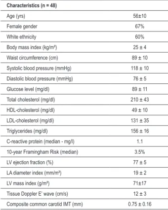
Table 1 - Clinical, laboratory and echocardiography characteristics of the study population
Characteristics (n = 48)
Age (yrs) 56±10
Female gender 67%
White ethnicity 60%
Body mass index (kg/m²) 25 ± 4
Waist circumference (cm) 89 ± 10
Systolic blood pressure (mmHg) 118 ± 10
Diastolic blood pressure (mmHg) 76 ± 5
Glucose level (mg/dl) 89 ± 11
Total cholesterol (mg/dl) 210 ± 43
HDL-cholesterol (mg/dl) 49 ± 10
LDL-cholesterol (mg/dl) 131 ± 35
Triglycerides (mg/dl) 156 ± 16
C-reactive protein (median - mg/l) 1.1
10-year Framingham Risk (median) 3.5%
LV ejection fraction (%) 77 ± 5
LA diameter index (mm/m²) 19 ± 2
LV mass index (g/m²) 71±17
Tissue Doppler E’ wave (cm/s) 12 ± 3
Composite common carotid IMT (mm) 0.75 ± 0.16
HDL - high-density lipoprotein; IMT - intima-media thickness; LA - left atrium; LDL - low-density lipoprotein; LV - left ventricle.
Table 2 - Common carotid intima-media thickness correlation with diastolic function parameters
Diastolic parameters (n = 48) Correlation coeficient p value
E/A ratio - 0.26 0.07
Deceleration time + 0.26 0.08
Isovolumetric relaxation time - 0.21 0.16
Tissue Doppler E’ wave - 0.32 0.03
Tissue Doppler E’/A’ ratio - 0.44 0.002 correlation coefficient and linear regression approach.
Secondly, diastolic variables were compared among quartiles of IMT by analysis of variance (ANOVA), followed by a comparison between two extreme groups according to the degree of atherosclerotic burden: those with composite common carotid IMT in the fourth quartile of our sample distribution (≥ 0.8 mm) vs those with IMT below the fourth quartile. Numeric diastolic parameters were compared by Student’s t test and those variables with a non-normal distribution were log-transformed before statistical comparison. Thirdly, baseline characteristics were compared between these two groups and those with a significant association with IMT (p < 0.05) were considered potentially confounding variables in the association between atherosclerosis and diastolic function. For these comparisons, Student’s t test was used for continuous variables and the chi-square test for categorical variables. Fourthly, in order to adjust the effect of atherosclerosis on diastolic function, an analysis of covariance (ANCOVA) was performed taking E’/A’ ratio as the continuous dependent variable, composite common carotid IMT in the fourth quartile as the predictive variable and potentially confounding variables as covariates (either continuous or categorical variables). Two-sided probability values < 0.05 were considered statistically significant. Data were presented as mean ± standard deviation for continuous variables and percentage for categorical variables. SPSS 13.0 for Windows (SPSS Inc, Chicago, Il) was the statistical software utilized for all data analysis.
Results
Forty-eight subjects were studied, aged 56 ± 10 years (range from 37 to 81 years), 67% females and 60% of white ethnicity. As depicted in table 1, blood pressure and glycemic levels were in normal range, while body mass index, total cholesterol and triglycerides were slightly elevated. The sample consisted of a low-risk population, with a median 10-year risk of death or myocardial infarction estimated on 3.5% based on the Framingham Risk Score. Echocardiography measurements of LV mass, ejection fraction and left atrium volume averaged normally. Composite common carotid IMT averaged 0.75 ± 0.16 mm, indicating a sample population with only initial degree of atherosclerosis. Tissue Doppler E’ wave mean was 12 ± 3.0 cm/s, indicating a predominantly normal population regarding diastolic function.
Composite common carotid IMT had negative correlations with E/A ratio, isovolumetric relaxation time, E’ wave and E’/A’ ratio and a positive correlation with mitral inflow deceleration time, indicating a negative association between atherosclerotic burden and diastolic function - Table 2. These associations were especially significant when tissue Doppler indices of diastolic function were utilized, such as E’ wave (r = - 0.32, p = 0.03) and E’/A’ ratio (r = - 0.437, p = 0.002 - Figure 1A). When E’/A’ ratio was compared between quartiles of IMT, the first (1.2 ± 0.29), second (1.2 ± 0.36) and third quartiles (1.1 ± 0.25) had similar values, while the fourth quartile was significantly lower (0.76 ± 0.25, p = 0.002 by ANOVA). This observation suggests that the influence of atherosclerosis on diastolic function has a threshold around the limit of the fourth quartile. Thus, we further compared the differences
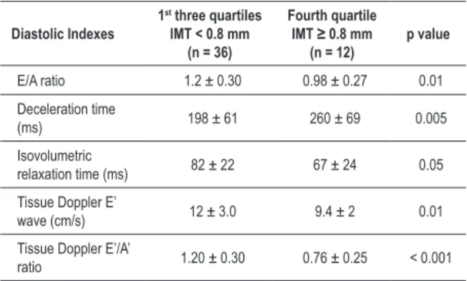
between the fourth quartile of IMT (≥ 0.8 mm) and the first three quartiles taken together (E’/A’ ratio: 0.76 ± 0.25 vs 1.2 ± 0.3; p < 0.001 - Figure 1b). Individuals in the fourth quartile had a lower mitral inflow E/A ratio (0.98 ± 0.27 vs 1.23 ± 0.30; p = 0.01), a longer deceleration time (260 ± 69 ms
vs 198 ± 61 ms; p = 0.005), while isovolumetric relaxation time had a trend towards being shorter in the fourth quartile, as compared to the first three quartiles (67 ± 24 ms vs 82 ± 22 ms; p = 0.05) - Table 3.
Fig. 1 -Panel A correlates common carotid intima-media thickness with tissue Doppler E’/A’ ratio. Panel B compares E’/A’ ratio between individuals in the fourth (≥ 0.8
mm) and irst three quartiles of intima-media thickness.
Table 3 - Comparison of diastolic indexes between individuals in the fourth (≥ 0.8 mm) and irst three quartiles of common carotid intima-media thickness
Diastolic Indexes 1
st three quartiles
IMT < 0.8 mm (n = 36)
Fourth quartile
IMT ≥ 0.8 mm
(n = 12)
p value
E/A ratio 1.2 ± 0.30 0.98 ± 0.27 0.01
Deceleration time
(ms) 198 ± 61 260 ± 69 0.005
Isovolumetric
relaxation time (ms) 82 ± 22 67 ± 24 0.05 Tissue Doppler E’
wave (cm/s) 12 ± 3.0 9.4 ± 2 0.01 Tissue Doppler E’/A’
ratio 1.20 ± 0.30 0.76 ± 0.25 < 0.001
IMT - Composite common carotid intima-media thickness.
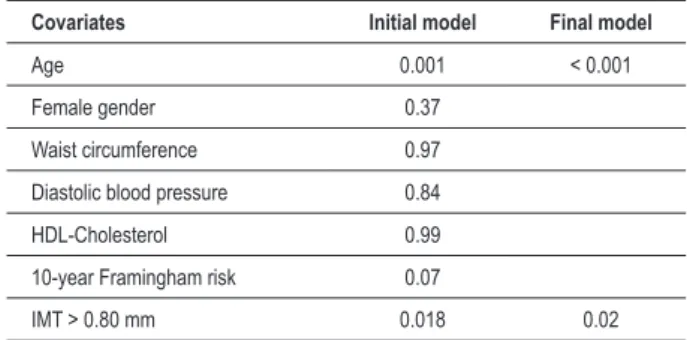
waist circumference, Framingham risk, HDL-cholesterol and diastolic blood pressure - Table 4. After adjustment for these confounding variables by analysis of covariance, common carotid IMT in the forth quartile (≥ 0.8 mm) remained a significant predictor of E’/A’ ratio (p = 0.02). Similarly, age was a predictor of the E’/A’ ratio regardless of atherosclerosis (p < 0.001) and Framingham risk tended to be a predictor (p = 0.07) - Table 5.
Discussion
This study provided evidence of a negative association of subclinical atherosclerosis with diastolic function in a highly selected sample of healthy subjects: mostly young adults, with absence of hypertension, very low cardiovascular risk according to Framingham assessment, no myocardial ischemia on stress test or any type of cardiac abnormality
on echocardiogram. Thus, the atherosclerosis studied here is usually not considered a pathological process in a clinical sense. Indeed, it is a very early stage of the disease, as demonstrated by our average values of IMT. Nonetheless, our data suggests that this subclinical condition, beyond a certain level, is associated with a relatively impaired diastolic function.
Because carotid IMT is related to age, we controlled the influence of aging on diastolic function as a potential confounder in the relationship between atherosclerosis and diastolic function. These analyses showed that after adjustment to age, the effect of atherosclerosis remained highly significant, demonstrating the independent nature of the association between atherosclerosis and diastolic function. In fact, the only two predictors of diastolic function were age and atherosclerotic burden. Considering that the first three quartiles of IMT did not have a gradient of diastolic function, which decreased only in the fourth quartile, atherosclerosis has a threshold to start impairing diastole, which was around 0.8 mm of IMT. This threshold value should not be considered a precise estimation, due to our small sample size. Thus, it must be confirmed in future studies. Nevertheless, it was interesting to note that it is the same cut-off that determines increase in cardiovascular risk according to most longitudinal studies19-22. At lower degrees
of IMT, the thickening may reflect an adaptive response to changes in shear stress, but beyond this level IMT most likely represents a disease process22.
Our findings are in line with recent data regarding the association between atherosclerosis and diastolic function23.
Fernandes et al24 addressed this issue in the
Table 4 - Comparison of clinical characteristics between individuals in the fourth (≥ 0.8 mm) and irst three quartiles of common carotid intima-media thickness
Characteristics
1st three quartiles
IMT < 0.8 mm (n = 36)
Fourth quartile
IMT ≥ 0.8 mm
(n = 12)
p value
Age (yrs) 54 ± 9 65 ± 11 0.001
Female gender 75% 42% 0.04
White ethnicity 83% 52% 0.09
Body mass index
(kg/m²) 25 ± 3 26 ± 6 0.47
Waist
circumference (cm)
87 ± 9.0 95 ± 11 0.02
Systolic blood
pressure (mmHg) 117 ± 10 123 ± 9.0 0.09 Diastolic blood
pressure (mmHg) 76 ± 5 79 ± 5 0.04
Current smoker 17% 0 0.32
Physical inactivity 39% 58% 0.32
10-year Framingham risk (median)
1.75% 4,28% < 0.001
Glucose level
(mg/dl) 88 ± 11 89 ± 7 0.08 C-reactive protein
(median - mg/l) 1.10 2.4 0.30 Total cholesterol
(mg/dl) 208 ± 42 217 ± 46 0.52 HDL-cholesterol
(mg/dl) 51 ± 10 44 ± 7 0.04 LDL-cholesterol
(mg/dl) 126 ± 32 143 ± 43 0.16 Triglycerides
(mg/dl) 137 ± 82 141 ± 52 0.88 Lipid-lowering
therapy 13% 25% 0.34
Estrogen therapy 19% 0 0.17
LV ejection
fraction (%) 77 ± 5 78 ± 5 0.22 LV mass index
(g/m²) 71 ± 17 70 ± 18 0.51
Table 5 - Multivariate analysis by ANCOVA, taking E’/A’ ratio as the dependent variable and all variables signiicantly associated with intima-media thickness at the univariate analysis as covariates
Covariates Initial model Final model
Age 0.001 < 0.001
Female gender 0.37
Waist circumference 0.97
Diastolic blood pressure 0.84
HDL-Cholesterol 0.99
10-year Framingham risk 0.07
IMT > 0.80 mm 0.018 0.02
HDL - high-density lipoprotein; IMT - composite common carotid intima-media thickness.
population were 10 years older in average, not totally free of cardiovascular risk factors (40% had hypertension, 18% had diabetes), the mean IMT was higher (0.86 ± 0.20 mm) and myocardial ischemia was not ruled out by provocative tests. Therefore, our data expand the MESA findings to a more normal and younger population, with no myocardial ischemia. Other preliminary data demonstrated similar finding in hypertensive patients6 and elderly individuals25.
Thus, the originality of the present study relies on the normal nature of a highly selected sample population, which prevents a complex interaction of confounding factors and emphasizes the impact of atherosclerosis in its
earliest stages. We must recognize that electrocardiography stress is not a highly sensitive test to rule-out significant coronary disease. On the other hand, the very low pre-test probability of coronary disease of our population leads to a very high negative predictive value of the stress test in our study. Therefore, the post-test probability of myocardial ischemia after the negative stress test must be very low in our sample population.
Mechanisms underlying the relationship of subclinical atherosclerosis and diastolic function are probably related to vascular function. Firstly, it is already known that thickened intima is an indicator of reduced myocardial flow reserve26.
This may be the result of coronary microvascular obstruction and/or reduced vasodilator reserve of epicardial vessels, due to endothelial dysfunction. These phenomena may take place in the absence of obstructive coronary disease and chronic ischemia. Secondly, atherosclerosis may enhance arterial stiffness of major arteries, such as the carotid artery and aorta, increasing afterload to the left ventricle, with a negative impact on diastolic function. Indeed, increased arterial stiffness is related not only to atherosclerosis but also to diastolic dysfunction27. Since endothelial function, coronary
flow reserve and arterial stiffness were not evaluated in the present study, a more comprehensive understanding of the pathophysiological process should be the focus of future research. Due to the cross-sectional nature of the study, one cannot guarantee that the observed association is the result of atherosclerosis impairing diastolic function. However, several scientific criteria of causation (such as consistency with previous literature, statistical strength, lack of alternate explanation and, mainly, biological plausibility) suggest that it is atherosclerosis that impairs diastolic function, and not the contrary.
surrogate for subclinical atherosclerosis in certain groups of patients. In other words, are individuals with isolated diastolic dysfunction at a higher risk for events related to atherosclerotic disease? Moreover, our findings may be the basis for future tests of the hypothesis that lipid-lowering therapy improves diastolic function. Regarding all these issues, studies of longitudinal nature will bring insights not provided by the cross-sectional design of our study.
In conclusion, the present study adds evidence to the independent and negative association between subclinical atherosclerosis and diastolic function and expands this knowledge by encompassing individuals at lower cardiovascular risk and lower degree of subclinical atherosclerosis.
Potential Conflict of Interest
No potential conflict of interest relevant to this article was reported.
Sources of Funding
There were no external funding sources for this study.
Study Association
This article is part of the thesis of master submitted by Maristela Magnavita Oliveira Garcia, from Escola Bahiana de Medicina e Saúde Pública.
References
1. Wong ND, Sciammarella MG, Polk D, Gallagher A, Miranda-Peats L, Whitcomb B, et al. The metabolic syndrome, diabetes, and subclinical atherosclerosis assessed by coronary calcium. J Am Coll Cardiol. 2003; 41 (9): 1547-53.
2. Bhuiyan AR, Srinivasan SR, Chen W, Paul TK, Berenson GS. Correlates of vascular structure and function measures in asymptomatic young adults: the Bogalusa Heart Study. Atherosclerosis. 2006; 189 (1): 1-7.
3. Zeiher AM, Drexler H, Wollschlager H, Just H. Modulation of coronary vasomotor tone in humans: progressive endothelial dysfunction with different early stages of coronary atherosclerosis. Circulation. 1991; 83 (2): 391-401.
4. Aroesty JM, McKay RG, Heller GV, Royal HD, Als AV, Grossman W. Simultaneous assessment of left ventricular systolic and diastolic dysfunction during pacing-induced ischemia. Circulation. 1985; 71 (5): 889-900.
5. Fernandes VR, Polak JF, Cheng S, Rosen BD, Carvalho B, Nasir K, et al. Arterial stiffness is associated with regional ventricular systolic and diastolic dysfunction: the Multi-Ethnic Study of Atherosclerosis. Arterioscler Thromb Vasc Biol. 2008; 28 (1): 194-201.
6. Parrinello G, Colomba D, Bologna P, Licata A, Pinto A, Paterna S, et al. Early carotid atherosclerosis and cardiac diastolic abnormalities in hypertensive subjects. J Hum Hypertens. 2004; 18 (3): 201-5.
7. Mizuguchi Y, Tanaka H, Oishi Y, Miyoshi H, Emi S, Ishimoto T, et al. Predictive value of associations between carotid arterial sclerosis and left ventricular diastolic dysfunction in patients with cardiovascular risk factors. J Am Soc Echocardiogr. 2007; 20 (7): 806-12.
8. Flegg H. An investigation of the determination of serum cholesterol by an enzymatic method. Ann Klin Biochem. 1973;10: 79-84.
9. Rifai N, Tracy RP, Ridker PM. Clinical efficacy of an automated high-sensitivity C-reactive protein assay. Clin Chem. 1999; 45 (12): 2136-41.
10. Grundy SM, Cleeman JI, Merz CN, Brewer HB Jr, Clark LT, Hunninghake DB, et al. Implications of recent clinical trials for the National Cholesterol Education Program Adult Treatment Panel III Guidelines. J Am Coll Cardiol. 2004; 44 (3): 720-32.
11. Gepner AD, Wyman RA, Korcarz CE, Aeschlimann SE, Stein JH. An abbreviated carotid intima-media thickness scanning protocol to facilitate clinical screening for subclinical atherosclerosis. J Am Soc Echocardiogr. 2007; 20 (11): 1269-75.
12. Gepner AD, Keevil JG, Wyman RA, Korcarz CE, Aeschlimann SE, Busse KL, et al. Use of carotid intima-media thickness and vascular age to modify cardiovascular risk prediction. J Am Soc Echocardiogr. 2006; 19 (9): 1170-4.
13. Kanters SD, Algra A, van Leeuwen MS, Banga JD. Reproducibility of in vivo carotid intima-media thickness measurements: a review. Stroke. 1997; 28 (3): 665-71.
14. Montauban van Swijndregt AD, De Lange EE, De GE, Ackerstaff RG. An in vivo evaluation of the reproducibility of intima-media thickness measurements of the carotid artery segments using B-mode ultrasound. Ultrasound Med Biol.
1999; 25 (3): 323-30.
15. Mogelvang R, Sogaard P, Pedersen SA, Olsen NT, Marott JL, Schnohr P, et al. Cardiac dysfunction assessed by echocardiographic tissue Doppler imaging is an independent predictor of mortality in the general population. Circulation. 2009; 119 (20): 2679-85.
16. Firstenberg MS, Levine BD, Garcia MJ, Greenberg NL, Cardon L, Morehead AJ, et al. Relationship of echocardiographic indices to pulmonary capillary wedge pressures in healthy volunteers. J Am Coll Cardiol. 2000; 36 (5): 1664-9.
17. Lang RM, Bierig M, Devereux RB, Flachskampf FA, Foster E, Pellikka PA, et al. Recommendations for chamber quantification. Eur J Echocardiogr. 2006; 7 (2): 79-108.
18. Devereux RB, Reichek N. Echocardiographic determination of left ventricular mass in man. Anatomic validation of the method. Circulation. 1977; 55 (4): 613-8.
19. Bots ML, Hoes AW, Koudstaal PJ, Hofman A, Grobbee DE. Common carotid intima-media thickness and risk of stroke and myocardial infarction: the Rotterdam Study. Circulation. 1997; 96 (4): 1432-7.
20. Chambless LE, Heiss G, Folsom AR, Rosamond W, Szklo M, Sharrett AR, et al. Association of coronary heart disease incidence with carotid arterial wall thickness and major risk factors: the Atherosclerosis Risk in Communities (ARIC) Study, 1987-1993. Am J Epidemiol. 1997; 146 (6): 483-94.
21. O’Leary DH, Polak JF, Kronmal RA, Manolio TA, Burke GL, Wolfson SK Jr. Carotid-artery intima and media thickness as a risk factor for myocardial infarction and stroke in older adults. Cardiovascular Health Study Collaborative Research Group. N Engl J Med. 1999; 340 (1): 14-22.
22. Salonen JT, Salonen R. Ultrasonographically assessed carotid morphology and the risk of coronary heart disease. Arterioscler Thromb. 1991; 11 (5): 1245-9.
23. Bots ML, Hofman A, Grobbee DE. Increased common carotid intima-media thickness. Adaptive response or a reflection of atherosclerosis? Findings from the Rotterdam Study. Stroke. 1997; 28 (12): 2442-7.
24. Fernandes VR, Polak JF, Edvardsen T, Carvalho B, Gomes A, Bluemke DA, et al. Subclinical atherosclerosis and incipient regional myocardial dysfunction in asymptomatic individuals: the Multi-Ethnic Study of Atherosclerosis (MESA). J Am Coll Cardiol. 2006; 47 (12): 2420-8.
25. Galetta F, Franzoni F, Femia FR, Bartolomucci F, Carpi A, Santoro G. Left ventricular diastolic function and carotid artery wall in elderly athletes and sedentary controls. Biomed Pharmacother. 2004; 58 (8): 437-42.
26. Sonoda M, Yonekura K, Yokoyama I, Takenaka K, Nagai R, Aoyagi T. Common carotid intima-media thickness is correlated with myocardial flow reserve in patients with coronary artery disease: a useful non-invasive indicator of coronary atherosclerosis. Int J Cardiol. 2004; 93 (2-3): 131-6.