Incidence of Pulmonary Complications in Myocardial
Revascularization
Leila D. N. Ortiz
1, Camila W. Schaan
1, Camila P. Leguisamo
1, Katiane Tremarin
1, Waldo L. L. D. Mattos
1,2, Renato
A. K. Kalil
1,2, Lucia C. Pellanda
1,2Instituto de Cardiologia do Rio Grande do Sul/Fundação Universitária de Cardiologia1; Universidade Federal de Ciências da Saúde de Porto Alegre2, Porto Alegre, RS - Brazil
Mailing address: Leila D. N. Ortiz •
Av. Princesa Isabel, 395 - Santana - Porto Alegre, RS - Brazil E-mail: [email protected], [email protected]
Manuscript received April 28, 2009; received manuscript revised August 31, 2009; accepted February 22, 2010.
Abstract
Background: Despite the increasingly careful attempts to reduce perioperative risks, pulmonary complications following surgery are still very common, leading to longer length of hospital stays or death.
Objective: To describe the incidence of pulmonary complications and identify their association with duration of extracorporeal circulation (ECC), surgery and ischemia, number of bypass grafts performed, location of drains and length of drainage following myocardial revascularization (MRV).
Methods: This contemporaneous cohort consisted of 202 patients undergoing elective myocardial revascularization (MRV) with saphenous vein graft and internal mammary artery graft and ECC, at a referral university cardiology hospital in Southern Brazil, from April 2006 to November 2007. The following outcomes were analyzed: duration of mechanical ventilation; pneumonia onset; atelectasis; pleural effusion; location of drains and time of removal; and length of hospital stay.
Results: Of the 202 patients, 90 developed some sort of pulmonary complication. The incidence of pleural effusion was 84%, whereas atelectasis was 65%. The following variables were associated with pulmonary complications: duration of ECC (p = 0.003), surgery (p = 0.040) and ischemia (p = 0.001); length of drainage (p = 0.050) and location of pleural drains (p = 0.033); age (p = 0.001); ejection fraction (p = 0.010); diagnosis of asthma (p = 0.047) and preoperative abnormal chest X-ray findings (p = 0.029).
Conclusion: Variables related to the complexity of the surgery and preexisting comorbidities are associated with a high incidence of postoperative pulmonary complications. These data reinforce the importance of having patients undergo perioperative clinical assessment to detect early respiratory complications after MRV. (Arq Bras Cardiol 2010; 95(4): 441-447)
Key words: Myocardial revascularization; postoperative complications; thoracic surgery; pleural effusion; perioperative care.
factors for pulmonary complications following MRV4-7.
Thus, it is important to look for factors potentially associated with pulmonary complications in order to devise preventive strategies that are more effective in reducing these complications.
Physical therapy can help prevent postoperative (PO) complications, especially when the physical therapist is aware of the changes that took place during the surgery and their consequences, thus making their work more effective with that specific patient8.
Therefore, the aim of this study was to evaluate the potential association between surgical variables and postoperative respiratory complications following MRV, at a referral university cardiology hospital.
Materials and methods
This contemporaneous cohort study was conducted at
Introduction
Advances in myocardial revascularization surgery (MRV), as well as in the development and enhancement of perioperative care, led to a drop in morbidity-mortality rates during the procedure, allowing MRV to be used to treat increasingly complex cases1-3. However, studies show that despite these
the Instituto de Cardiologia do Rio Grande do Sul/Fundação Universitária de Cardiologia - ICFUC (Rio Grande do Sul State Institute of Cardiology/University Cardiology Foundation), in Porto Alegre, Brazil, and was approved by the institution’s Research Ethics Committee. Data collection was performed by qualified investigators from April 2006 to November 2007. Data were collected from 202 patients undergoing elective MRV surgery with saphenous vein grafting (SVG) and internal mammary artery grafting (IMG) and ECC; these patients signed an informed consent form and were followed from the preoperative phase through discharge from the hospital. Some variables could not be used since information was lacking in the patient’s medical chart. Exclusion criteria were: patients who refused to participate in the study, emergency surgeries, surgeries performed without ECC and MRV with concomitant valve surgery.
Preoperative evaluation
Data were collected on two days of the week, which were selected through a table with random numbers. Patient data were collected from medical charts and recorded in detail in an evaluation chart containing: identification; admission date; date of surgery; weight and height; calculation of body mass index (BMI): weight/height2, considering as normal =
24.9 kg/m2, overweight = 25 to 29.9 kg/m2 and obese ≥
30 kg/m2; risk factors for coronary disease (systemic arterial
hypertension, diabetes mellitus; previous associated diseases such as cerebrovascular accident (CVA), angina, acute myocardial infarction (AMI), chronic obstructive pulmonary disease (COPD)). The routine procedures performed by the physician as preoperative evaluation were also recorded: New York Heart Association (NYHA) Functional Classification; ejection fraction (EF); arterial gasometry; complete blood count; chest X-ray (all reports showing abnormal findings as evaluated by the radiologist). The patient was asked about his/ her alcohol consumption and smoking status and was classified as: ex-smoker - patient who used to smoke and quit smoking up to 6 months before surgery; smoker - patient who currently smoked or used to smoke up to 6 months before MRV, and non-smoker - patient who never smoked.
Perioperative evaluation
MRV was performed via median sternotomy, using saphenous vein graft (SVG) and internal mammary artery graft (IMG). All patients underwent general anesthesia, orotracheal intubation, controlled mechanical ventilation and ECC. During the surgical procedure, the following information was recorded on the medical chart: duration of surgery, ECC and ischemia, number and type of grafts used, and drain placement.
Postoperative evaluation
At the postoperative unit, the following data on patient’s clinical status were recorded on the medical chart: weaning from mechanical ventilation; onset of pathological conditions such as pneumonia (defined as the presence of a recent or chronic pulmonary infiltrate); high temperature; leukocytosis; purulent tracheal secretion and medical diagnosis from the medical chart9; atelectasis (defined as insufficient lung
aeration in a part of the lung or the whole lung)10 based on
the radiological report; pleural effusion (characterized by the collection of excess fluid in the pleural cavity, as detected by anteroposterior and lateral chest radiographs)10,11; acute
respiratory distress syndrome (ARDS) (defined as being of acute onset, oxygenation with a partial pressure arterial oxygen/ fraction of inspired oxygen ratio of < 200 mmHg despite positive end-expiratory pressure (PEEP)); bilateral pulmonary infiltrate detected by chest radiographs, pulmonary artery wedge pressure < 18 mmHg or no clinical evidence of left atrial hypertension12; length of hospitalization, and death
during this period. During the postoperative period, patients underwent routine tests again: chest X-ray, arterial gasometry and complete blood count.
Statistical analysis
The sample size was based on data from literature with an approximate 30% incidence of complications following MRV5-7, a confidence interval amplitude of 10, alpha 0.05,
and statistical power of 80%.
The analyses were performed using the version 15.0 of the SPSS statistical software.
The quantitative variables were presented as means and standard deviations or medians and interquartile ranges (25th
to 75th percentile). The qualitative variables were presented
as distribution of absolute and relative frequencies.
The Spearman’s Coefficient of Rank Correlation was used to evaluate the correlation between the quantitative outcomes and the quantitative predictive variables or quantitative clinical profile. In order to evaluate the difference between the quantitative variables and qualitative variables (dichotomous) or qualitative clinical profile (dichotomous), the Student’s t test was used whenever the outcomes had a normal distribution; for the other cases, the nonparametric Mann-Whitney test was used.
The chi-square test was used to evaluate the relationship between qualitative outcomes and the qualitative predictive variables or qualitative clinical profile. The nonparametric Kruskal-Wallis test was used to evaluate the quantitative outcomes relative to the patients’ smoking status.
A p value of < 0.05 was considered statistically significant. Multiple Linear Regression Analysis was used with automatic stepwise selection to evaluate the relationship between the length of hospital stay/duration of mechanical ventilation and the clinical profile/surgical predictive variables statistically significant on bivariate analysis.
Results
The sample consisted of 202 patients undergoing MRV, and their characteristics are shown on Table 1. Their mean age was 62 years, 70% of them were men, 45% were ex-smokers and 13% had a previous history of chronic obstructive pulmonary disease (COPD).
Table 2 - Surgical characteristics: number of grafts, duration of surgery and location of drain tubes
Surgical variables
Number of grafts 2.8 ± 0.7
Duration of EEC § 79.1 ± 25.5
Duration of surgery* 4.3 ± 0.5
Duration of ischemia § 56 ± 18.3
Length of drainage * 51.9 ± 22.2
Mediastinal drain (n = 191) 107 (56%)
Mediastinal and pleural drains (n = 191) 84 (44%) ECC - extracorporeal circulation; * time in hours; § time in minutes.
Table 1 - Patient characteristics
Variables
Age 62.50 ± 9.52
Male 143 (70.7%)
Female 59 (29.3%)
BMI
Normal 58 (28.8%)
Overweight 100 (49.7%)
Obese 43 (21.3%)
Current smoker 40 (19.80%)
Ex-smoker 91 (45.05%)
Never smoked 71 (35.15%)
History of alcohol consumption 22 (10.7%)
Abnormal chest X-ray inding 55 (27.2%)
Ejection fraction 62.63 ± 14.51
NYHA*
1 19 (9.4%)
2 49 (24.3%)
3 22 (10.9%)
4 5 (2.5%)
Previous history
COPD 27 (13.3%)
Asthma 9 (4.5%)
DM 54 (26.7%)
CVA 21 (10.3%)
SAH 158 (78.2%)
Angina 163 (80.7%)
AMI 87 (43%)
CVA - cerebrovascular accident; DM - diabetes mellitus; COPD - chronic obstructive pulmonary disease; SAH - systemic arterial hypertension; AMI - acute myocardial infarction; BMI - body mass index; NYHA - New York Heart Association; percentage on n = 95, 107 patients with no NYHA classiication.
Table 3 shows the incidence of postoperative complications that occurred during hospitalization. Of the 202 patients, 90 developed some sort of pulmonary complication. The frequency of pleural effusion was 84%, whereas atelectasis was 65%. Patients received mechanical ventilation for a median of 11 hours (8-16) and their average length of hospital stay was 12.7 days. Eleven patients died after MRV (5.4%).
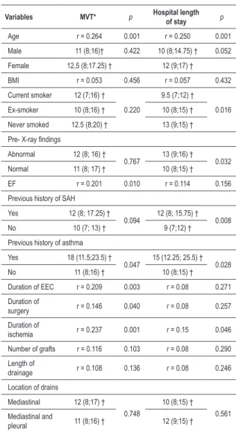
Table 4 shows the correlations between the duration of mechanical ventilation (DMV) and length of hospital stay and pre- and perioperative variables. Statistically significant associations have been observed among the duration of mechanical ventilation, ejection fraction, previous history of asthma, and duration of ECC and ischemia, and time in the operating room. As to the length of hospital stay, associations with age, ex-smoker status, abnormal chest X-ray findings, previous history of hypertension and asthma, and duration of ischemia were significant. The correlations between duration of mechanical ventilation and length of hospital stay were weak; however, the correlations between clinical characteristics and surgical variables were significant.
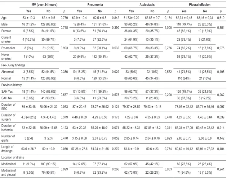
Table 5 shows bivariate analyses between mechanical ventilation over 24 hours, pneumonia, atelectasis and pleural effusion and pre- and perioperative variables. Associations were observed between mechanical ventilation over 24 hours and length of drainage, pneumonia and preoperative abnormal chest X-ray findings, atelectasis and location of drains, and pleural effusion and time in the operating room.
Table 6 shows the results after the adjustments made to the multiple linear regression model, showing that age, preoperative abnormal chest X-ray findings, asthma, SAH and previous CVA remained independently associated with the length of hospital stay.
Discussion
In this contemporaneous cohort, pulmonary complications were frequent. More than 87% of the patients experienced at least one pulmonary complication during hospitalization. The occurrence of postoperative pulmonary complications is closely related to the presence of risk factors inherent to the surgical procedure, anesthesia and preexisting comorbidities4,5.
Consistent with the literature, an association was observed
Table 3 - Incidence of pulmonary complications and deaths during the postoperative phase of myocardial revascularization
Outcomes
Pneumonia (n = 202) 20 (9.9%)
Atelectasis (n = 194) 126 (64.9%)
Pleural effusion (n = 194) 156 (80.4%)
Prolonged MV (> 24 h) (n = 200) 19 (9.6%)
Deaths (n = 202) 11 (5.4%)
Table 4 - Correlations between duration of mechanical ventilation (MVT) and length of hospital stay and pre- and perioperative variables
Variables MVT* p Hospital length of stay p
Age r = 0.264 0.001 r = 0.250 0.001
Male 11 (8;16)† 0.422 10 (8;14.75) † 0.052
Female 12,5 (8;17.25) † 12 (9;17) †
BMI r = 0.053 0.456 r = 0.057 0.432 Current smoker 12 (7;16) †
0.220
9.5 (7;12) †
0.016 Ex-smoker 10 (8;16) † 10 (8;15) †
Never smoked 12.5 (8;20) † 13 (9;15) †
Pre- X-ray indings
Abnormal 12 (8; 16) †
0.767 13 (9;16) † 0.032 Normal 11 (8; 17) † 10 (8;15) †
EF r = 0.201 0.010 r = 0.114 0.156
Previous history of SAH
Yes 12 (8; 17.25) †
0.094 12 (8; 15.75) † 0.008
No 10 (7; 13) † 9 (7;12) †
Previous history of asthma
Yes 18 (11.5;23.5) †
0.047 15 (12.25; 25.5) † 0.028
No 11 (8;16) † 10 (8;15) †
Duration of EEC r = 0.209 0.003 r = 0.08 0.271
Duration of
surgery r = 0.146 0.040 r = 0.08 0.257
Duration of
ischemia r = 0.237 0.001 r = 0.15 0.046
Number of grafts r = 0.116 0.103 r = 0.08 0.290
Length of
drainage r = 0.108 0.136 r = 0.08 0.246
Location of drains
Mediastinal 12 (8;17) †
0.748
10 (8;15) †
0.561
Mediastinal and
pleural 11 (8;16) † 12 (9;15) †
EEC - extracorporeal circulation; SAH - systemic arterial hypertension; BMI - body mass index; EF - ejection fraction; p<0,05; r - Spearman’s coeficient; Pre- X-ray - preoperative radiographic exam. *MVT - duration of mechanical ventilation in
hours; ** hospital length of stay in days; † Median (Percentile 25th; Percentile
75th), Mann-Whitney test.
between postoperative complications, complexity of the surgical procedure, and previous comorbidities in MRV.
It is noteworthy that the criteria used for the establishment of a diagnosis of pulmonary complications were quite comprehensive, without necessarily considering their clinical relevance.
In this study, a mortality rate of 5.4% was recorded after MRV, similar to that reported in literature for elective surgeries at a referral center7.
Independent of other factors, general anesthesia combined with the surgical procedure accounts for the changes in
pulmonary function that take place in the postoperative period. Due to the fact that general anesthesia depresses the function of respiratory centers, the longer the sedation time the higher the risk of pulmonary impairment13,14.
Use of ECC triggers physiological changes secondary to the exposure of blood to the plastic surface of tubes, oxygenators and filters. This, in turn, leads to an increase in extravascular fluid volume in pulmonary circulation, resulting in the filling of the alveoli by inflammatory cells and inactivation of pulmonary surfactant and collapse of some areas. This picture may result in changes in the ventilation/pulmonary perfusion relationship, reduction of compliance, and changes in respiratory work during the postoperative period, making it difficult to wean the patient and increasing the time on mechanical ventilation15.
As per our findings, prolonged mechanical ventilation (over 24 hours) was associated with length of drainage. There are reports in the literature about the damages chest drains can cause to pulmonary function, especially those inserted in the intercostal region, which can change the ventilatory mechanics and lead to pulmonary hypoventilation. These patients are thus more susceptible to developing pulmonary complications, as well as experiencing pain caused by the drain itself, potentially prolonging mechanical ventilation15,16.
The use of pleural drain tubes may be required due to potential injuries caused to the pleura during the removal of the internal mammary artery. However, the use of drains may impair the pulmonary function, since it increases the respiratory work through alterations in gas exchanges and lung mechanics, reducing lung volumes, allowing secretions to collect, leading to potential airflow obstruction and, thus, development of atelectasis4,16-18. This study observed
an association between the location of drain tubes and the development of atelectasis. This complication was further worsened in patients who had two drains inserted both in the mediastinum and in the pleural cavity, compared to patients who had a drain tube inserted only in the mediastinum. This data also show that the greater the number of chest drains inserted the higher the potential for pulmonary complications.
Besides the preexisting comorbidities, a long surgery may be directly related to the complexity of the case, since it presupposes that a large number of procedures was performed. Thus, surgeries taking longer than 210 minutes are considered an important risk factor for the development of postoperative pulmonary complications14. Consistent with
literature data, we observed that in our sample longer surgeries were associated with a higher incidence of pleural effusions, possibly due to the pleural inflammatory responses triggered by the surgical procedure itself11,18. In our sample, the high
incidence of pleural effusion observed was probably due to the use of strictly radiological criteria, independently of their dimension and clinical repercussion. In other studies, the criteria employed were also based on radiographic exams, however with classifications according to the degree of the pulmonary impairment11,18.
Table 5 - Association between mechanical ventilation > 24 hours, pneumonia, atelectasis and pleural effusion, and pre- and perioperative variables
MV (over 24 hours) Pneumonia Atelectasis Pleural effusion
Yes No p Yes No p Yes No p Yes No p
Age 63 ± 10.3 62.4 ± 9.5 0.779 62.9 ± 10.4 62.5 ± 9.5 0.842 61.73± 9.20 63.85 ± 9.7 0,134 62,31 ± 9,45 63,16 ± 9,34 0,619
Male 16 (11.2%) 127 (88.8%)
0.748 12 (8.4%) 131 (91.6%) 0.390 90 (65.2%) 48 (34.8%) 1,000 110 (79,7%) 28 (20,3%) 0,851 Female 5 (8.5%) 54 (91.5%) 8 (13.6%) 51 (86.4%) 36 (64.3%) 20 (35.7%) 46 (82,1%) 10 (17,9%)
Current
smoker 4 (10.3%) 35 (89.7%)
0.993
3 (7.5%) 37 (92.5%)
0.532
24 (64.9%) 13 (35.1%)
0,756
29 (78,4%) 8 (21,6%)
0,975
Ex-smoker 8 (9%) 81 (91%) 9 (9.9%) 82 (90.1%) 60 (66.7%) 30 (33.3%) 74 (82,2%) 16 (17,8%)
Never
smoked 7 (10%) 63 (90%) 20 (9.9%) 182 (90.1%) 42 (62.7%) 25 (37.3%) 53 (79,1%) 14 (20,9%)
Pre- X-ray indings
0.350 0.029 0,572 0,195
Abnormal 3 (5.5%) 52 (94.5%) 10 (18.2%) 45 (81.8%) 33 (60%) 22 (40%) 41 (74,5%) 14 (25,5%)
Normal 15 (11.1%) 120 (88.9%) 9 (6.5%) 129 (93.5%) 86 (65.6%) 45 (34.4%) 110 (84%) 21 (16%)
Previous history
SAH Yes 18 (11.4%) 140 (88.6%)
0.577 17 (10.8%) 141 (89.2%) 0.575 96 (62.7%) 57 (37.3%) 0,290 120 (78,4%) 33 (21,6%) 0,262 SAH No 3 (6.8%) 41 (93.2%) 3 (6.8%) 41 (93.3%) 30 (73.2%) 11 (26.8%) 36 (87,8%) 5 (12,2%)
Duration of
EEC 89 ± 33.48 78.06 ± 24.32 0.063 87 ± 20.46 78.27 ± 25.92 0.124 79.37 ± 28.52 79.93 ± 19.13 78,06 ± 22,42 85,74 ± 35,46 0,097
Duration of
surgery 4.3 (4.02;5) 4.3 (4; 4.45) 0.379 4.48 ± 0.59 4.29 ± 0.56 0.173 4.29 ± 0.6 4.35 ± 0.53 0,470 4,27 ± 0,55 4,48 ± 0,64 0,039 Duration of
ischemia 62 ± 22.45 55.09 ± 17.58 0.123 63 ± 20.33 55.29 ± 18.01 0.074 55.22 ± 18.31 57.85 ± 18.2 0,341 55,34 ± 17,09 59,45 ± 22,42 0,214 Number of
grafts 3 (2;4) 3 (2;3) 0.470 3.15 ± 0.58 2.81 ± 0.75 0.052 2.85 ± 0.74 2.84 ± 0.76 0,923 2,88 ± 0,73 2,68 ± 0,8 0,142 Length of
drainage 63.6 ± 26.7 50 ± 19.9 0.050 57.26 ± 27.6 51.34 ± 21.55 0.270 51.6 ± 19.9 50.6 ± 23 0,774 50,62 ± 19,12 53,91 ± 27,92 0,404 Location of drains
Mediastinal 11 (9.9%) 100 (90.1%)
0.999
14 (12.6%) 97 (87.4%)
0.266
62 (57.9%) 45 (42.1%) 0,033
82 (76,6%) 25 (23,4%) 0,241 Mediastinal
and pleural 8 (9.5%) 76 (90.5%) 6 (6.8%) 82 (93.2%) 62 (73.8%) 22 (26.2%) 71(84,5%) 13 (15,5%)
EEC - extracorporeal circulation; SAH - systemic arterial hypertension; PRE- X-RAY - preoperative radiographic exam; MV - mechanical ventilation.
Table 6 - Multiple linear regression analysis for length of hospital stay
Length of stay b ( 95% CI) p
Age 0.139 (0.050; 0.227) 0.002
Pre- X-ray 2.829 (0.992; 4.666) 0.003
SAH 2.670 (0.680; 4.659) 0.009
CVA 3.281 (0.440; 6.123) 0.024
Asthma 6.398 (2.369; 10.427) 0.002
CVA - cerebrovascular accident; b - angular coeficient; SAH - systemic arterial hypertension; p < 0.05; PRE- R-X - abnormal preoperative radiographic exam.
comorbidities and the patients’ clinical status before the surgery, which prolonged the length of hospital stay due to their higher susceptibility to pre- and postoperative complications5,20.
The limitations of this study are inherent to its design, since cohort studies are prone to selection and analysis bias. The fact that different professionals collected the data and recorded them on the patient medical charts was a limitation for the investigator, since it meant that he/she had to rely on the recorded information, rather than directly accompanying patient progress during the postoperative phase. Moreover, observational studies are prone to residual confounding factors not considered in the multivariate analysis. Nevertheless, a detailed review of the medical chart was conducted in an attempt to extract the best quality information possible.
All pulmonary complications observed in this study were taken into account independently of the degree of the impairment, through radiographic reports and diagnoses made by the medical team during the progression of the patient, abnormal findings were taken into account, whether or not
they were clinically relevant, and this can represent a bias for the evaluation of this variable.
which can justify the high number of outcomes.
In conclusion, over the past several decades, MRV and pre-, peri- and postoperative care have advanced greatly, reducing the occurrence of postoperative complications. However, surgeries are being performed on increasingly complex cases, thus leading to the development of complications following MRV, especially in the respiratory tract, such as those reported in this and in other studies20-22.
Therefore, concomitantly with technological advances, there is a need to conduct better clinical assessments in light of the potential risk factors, seeking to further improve the status of the patient in the postoperative phase.
Thus, there are many aspects to be studied in this area, in the search for effective physiotherapeutic interventions to further reduce the clinical outcomes which are significant to the patients. Other studies have shown that physical therapy can play an important role both in preventing and reducing
postoperative complications, through techniques such as pulmonary expansion and ventilation, bronchial hygiene and respiratory muscle training, among other resources capable of ensuring better recovery after MRV23,24.
Potential Conflict of Interest
No potential conflict of interest relevant to this article was reported.
Sources of Funding
There were no external funding sources for this study.
Study Association
This article is part of the thesis of master submitted by Leila D. N. Ortiz, from Instituto de Cardiologia do RS/FUC.
References
1. Ishitani, LH, Franco GC, Perpetuo IHO, França E. Desigualdade social e mortalidade precoce por doenças cardiovasculares no Brasil. Rev Saúde Publica. 2006; 40 (4): 684-91.
2. Anis Rassi Jr. Otimização do tratamento medicamentoso na doença arterial coronariana: tarefa para o subespecialista? Arq Bras Cardiol. 2004; 83 (3): 187-8.
3. Mack MJ, Pfiser A, Bachand D, Emery R, Magee M, Connolly M, et al. Comparison of coronary bypass surgery with and without cardiopulmonary bypass in patients with multivessel disease. J Thorac Cardiovasc Surg. 2004; 127 (1): 167-73.
4. Guizilini S, Gomes WJ, Faresin SM, Bolzan DW, Alves FA, Catani R, et al. Avaliação da função pulmonar em pacientes submetidos à cirurgia de revascularização do miocárdio com e sem circulação extracorpórea. Rev Bras Cir Cardiovasc. 2005; 20 (3): 310-6.
5. Filardo FA, Faresin SM, Fernandes ALG. Validade de um índice prognóstico para ocorrência de complicações pulmonares no pós-operatório de cirurgia abdominal alta. Rev Assoc Med Bras. 2002; 48 (3): 209-16.
6. Bellinetti LM, Thomson JC. Avaliação muscular respiratória nas toracotomias e laparotomias superiores eletivas. J Bras Pneumol. 2006; 32 (2): 99-105.
7. Bianco ACM, Timerman A, Paes AT, Gun C, Ramos RF, Freire RBP, et al. Análise prospectiva de risco em pacientes submetidos à cirurgia de revascularização miocárdica. Arq Bras Cardiol. 2005; 85 (4): 254-61.
8. Sampaio RF, Mancini MC, Fonseca ST. Produção cientifica e atuação profissional: aspectos que limitam essa integração na fisioterapia e na terapia ocupacional. Rev Bras Fisioter. 2002; 6 (3): 113-8.
9. Sociedade Brasileira de Pneumologia e Tisiologia (SBPT). Diretrizes brasileiras para tratamento das pneumonias adquiridas no hospital e das associadas à ventilação mecânica. J Bras Pneumol. 2007; 33 (1): 1-30.
10. Sutton D. Doenças das vias aéreas, colapso e consolidação: tratado de radiologia e diagnóstico por imagem. 6ª ed. Rio de Janeiro: Revinter; 2003. p. 464-72.
11. Light RW, Rogers JT, Moyers JP, Lee YCG, Rodriguez RM, Alford Jr WC, et al. Prevalence and clinical course of pleural effusions at 30 days after coronary artery and cardiac surgery. Am J Respir Crit Care Med. 2002; 166: 1567-71.
12. Milot J, Perron J, Lacasse Y, Lètourneau L, Cartier PC, Maltais F. Incidence and predictors of ARDS after cardiac surgery. Chest. 2001; 119: 884-8.
13. Auler Junior JOC, Galas FRBG, Hajjar LA, Franca S. III Consenso brasileiro de
ventilação mecânica. J Bras Pneumol. 2007; 33 (2): 14-47.
14. Machado LB, Chiaroni S, Vasconcelos Filho PO, Auler Junior JOC, Carmona MJC. Incidência de cirurgia cardíaca em octogenários: estudo retrospectivo. Rev Bras Anestesiol. 2003; 53 (5): 646-56.
15. Nozawa E, Kobayashi E, Matsumoto ME, Feltrim MIZ, Carmona MJC, Auler Junior JOC. Avaliação dos fatores que influenciam no desmame de pacientes em ventilação mecânica prolongada após cirurgia cardíaca. Arq Bras Cardiol. 2003; 80 (3): 301-5.
16. Guizilini S, Gomes WJ, Faresin SM, Carvalho ACC, Jaramillo JI, Alves FA, et al. Efeitos do local de inserção do dreno pleural na função pulmonar no pós-operatório de cirurgia de revascularização do miocárdio. Rev Bras Cir Cardiovasc. 2004; 19 (1): 47-54.
17. Goiksin I, Baltalarli A, Sacar M, Sungurtekin H, Ozcan V. Preservation of pleural integrity in patients undergoing coronary artery bypass grafting: effect on postoperative bleeding and respiratory function. Acta Cardiol. 2006; 61 (1): 89-94.
18. Iyem H, Islamoglu F, Yagdi T, Sargin M, Berber O, Hamulu A, et al. Effects of pleurotomy on respiratory sequelae. Tex Heart Inst J. 2006; 33 (2): 116-21.
19. Neto LJ, Thompson JC, Cardoso JR. Complicações respiratórias no pós-operatório de cirurgias eletivas e de urgência e emergência em um hospital universitário. J Bras Pneumol. 2005; 31: 41-7.
20. Ambrozin AR, Cataneo AJM. Aspectos da função pulmonar após revascularização do miocárdio relacionados com risco pré-operatório. Rev Bras Cir Cardiovasc. 2005; 20 (4): 408-15.
21. Nakagawa M, Tanaka H, Tsukuma H, Kishi Y. Relationship between the duration of the preoperative smoke-free period and the incidence of postoperative pulmonary complications after pulmonary surgery. Chest. 2001; 120: 705-10.
22. Feier FH, Sant’anna RT, Garcia E, De Bacco FW, Pereira E, Santos MF, et al. Modificações no perfil do paciente submetido à operação de revascularização do miocárdio. Rev Bras Cir Cardiovasc. 2005; 20 (3): 317-22.
23. Hulzebos EHJ, Helders PJM, Favié NJ, De Bie RA, Riviere AB, Meeteren NLUV. Preoperative intensive inspiratory muscle training to prevent postoperative pulmonary complications in high-risk patients undergoing CABG surgery JAMA. 2006; 296 (15): 1851-7.