Periodontal Disease in Pregnant Patients with Rheumatic Valvular
Disease - Clinical and Microbiological Study
Walkiria Samuel Ávila
1, Lilia Timerman
2, Giuseppe Alexandre Romito
3, Sílvia Linard Marcelino
3, Itamara Lúcia
Itagiba Neves
1, Marcelo Zugaib
4, Max Grinberg
1Instituto do Coração do Hospital das Clínicas da Faculdade de Medicina da Universidade de São Paulo1; Instituto Dante Pazzanese de Cardiologia2; Disciplina de Periodontia do Departamento de Estomatologia da Faculdade de Odontologia da Universidade de São Paulo3; Departamento de Obstetrícia e Ginecologia do Hospital das Clínicas da Faculdade de Medicina da Universidade de São Paulo4 - São Paulo, SP - Brazil
Mailing address: Walkiria Samuel Ávila •
Rua Apiacás, 688/31 - Perdizes - 05017-020 - São Paulo, SP - Brazil E-mail: [email protected], [email protected]
Manuscript received February 24, 2010; revised manuscript received July 26, 2010; accepted July 28, 2010.
Abstract
Background: The periodontal disease during pregnancy of women with rheumatic valve disease imply infective endocarditis risks and higher rate of preterm birth and low birth weight.
Objective: To study the periodontal disease rate of women with rheumatic valve disease during pregnancy.
Methods: We studied 140 pregnant women who included 70 patients with rheumatic valve disease and 70 healthy
women. The periodontal examination included: 1) periodontal clinical exam regard the follow variables: a) probing
depth; b) gingival margin; c) clinical attachment level; d) bleeding on probing; e) plaque index and f) gingival index; and
2) microbiological test was performed in samples serum and gingival crevicular fluid and considered positive controls to Porphyromonas gingivalis, Tannerella forsithia e Aggregobacter actinomycetemcomitans.
Results: Age and parity were similar between groups; as single or combined the mitral valve disease was prevalent
among the rheumatic valve lesion in 45 (32.1%) e 20 (28.5%) cases, respectively. Among the periodontal variables gingival margin (p=0.01) and plaque index (p=0.04) were different between groups. The periodontal disease was identified in 20 (14,3%) pregnant women, seven (10%) of them were patients with valve rheumatic disease and the remain 13 (18,6%) were healthy women, its percentual was not different between groups (p=0,147). Microbiological
analyses of oral samples showed higher percentual of P. gingivalis in healthy pregnant women (p=0.004).
Conclusion: The clinical and microbiological study during pregnancy showed comparable incidence of periodontal
disease between women with rheumatic valve disease and healthy women. (Arq Bras Cardiol 2011;96(4):307-311)
Keywords: Periodontitis; gengivitis; heart valve diseases; rheumatic disease; pregnancy.
especially the bacteria Porphyromonas gingivalis (P. gingivalis), Tannerella forsythia (T. forsythia) and Aggregatibacter actinomycetemcomitans (A. actinomycetemcomitans)4.
During pregnancy, the permanent inflammatory and infectious state of the oral cavity5 represents a potential risk, either by favoring the occurrence of infectious endocarditis or by causing obstetric complications, such as miscarriage and prematurity6.
In this sense, the objective of the present study was to study the frequency of periodontal disease in women with rheumatic valvular disease during pregnancy.
Methods
From January2005 to July 2008, a cross-sectional study was carried out, which selected 140 pregnant women aged 18 to 35 years, consecutively treated at the Brazilian Public Health System (SUS). The group of women with rheumatic valvular disease consisted of 70 pregnant women treated at the Cardiopathy and Pregnancy Outpatient Clinic of the Valvular Cardiopathy Unit of Instituto do Coração (InCor). The control group consisted of 70
Introduction
Periodontal disease comprehends a broad spectrum of alterations in periodontal tissue, which has an inflammatory and/or infectious origin. These alterations can take place during pregnancy, when physiological alterations in the oral cavity, attributed to hormonal mechanisms that occur during pregnancy, result in what is called pregnancy gingivitis1, characterized by inflammation of the gingiva, cement and periodontal ligament and that predisposes the pregnant woman to periodontitis.
healthy pregnant women followed at the Prenatal Outpatient Clinic of Centro de Saúde Geraldo de Paula Souza, from the School of Public Health of the University of São Paulo (USP).
When included in the study, the groups of healthy and rheumatic pregnant women were paired for age, socioeconomic and cultural level and gestational age. All patients followed a protocol that included periodic clinical evaluation and dental clinical assessment, aiming at the identification of periodontal disease and the microbiological study of the oral cavity. In the group of rheumatic pregnant women, among the medications used for the cardiological treatment, the intramuscular injections of G benzathine (1,200,000 IU every 21 days) were maintained, for secondary prophylaxis of the rheumatic disease. Women with other comorbidities, such as arterial hypertension, diabetes mellitus, infectious diseases or those that had received antibiotic therapy during the previous three months, were excluded from the study.
The anamnesis included questions on oral hygiene habits. The patients were submitted to periodontal clinical assessment, which used the dental mirror and a probe with millimeter markings to record the following parameters: a) probing pocket depth (PPD), that corresponds to the distance (mm) between the gingival margin and the depth of the gingival sulcus or periodontal pocket; b) distance of the enamel-cement junction to gingival margin (ECJ/GM); c) clinical attachment level (CAL), which is the arithmetic difference of the values of PPD and ECJ/GM; d) the bleeding index (BI) on the four sides of each assessed tooth; e) bacterial plaque index (BPI); and f) furcation involvement.
The diagnosis of periodontal disease was based on the criteria by Tonetti and Claffey7. The microbiological analysis of saliva and cone samples used DNA extraction and polymerase chain reaction (PCR) techniques. The positive controls were cultivated strains of the bacteria P. gingivalis, T. forsythia and A. actinomycetemcomitans, according to Ashimoto’s technique8.
The statistical analysis of the results used absolute and relative frequencies of data, central trend measurements (means and medians) and the dispersion of values (minimum and maximum values and standard deviations). The association tests (Pearson’s Chi-square and Fisher’s exact test) were calculated to assess the association between the presence of periodontal disease and the qualitative variables. The assessment of the periodontal disease took into account the clinical parameters: PPD, CAL, ECJ/GM, BI and BPI. The Mann-Whitney nonparametric and Pearson’s
Chi-square tests were used to compare the rheumatic and the healthy pregnant women. The level of significance was set at 5.0% for the rejection of the null hypothesis.
The study was approved by the Committee of Ethics in Research of Hospital das Clínicas of the School of Medicine (CAPPesp - process #199/05) and of InCor (process # 2557-04/177) of the University of São Paulo (USP) and by the Committee of Ethics in Research of Instituto Dante Pazzanese de Cardiologia. The patients were included in the protocol after signing the Free and Informed Consent Form, according to the norms established by Resolution # 196 of October 10,1996, of the National Council of Health.
Results
A total of 61 patients (43.6%) were primiparas and the mean maternal age was not different (p < 0.451) between the groups, being 27 ± 4.6 years in rheumatic patients and 27 ± 5.1 in the healthy ones. The analysis of the distribution of valvular lesion in the rheumatic group showed that 45 (32.1%) of them presented mitral valve lesion, 5 (3.6%) presented aortic valve lesion and 20 (28.5%) presented a combination of aortic and mitral valve lesion. Fifteen of the 70 rheumatic patients had been submitted to cardiac surgery for biological valve replacement.
Clinical-odontological assessment
The analysis of the information on the oral hygiene showed that flossing was more common (p = 0.02) in the group of rheumatic pregnant women (48 = 68.5%) when compared to the healthy women (32 = 45.7%), whereas rinsing the mouth with mouthwash was 2.3-fold more common among the healthy pregnant women. The odontological and microbiological assessments were carried out at similar gestational ages (p = 0.801), generally at the 27.8th ± 6.6 week of gestation in the rheumatic group and at the 29.8th ± 7.6 week in the healthy group.
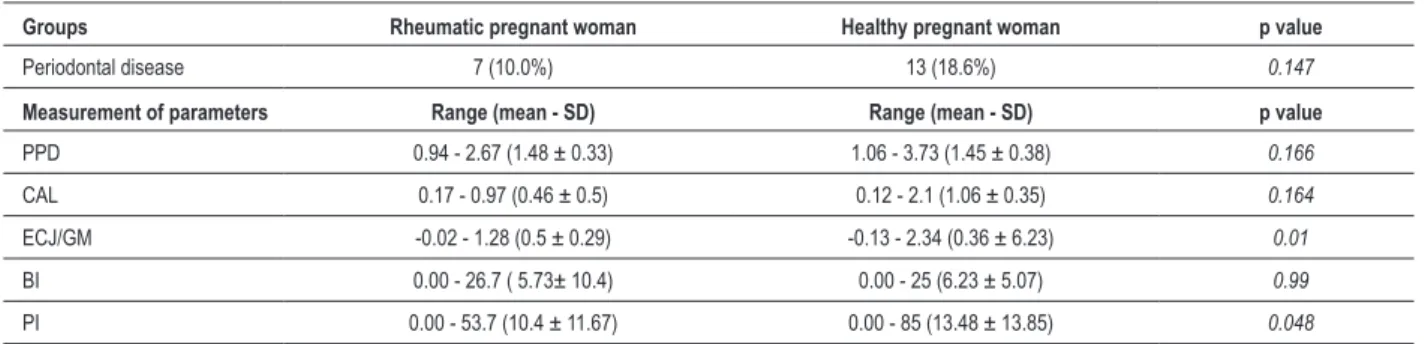
The mean number of preserved teeth was 24.1 teeth in the rheumatic group vs 24.0 in the healthy group. Periodontal disease was identified in 20 (14.3%) pregnant women, being 7 (10%) rheumatic and 13 (18.6%) healthy ones, with no statistical difference (p = 0.147) when comparing the two groups. The analysis of the periodontal parameters shown in Table 1 demonstrated differences between the groups regarding ECJ/GM and PI (p = 0.04).
Table 1 - Frequency of periodontal disease and measurements of odontological parameters
Groups Rheumatic pregnant woman Healthy pregnant woman p value
Periodontal disease 7 (10.0%) 13 (18.6%) 0.147
Measurement of parameters Range (mean - SD) Range (mean - SD) p value
PPD 0.94 - 2.67 (1.48 ± 0.33) 1.06 - 3.73 (1.45 ± 0.38) 0.166 CAL 0.17 - 0.97 (0.46 ± 0.5) 0.12 - 2.1 (1.06 ± 0.35) 0.164 ECJ/GM -0.02 - 1.28 (0.5 ± 0.29) -0.13 - 2.34 (0.36 ± 6.23) 0.01
BI 0.00 - 26.7 ( 5.73± 10.4) 0.00 - 25 (6.23 ± 5.07) 0.99
Table 2 -Proportion of bacteria identiied in saliva and cone samples in the groups
Bacteria Sample Rheumatic pregnant woman Healthy pregnant woman Total p value
n (%) n (%) n (%)
Pg Saliva 21 (30.0) 38 (54.3) 56 (42.1) 0.004
Cone 19 (27.1) 26 (37.1) 45 (32.1) 0.205
Tf Saliva 24 (34.3) 19 (27.1) 43 (30.7) 0.36
Cone 31 (44.3) 23 (32.9) 54 (38.6) 0.165
Aa Saliva 8 (11.4) 5 (7.1) 13 (9.3) 0.382
Cone 4 (5.7) 4 (5.7) 8 (5.7) 1
Pg - P. gingivalis; Tf - T. forsythia; Aa - A. actinomycetemcomitans.
Table 3 - Distribution of the bacteria Porphyromonas gingivalis,
Tannerella forsythia, Aggregatibacter actinomycetemcomitans
in saliva and cone samples of pregnant women with periodontal disease (*p = 0.001)
Bacteria P gingivalis T. forsythia A. actinomycetemcomitans
Saliva 11 (55.0%)* 4 (20.0%) 1 (5.0%) Cone 7 (35.0%) 8 (40.0%)* 1 (5.0%) Absent 2 (10.0%) 8 (40.0%) 18 (90.0%)
Microbiological assessment
The comparative analysis between the groups showed that the proportion of P. gingivalis was significantly higher in the saliva of pregnant women, when compared to the rheumatic ones (p = 0.004), but there was no difference regarding the bacteria T. forsythia and A. actinomycetemcomitans, in both saliva and cone samples (Table 2). The analysis of the distribution of types of bacteria among the 20 pregnant women that presented periodontal disease showed that the proportion of P. gingivalis and A. actinomycetemcomitans was significantly higher in the saliva and cone samples, respectively (Table 3).
Obstetric data
There were no maternal obstetric complications during gestation and delivery. The gestational age at delivery, calculated as weeks of gestation, ranged from 36 to 42 (mean = 38.54 ± 6.65) weeks in the rheumatic and between 32 and 42 (mean = 39.28 ±7.65) weeks in the healthy pregnant women. Newborn weight ranged between 3,017 and 4,488 g (mean = 3,069 g ± 455) in the rheumatic group and between 3,290 and 4,260 g (mean = 3,286 g ± 455) in the healthy group. The comparative analysis showed that the gestational age at delivery and newborn weight were lower in the rheumatic group, showing statistical significance (respectively, p = 0.0036 and p = 0.034).
Discussion
The present study provides original data on the effect of periodontal disease in two special situations: pregnancy and rheumatic valvular disease.
Hormonal alterations during gestation, changes in eating habits and poor oral hygiene habits are factors that favor the manifestation of gestational gingivitis, common during the pregnancy-puerperium cycle9,10. That is further complicated by the lack of access to dental care and the myth about the safety of dental treatment, which predispose to periodontal disease during pregnancy11.
Although the population considered in this study was exposed to all these factors, the incidence of 14.0% of periodontal disease was not different from the percentage found in the general population12. Among the explanations for this finding are the sample’s characteristics: young patients
(mean of 27 years) who are known13 to present a lower risk of periodontal disease when compared to older patients and the exclusion of smokers and patients with diabetes mellitus, conditions known to elevate the risk for periodontal disease in the general population14-15. It is noteworthy, however, that studies on the incidence of periodontal disease in cardiopathic patients with rheumatic valvular disease are scarce.
The present study showed that the proportion of 13 (18.6%) cases of periodontal disease in the healthy group and 7 (10.0%) cases in the rheumatic group did not present statistical difference. However, at the clinical examination, parameters such as distance of the enamel-cement junction (ECJ) to gingival margin (GM) and the bacterial plaque index (PI) were different in the healthy pregnant women.
Such findings might be explained by the better oral hygiene performed by the rheumatic pregnant women, who flossed more regularly than the healthy ones. Although they presented similar socioeconomic levels, the women with valvular disease are probably better informed on the potential cardiac risks associated with poor oral hygiene, the result of the continuing education received throughout the cardiological follow-up.
and periodontal destruction in periodontal disease19, with a prevalence of gram-negative anaerobic bacilli and among them, A. actinomycetemcomitans (Hacek group), P. gingivalis and P. intermedia, with a predominance in gingival samples20.
The identification of a higher proportion of P. gingivalis in the saliva of healthy pregnant women in comparison with the rheumatic ones (54.3% vs 29.20%; p = 0.004) reinforces the explanation of a better oral health status of the cardiopathic patients, when compared to the healthy ones.
However, the positive influence of benzathine penicillin in the rheumatic group should be considered. Thus, the recommendation for maintaining the administration of this antibiotic every 21 days throughout pregnancy with the objective of preventing new rheumatic episodes might represent an additional benefit for the improvement of oral health and the prevention of periodontal disease during pregnancy.
In the present study, the prevalence of P. gingivalis in the saliva samples of patients that had periodontal disease corroborates the reports by Dietrich and Takeuchi19, who demonstrated that P. gingivalis was a very virulent pathogen and that its detection must be used as a clinical indicator for periodontal disease.
As for the obstetric evolution, our results did not allow us to correlate periodontal disease with the adverse effects in pregnancy. Although the literature has called attention to the association between periodontal disease with low birth weight and prematurity, due to the systemic inflammatory state, there is not enough evidence to allow the correlation of the effects of periodontal disease with com adverse obstetric outcomes21. On the other hand, the significant proportion of low-weight newborns and the younger gestational age at birth verified in the group of rheumatic pregnant women are explained by the maternal cardiac output restrictions and beta-blocker use by the rheumatic pregnant women.
Regarding the maternal scenario, the absence of complications during the pregnancy-puerperium cycle is attributed to the non-inclusion of patients with arterial hypertension, diabetes mellitus and other morbidities, associated with the rigorous prenatal care provided by the multidisciplinary team.
Conclusion
The clinical-microbiological study of the oral cavity during pregnancy showed that the frequency of periodontal disease in women with rheumatic valvular disease was not different when compared to healthy pregnant women.
Acknowledgements
The research was supported by Fundação de Amparo à Pesquisa do Estado de São Paulo (process # 57931-2).
To the Prenatal Care Team of Centro de Saúde Geraldo de Paula Souza of the Public Health School of the University of São Paulo, for their support in referring the patients that constituted the sample, which included the healthy pregnant women.
Potential Conflict of Interest
No potential conflict of interest relevant to this article was reported.
Sources of Funding
This study was funded by FAPESP.
Study Association
This article is part of the thesis of doctoral submitted by Lilia Timerman, from Incor.
References
1. Raber-Durlacher JE, van Steengergen TJ, Van Der Velden U, de Graaff J, Abracham-Inpijn L. Experimental gingivitis during pregnancy and post-partum: clinical, endocrinological and microbiological aspects. J Clin Periodontal. 1994;21(8):549-58.
2. Abegg C. Oral hygiene habits among Brazilian adults in an urban area of southern Brazil. Rev Saúde Pública. 1997;31(6):586-93.
3. Gjermo P, Rosing CK, Susin C, Oppermann R. Periodontal diseases in Central and South America. Periodontol 2000;2002;29:70-8.
4. Sanz M, Lau L, Herrera D, Morillo JM, Silva A. Methods of detection of Actinobacillus actinomycetemcomitans, Porphyromonas gingivalis and Tannerella forsythensis in periodontal microbiology, with special emphasis on advanced molecular techniques: a review. J Clin Periodontol. 2004;31(12):1034-47.
5. Mombelli A. Periodontitis as an infectious disease: specific features and their implications. Oral Dis. 2003;9(Suppl. 1):6-10.
6. Agueda A, Ramon JM, Manau C, Guerrero A, Echeverria JJ. Periodontal disease as a risk factor for adverse pregnancy outcomes: a prospective cohort study. J Clin Periodontol. 2008;35(1):16-22.
7. Tonetti MS, Claffey N. Advances in the progression of periodontitis and proposal of definitions of a periodontitis case and disease progression for use
in risk factor research. In: 5th European Workshop in Periodontology. J Clin Periodontol. 2005;32(Suppl. 6):210-3.
8. Ashimoto A, Chen C, Bakker I, Slots J. Polymerase chain reaction detection of 8 putative periodontal pathogens in subgingival plaque of gingivitis and advanced periodontitis lesions. Oral Microbiol Immunol. 1996;11(4):266-73.
9. Kinane DF PM, Peterson M, Stathopoulou PG. Environment and other modifying factors of the periodontal disease. J Periodontol. 2006;40:107-19.
10. Yalcin FEE, Soydinc M, Basegmez C, Issever H, Isik G, Berber L, et al. The effect of sociocultural status on periodontal conditions in pregnancy. J Periodontol. 2002;73(2):178-82.
11. Boggess, KA. Maternal oral healthy in pregnancy. Obstetric Gynecol. 2008;111(4):976-86.
12. Oliver RC, Brown LJ, Loe H. Periodontal diseases in the United States population. J Periodontol. 1998;69(2):269-78.
13. Dietrich T, Jimenez M, Krall Kaye EA, Vokonas PS, Garcia RI. Age-dependent associations between chronic periodontitis/edentulism and risk of coronary heart disease. Circulation. 2008;117(13):1668-74.
15. Kinane DF, Chestnutt IG. Relationship of diabetes to periodontitis. Curr Opin Periodontol. 1997;4:29-34.
16. Hull MW, Chow AW. An approach to oral infections and their management. Curr Infect Dis Rep. 2005;7(1):17-27.
17. Holmstrup P, Poulsen AH, Andersen L, Skuldbol T, Fiehn NE. Oral infections and systemic diseases. Dent Clin North Am. 2003;47(3):575-98.
18. Darveau RJP, Tanner A, Page RC. The microbial challenge in periodontitis. Periodontol 2000;1997;14:12-32.
19. Takeuchi Y, Umeda M, Sakamoto M, Benno Y, Huang Y, Ishikawa I. Treponema socranskii, Treponema denticola, and Porphyromonas gingivalis
are associated with severity of periodontal tissue destruction. J Periodontol. 2001;72(10):1354-63.
20. Yano-Higuchi K, Takamatsu N, He T, Umeda M, Ishikawa I. Prevalence of Bacteroides forsythus, Porphyromonas gingivalis and Actinobacillus actinomycetemcomitans in subgingival microflora of Japanese patients with adult and rapidly progressive periodontitis. J Clin Periodontol. 2000;27(8):597-602.