Arq Bras Cardiol volume 72, (nº 6), 1999
Zimerman et al AV block during tachycardia ablation
713 From the Division of Cardiology, Hospital de Clínicas de Porto Alegre, RS
Mailing adress: Leandro Zimerman - Av. Iguassu, 176/402 - 90470-430 - Porto Alegre, RS - Brazil
Leandro Zimerman, Claudio Medeiros, Gustavo Lima
Porto Alegre, RS - Brazil
Catheter-Induced 3:1 Second Degree Atrioventricular Nodal
Block During Atrioventricular Nodal Reentrant Tachycardia
Case Report
Second-degree atrioventricular (AV) nodal block with 2:1 AV conduction during atrioventricular nodal reentrant tachycardia (AVNRT) has been previously described. We report a patient who developed AVNRT during which 3:1 intra-infra-Hisian block due to catheter manipulation was observed. This was followed by gradual, incremental recovery of AV conduction.
AVNRT accounts for approximately 50% of cases of supraventricular tachycardia. This mechanism of tachy-cardia uses a reentrant circuit in the region of the AV Node which usually contains distinct slow and fast pathways. Radiofrequency ablation of the slow pathway is an effec-tive and safe treatment for AVNRT 1.
In this report, we present a patient with AVNRT of the common type who developed 3:1 intra-infra-Hisian block during AVNRT as a result of catheter manipulation. Before resumption of normal 1:1 conduction, we observed split His potentials in normal sinus rhythm, followed by atrioven-tricular (AV) nodal echo beats with intra-infra-Hisian block.
Case Report
An 81-year-old white female was referred for evalua-tion of medically refractory supraventricular tachycardia. At electrophysiologic study, catheters were advanced to the right ventricular apex, His bundle area, high right atrium and the coronary sinus without difficulty. After obtaining basic intervals (PA:47msec; AH: 40msec; HV: 53msec), right atrial and right ventricular premature stimulation and decre-mental pacing were performed without induction of tachycardia. With a basic drive train of 600msec, the AV nodal efective refractory period was <340ms.
Administration of isoproterenol 3µg/min and atropine 0.6mg intravenously caused the heart rate to increase from 57bpm to 75bpm. Programmed atrial extrastimulation with basic drive of 600msec showed that the effective refractory period of the fast and slow AV nodal pathways were 380msec and <330msec respectively. Right atrial burst pacing subsequently induced sustained AVNRT of the
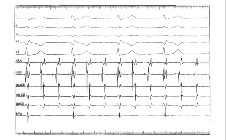
common type with 3:1 intra-infra-Hisian block (fig. 1). The atrial cycle length during tachycardia was 280msec, while the ventricular cycle length was 840msec. Evidence suppor-ting AVNRT as the mechanism of tachycardia are as follows: 1) dual AV nodal physiology was present; 2) retrograde atrial activation was concentric during ventricular pacing and during tachycardia; 3) premature ventricular beats introduced when the His bundle was refractory did not preexcite the atrium; 4) the tachycardia was induced with a critical AV delay; 5) the surface V to His A interval during reciprocating tachycardia was -15msec; 6) tachycardia persisted during 3:1 conduction, showing that the ventricle was not a critical component of the circuit; 7) application of radiofrequency current to the posteroseptal right atrium resulted in noninducibility of the tachycardia. Isoproterenol was discontinued, and the tachycardia was terminated with right ventricular burst pacing.
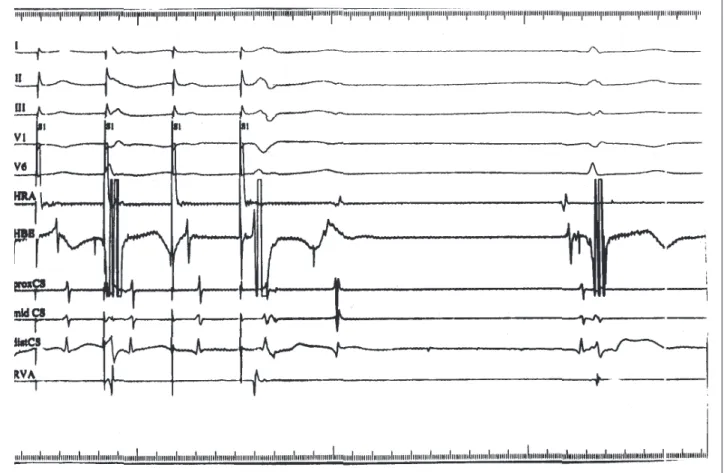
During normal sinus rhythm,1:1 AV nodal conduction returned. However, the His bundle electrogram was notable for a split His depolarization (Figure 2). The basic intervals were as follows: PA, 45msec; AH, 47msec; HH’, 34msec; H’V, 38msec. The split His potentials resolved after one hour, at which point right atrial burst pacing induced AV nodal echo beats with intra-infra-Hisian block(Figure 3). Subsequently, right atrial extrastimuli and decremental pacing were repeated, and no abnormalities in His bundle conduction were noted. AVNRT with 1:1 HV conduction was then induced with right atrial pacing. Six applications of radiofrequency current were then delivered to the posteroseptal right atrium, and junctional rhythm was noted during the final application. AVNRT was no longer inducible, and AV nodal conduction time was normal during sinus rhythm (PA: 49msec; AH: 49msec; HV: 50msec).
Discussion
In previous reports, 2:1 infranodal conduction block during AVNRT has been rarely observed, usually appearing immediately after induction of AVNRT and lasting for a few beats 1-5. All but one 5 of the cases of second degree AV
block 1-5 and atrioventricular dissociation 3 during AVNRT
occurred during AVNRT of the common type. Sites of block in previous reports have been above the AV node 1,3, above
714
Zimerman et al
AV block during tachycardia ablation
Arq Bras Cardiol volume 72, (nº 6), 1999
Fig. 1 - AVNRT of the common type with 3:1 conduction block to the ventricles and 1:1 concentric retrograde atrial activation. Despite the presence of AVN block, the tachycardia is sustained. I, II, III, V1 and V6- surface leads; HRA- high right atrium; HBE- His bundle; prox CS- proximal coronary sinus; mid CS- middle coronary sinus; dist CS- distal coronary sinus; RVA- right ventricular apex.
Arq Bras Cardiol volume 72, (nº 6), 1999
Zimerman et al AV block during tachycardia ablation
715 persisting for almost one hour with subsequent incremental
recovery has not been previously reported. We are also unaware of any previous reports of high-degree 3:1 AV conduction block during AVNRT.
This patient developed a 3:1 intra-infra-Hisian block during AVNRT of the common type that was present for one hour. The most probable cause of the block in this case was catheter manipulation. Conduction block during AVNRT attributed to His bundle catheter manipulation has been previously described 6, but the site of block was above the
His bundle. During AVNRT with 3:1 block, we observed an H potential on the His bundle recordings, with a fixed HA
interval. This is suggestive of an infra-Hisian point of block. However, since a split His depolarization was noted after restoration of normal conduction, intra-Hisian, rather than infra-Hisian, block is more likely. Interestingly, the tachy-cardia with 3:1 block was terminated with right ventricular burst pacing, suggesting that the conduction block was antegrade only.
In summary, this case is notable for the novel obser-vation of prolonged 3:1 infranodal conduction block during AVNRT. The site of block was either intra or infra-Hisian and possibly only antegrade. Block was followed by incremental recovery of conduction.
Fig. 3 - Atrioventricular nodal reentry echo beat with the same retrograde atrial activation seen in figure 1 (during tachycardia) and with infra-Hisian block observed at the end of atrial burst pacing with a cycle length of 350ms. This confirms that the atrioventricular conduction is still abnormal, despite the absence of split His in the sinus beat. S1: atrial stimuli. I, II, III, V1 and V6: surface leads; HRA- high right atrium; HBE- His bundle; prox CS- proximal coronary sinus; mid CS- middle coronary sinus; dist CS- distal coronary sinus; RVA- right ventricular apex.
References
1. Haissaguerre M, Gaita F, Fischer B, et al - Elimination of atrioventricular reentrant tachycardia using discrete slow potentials to guide application of radiofre-quency energy. Circulation 1992; 85: 2162-75
2. Wellens HJ, Wesdorp JC, Duren DR, Lie KI - Second degree block during reciprocal atrioventricular nodal tachycardia. Circulation 1976; 53: 595-9. 3. Vassalo JA, Cassidy DM, Josephson ME - Atrioventricular nodal
supra-ventricular tachycardia. Am J Cardiol 1985; 56: 193-5.
4. Portillo B, Mejias J, Leon-Portillo N, et al - Entrainment of atrioventricular
nodal reentrant tachycardias during overdrive pacing from high right atrium and coronary sinus - with special reference to atrioventricular dissociation and 2:1 retrograde block during tachycardias. Am J Cardiol 1984; 53: 1570-5. 5. DiMarco JP, Sellers TD, Belardinelli L - Paroxysmal supraventricular
tachycardia with Wenckebach block: evidence for reentry within the upper portion of the atrioventricular node. J Am Coll Cardiol 1984; 3: 1551-5. 6. Ko PT, Nacarelli GV, Gulamhusein S, et al - Atrioventricular dissociation during