LETTER TO THE EDITOR
Cheyne-stokes respiration associated with
hypertrophic cardiomyopathy and normal left
ventricular ejection fraction
Rodrigo P. Pedrosa,ILuciano F. Drager,IMurillo O. Antunes,IIEdmundo Arteaga,II Geraldo Lorenzi-FilhoI ISleep Laboratory, Pulmonary Division, Heart Institute (InCor), Hospital das Clı´nicas, Faculdade de Medicina, Universidade de Sa˜o Paulo, Sa˜o Paulo, SP,
Brazil.IICardiomyopathy Medical Unit, Heart Institute (InCor), Hospital das Clı´nicas, Faculdade de Medicina, Universidade de Sa˜o Paulo, Sa˜o Paulo, SP,
Brazil.
Email: [email protected] Tel.: 55 11 3069-5486
INTRODUCTION
Cheyne-Stokes respiration (CSR) is a form of periodic breathing in which central apneas and hypopneas alternate with periods of hyperventilation, producing a waxing and waning pattern of tidal volume. This brief case described an interesting association of CSR and hypertrophic cardiomyo-pathy (HCM) in a patient with normal left ventricular ejection fraction, providing physiological insights for the genesis of CSR.
CASE DESCRIPTION
A 64-year-old male patient was admitted for a routine cardiac evaluation with complaints of witnessed apnea during sleep. He had previous history of HCM and permanent atrial fibrillation. He was on regular use of carvedilol 50 mg/d; enalapril 40 mg/d, furosemide 80 mg/ d and warfarin 5 mg/d and was on NYHA stage III heart failure. Echocardiography demonstrated interventricular septum wall thickness of 20 mm and posterior wall thickness of 11 mm, without left ventricle outflow tract obstruction. The left ventricle ejection fraction was in the normal range (69%). The right ventricle systolic pulmonary pressure was 35 mmHg. Echocardiographic diastolic func-tion evaluafunc-tion was impaired due to atrial fibrillafunc-tion. Technetium (99mTc) radionuclide ventriculography showed
normal biventricular systolic function and a median pulmonary transit time of 13 seconds (normal,10 seconds), denoting diastolic dysfunction. Cardiac nuclear magnetic resonance also demonstrated normal global biventricular function. Arterial blood gas while awake showed pH:7.41; pCO2:33 mm Hg; paO2: 78 mm Hg; Sat: 97%; HCO3:
20.6 mMol/L; BE: -2.6 mMol/L. A full-overnight polysom-nography indicated a sleep efficiency of 95% with pre-dominantly light sleep (9.9% of stage 1, 75.9% stage 2, 4% of delta sleep (non REM stage 3/4) and 10.2% stage REM). The study showed an apnea-hypopnea index of 34.3 events per hour divided in: obstructive apneas = 8; central apneas = 12.1; mixed apneas = 5.3 and hypopneas = 8.9 events/
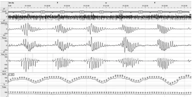
hour. CSR pattern of breathing was present during the entire night (Figure 1). The respiratory pattern character-istics were averaged over 10 consecutive cycles of apnea at stage 2 sleep and the results are presented as mean¡SD.
Hyperpnea duration was 42¡3 seconds (calculated as the
time between the beginning of inspiration during the ventilatory cycle and the onset of the subsequent apnea). Apnea duration was 19¡3 seconds (defined as the time
between the end of inspiration of the breath preceding a central apnea and the onset of inspiration of the breath terminating the apnea) resulting in a cycle duration of 62¡5
seconds (calculated as the sum of the hyperpnea and apnea durations). The circulation time from the lungs to the peripheral chemoreceptors was increased (29¡2
seconds-estimated by measuring the lung-to-ear circulation time [LECT]), which is the time from the first breath after a central apnea to the subsequent nadir of SaO2detected at
the ear by an oximeter. This measure reflects the circulatory delay due to cardiac dysfunction.
DISCUSSION
HCM is the most common hereditary cardiac disease and is characterized by left ventricular hypertrophy that may lead to diastolic heart failure. The annual rate of sudden cardiac death, the most feared complication of HCM, ranges from 0.5% to 1.5% per year for most age groups.1We have recently reported a high prevalence of obstructive sleep apnea in 80 consecutive patients with HCM.2 Obstructive
sleep apnea was independently associated with heart remodeling and atrial fibrillation in this population.
In patients with systolic heart failure, CSR is an independent marker of poor prognosis3 and may be part of a vicious cycle, further stressing the failing heart in patients with congestive heart failure. CSR is characterized by a prolonged waxing and waning pattern of breathing interposed by central apneas. CSR has been extensively described among patients with systolic congestive heart failure with low ejection fraction. The prevalence of CSR among patients with systolic congestive heart failure is strikingly high and ranges from 30% to 50%.3One of the key features of CSR is hyperventilation and low arterial PaCO2
that, when it falls below the apnea threshold during sleep, triggers central apneas.4 Patients with CSR present a low PaCO2both awake and asleep. Our patient also presented a
low awake PaCO2(33 mmHg). The mechanisms leading to
Copyrightß2010CLINICS– This is an Open Access article distributed under the terms of the Creative Commons Attribution Non-Commercial License (http:// creativecommons.org/licenses/by-nc/3.0/) which permits unrestricted non-commercial use, distribution, and reproduction in any medium, provided the original work is properly cited.
CLINICS 2010;65(9):927-929 DOI:10.1590/S1807-59322010000900017
hyperventilation are not completely understood. Increased pulmonary capillary vascular pressures may trigger hyper-ventilation through stimulation of pulmonary vagal affer-ent-nerve activity. Left ventricular systolic dysfunction has been suggested as the cause of pulmonary congestion, but diastolic dysfunction could also lead to pulmonary congestion. Diastolic dysfunction has also been associated with increased hypercapnic ventilatory responsiviness.5 Increased chemorreflex could contribute to respiratory instability due to increased loop gain.3 A previous study that evaluated 20 patients with diastolic dysfunction found that 2 patients presented central sleep apnea and CSR pattern of breathing.6The present case report extends these
findings by showing that CSR may also be present in patients with HCM. To the best of our knowledge, this is the first time this association was presented in a patient with hypertrophic cardiomyopathy and normal systolic function. It is important to stress, however, that the presence of atrial fibrillation is a marker for loss of atrial contraction and poor cardiac pumping function and may contribute to the genesis of CSR in the present case.7Atrial fibrillation is 8 times more common among patients with central sleep apnea than among normal controls.8 Another important finding en-countered in this patients that helps explain the respiratory pattern is that mean pulmonary transit time was prolonged, denoting increased circulation time, a hallmark of CSR, previously described in association with systolic dysfunc-tion. It also has been found that the duration of hyperpnea is directly proportional to the lung to peripheral chemorecep-tor circulation time, and inversely proportional to cardiac output. Patients with heart failure and CSR present with the periodic cycle duration averaging approximately 60 seconds, compared with only 35 seconds in patients with
idiopathic central sleep apnea or high-altitude periodic breathing without heart failure.9 Hall et al.10 compared
central apnea in patients with normal systolic function versus patients with systolic dysfunction. While the dura-tion of apnea was similar, patients with normal heart function presented a significantly shorter hyperpnea and cycle length time than patients with systolic dysfunction. The investigators concluded that prolonged periodic breath-ing cycle and hyperpnea durations, typical of CSR, are a consequence of left ventricular systolic dysfunction, low cardiac output, and prolonged circulation time. However, the patient described presented values of cycle length, apnea length, and LECT similar to those described in patients with CSR and heart failure due to systolic dysfunction:11hyperpnea time (42¡ 3 vs 35 ¡ 7), apnea
time (19¡3 vs 27¡3), cycle length (62¡5 vs 62¡10) and
LECT (29¡2 vs 27¡3). Therefore, we extend the concept
proposed by Hall et al.10 by showing that prolonged
circulation time can also occur in patients with normal left ventricular systolic function but impaired left ventricular diastolic function. We conclude that patients with normal left ventricular function but with severe diastolic dysfunc-tion and atrial fibrilladysfunc-tion may present the typical CSR.
REFERENCES
1. Arteaga E, Ianni BM, Fernandes F, Mady C. Benign outcome in a long-term follow-up of patients with hypertrophic cardiomyopathy in Brazil. Am Heart J. 2005;149:1099-105, doi: 10.1016/j.ahj.2004.09.049.
2. Pedrosa RP, Drager LF, Genta PR, Amaro AC, Antunes MO, Matsumoto AY, et al. Obstructive sleep apnea is common and independently associated with atrial fibrillation in patients with hypertrophic cardio-myopathy. Chest. 2010;137:1078-84, doi: 10.1378/chest.09-2335. 3. Lorenzi-Filho G, Genta PR, Figueiredo AC, Inoue D. Cheyne-Stokes
respiration in patients with congestive heart failure: causes and consequences. Clinics. 2005;60:333-44.
Figure 1 -Polysomnographic recording (5 minutes tracing) of an HCM patient with normal left ejection fraction presenting a Cheyne-Stokes respiration during sleep. The patient was on stage 2 sleep; top channel is related to sleep recordings.rThe following channels, from top to bottom, trac respiration by monitoring motion of the thorax, abdomen, respiratory flow (nasal canulla), ear lobe oxymetry, and derived heart rate. Black arrows show how the circulation time is measured (time from the first breath after a central apnea to the subsequent nadir of SaO2detected at the ear by an oximeter).
Cheyne-Stokes in hypertrophic cardiomyopathy
Pedrosa et al. CLINICS 2010;65(9):927-929
4. Lorenzi-Filho G, Azevedo E, Parker JD, Bradley TD. Reletionship of PaCO2to pulmonary wedge pressure in heart failure. Eur Respir J.
2002;19:37-40, doi: 10.1183/09031936.02.00214502.
5. Solin P, Jackson DM, Roebuck T, Naughton MT. Cardiac diastolic function and hypercapnic ventilatory responses in central sleep apnoea. Eur Respir J 2002;20:717-23, doi: 10.1183/09031936.02.00742002. 6. Chan J, Sanderson J, Chan W, Lai C, Choy D, Ho A, Leung R. Prevalence
of sleep-disordered breathing in diastolic heart failure. Chest. 1997;111:1488-93, doi: 10.1378/chest.111.6.1488.
7. Schlant RC. 1986. Normal physiology of the cardiovascular system. In JW Hurst, editor. The Heart. McGraw-Hill, New York. 37-72.
8. Leung RS, Huber MA, Rogge T, Maimon N, Chiu KL, Bradley TD. Associa-tion between atrial fibrillaAssocia-tion and central sleep apnea. Sleep. 2005;28:1543-6. 9. Yumino D, Bradley TD. Central Sleep Apnea and Cheyne-Stokes Respira-tion.Proc Am Thorac Soc. 2008;5:226-36, doi: 10.1513/pats.200708-129MG. 10. Hall MJ, Xie A, Rutherford R, Ando S, Floras JS, Bradley TD. Cycle length of periodic breathing in patients with and without heart failure. Am J Respir Crit Care Med. 1996;154:376-81.
11. Nopmaneejumruslers C, Kaneko Y, Jajek V, Zivanovic V, Bradley TD. Cheyne-Stokes Respiration in Stroke – Relationship to Hypocpnia and Occult Cardiac Dysfunction. Am J Respir Crit Care Med. 2005;171:1048-52, doi: 10.1164/rccm.200411-1591OC.
CLINICS 2010;65(9):927-929 Cheyne-Stokes in hypertrophic cardiomyopathy
Pedrosa et al.