ARQGA/1765
COMUNIC
AÇ
ÃO BREVE / BRIEF COMMUNIC
ATION
72 Arq Gastroenterol v. 52 no. 1 - jan./mar. 2015
INTRODUCTION
Gastrojejunostomy or Percutaneous Endoscopic Gastrostomy with jejunal extension (PEG-J) is a tube feeding technique which allows post-pyloric deliver-ance of nutrients or drugs. It has been used as an enter-ic feeding technique in several clinenter-ical settings, mostly in PEG patients with gastro-esophageal relux and high risk of respiratory aspiration and infection(2, 10),
and occasionally in patients with acute(7) or chronic(5)
pancreatitis. It has also been successfully used as a drug delivery system in severe cases of Parkinson’s disease(1, 6, 8).
The gold standard for feeding palliation of upper GI tract cancer is stenting the obstructed GI segment
PERCUTANEOUS ENDOSCOPIC
GASTROSTOMY WITH JEJUNAL EXTENSION
PLUS PERCUTANEOUS ENDOSCOPIC
GASTROSTOMY (PEG-J PLUS PEG) IN
PATIENTS WITH GASTRIC/DUODENAL CANCER
OUTLET OBSTRUCTION
Jorge
FONSECA
1,2and Carla Adriana
SANTOS
1ABSTRACT – Background – Stent palliation is the gold standard for gastric/duodenal cancer outlet obstruction. When stenting is impos-sible, feeding may be achieved through a gastrojejunostomy (PEG-J), but displacement of jejunal tube is frequent due to manipulation for feeding and drainage. Gastric outlet obstruction results on increased gastroesophageal relux or extra-tube leakage. In order to reduce the jejunostomy tube manipulation and the gastric residuum, we created a second gastrostomy (PEG) dedicated to gastric drainage, reducing the PEG-J handling. Objective – Our aim was evaluating of the usefulness of an added second gastrostomy in a PEG-J patient, for: 1. controlling symptomatic relux and extra-tube leakage; 2. preventing jejunal tube dislocation. Methods – We retrospectively evaluated patients were stent palliation of gastric/duodenal cancer outlet obstruction was not achieved, who were referred and underwent PEG-J. We selected four of these patients who needed a second PEG dedicated to gastric drainage, which was performed a few centimetres apart from the gastrojejunostomy. In order to achieve an eficient gastric drainage and provide the maximum comfort to the patient, the drainage PEG tube could be linked to an ileostomy bag. Results – The four PEG-J cancer patients with longer survival developed symptoms associated with an important gastric residuum. After the drainage gastrostomy, symptoms subsided or vanished and there were no jejunal tube dislocations. Conclusions – When stenting is not possible in patients with gastric/duodenal outlet obstruction due to cancer growing, feeding PEG-J plus drainage PEG may be an alternative, allowing duodenal/jejunal feeding and gastric drainage with minimal manipulation of the jejunal tube.
HEADINGS – Gastrostomy. Jejunostomy. Gastric outlet obstruction. Stomach neoplasms.
Declared conflict of interest of all authors: none Disclosure of funding: no funding received
1 Hospital Garcia de Orta – Serviço de Gastrenterologia – GENE – Grupo de Estudo de Nutrição Entérica; 2 Instituto Superior de Ciências da Saúde Egas Moniz. Almada. Portugal.
Correspondence: Prof. Jorge Fonseca. Hospital Garcia de Orta – Serviço de Gastrenterologia – GENE – Grupo de Estudo de Nutrição Entérica. Av. Prof. Torrado da Silva, 2800 – Pragal, Almada, Portugal. E-mail: [email protected]
with a self-expanding metallic stent, allowing feeding with an almost normal oral diet. The use of PEG-J as a tube feeding technique for palliation in upper GI tract malignancy has been seldom reported. To the best of our knowledge, the irst report of the use of PEG-J for feeding an obstructive cancer patient comes from a Japanese pancreatic cancer case(4).
Ac-tually, PEG-J should not be used routinely in gastric cancer obstruction, as it has been proved to be asso-ciated with shorter survival and device patency then stent palliation(9). Nevertheless, PEG-J may be useful
in gastric/duodenal cancer outlet obstruction when stenting is impossible due to technical issues or other reasons. These cases include gastric cancers from the antrum and duodenal malignancies arising from the
Fonseca J, Santos CA. Percutaneous endoscopic gastrostomy with jejunal extension plus percutaneous endoscopic gastrostomy (PEG-J plus PEG) in patients with gastric/duodenal cancer outlet obstruction
v. 52 no. 1 - jan./mar. 2015 Arq Gastroenterol 73
pancreas or from the duodenal wall. In these cases, feeding may be achieved using a PEG-J, but complications occur. Dislocation of jejunal tube is frequent due to manipulation for feeding and gastric drainage. We have a large experience of creating a gastrojejunostomy using a 24 French Kimber-ly-Clark® gastrostomy kit and, subsequently, passing a 12
French Bard® jejunal feeding/gastric decompressing tube
whose distal tip is placed in the distal duodenum or in the jejunum. Later, as the gastrostomy istula matures, we may replace this two tubes system by a single 22 French Kim-berly-Clark® jejunal feeding tube with a gastric drainage. But using any of these two all-in-one systems demands manipulation of the feeding tube whenever it is necessary drainage of gastric contents. Also, gastric outlet obstruc-tion may result on an important gastric residuum, leading to increased gastroesophageal relux or extra-tube leakage through the gastrostomy, with persistent skin lesion. In or-der to reduce the jejunal tube manipulation and to improve the reduction of gastric residuum and stasis symptoms, we developed a technical solution: performing a second endoscopy gastrostomy (PEG) and placing a PEG tube devoted to gastric drainage. To the best of our knowledge, this technical solution is unprecedented.
The aim of our study was the retrospective evaluation of the usefulness of an added second gastrostomy in a PEG-J patient, when gastric stasis symptoms develop, namely symptomatic gastroesophageal relux or extra-tube leakage through the gastrostomy istula. An additional aim was the evaluation of the usefulness of this procedure in order to prevent jejunal tube dislocation from the small bowel into the stomach.
METHODS
Patients with advanced gastric antrum or duodenal ma-lignancies, candidates for palliation with gastric/duodenal stents, were referred to PEG-J when stenting was also unsuit-able. After a successful gastrojejunostomy, patients started continuous enteral feeding with an increasing infusion rate. After achieving an infusion rate of 80cc/hour, feeding was changed to a bolus every hour, and was gradually increased according with patient’s tolerance. Increasing bolus volume was accompanied with increasing time gaps between boluses, allowing the patients to live with a greater independence from the feeding procedure. Continuous enteral feeding infusion was kept during the night only if needed to achieve patient’s needs. When bolus feeding balance was achieved, patients were discharged and followed as outpatients, initially with appointments on a weekly basis.
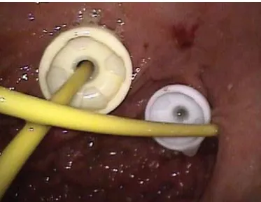
During hospitalization or after discharge, some patients develop stasis-associated symptoms, as relux symptoms, mainly regurgitation, or extra-tube leakage causing skin le-sions. To these patients, a second endoscopic gastrostomy is proposed, in order to drain the gastric residuum. This second gastrostomy may be performed in an outpatient basis, and the tube is placed a few centimetres apart from the gastro-jejunostomy using the same stomach area, already in close
contact with the abdominal wall (Figure 1). The drainage PEG tube (Figure 2) may be linked to a urine drainage bag or, more practical for some patients, to a skin level ileostomy bag. The ileostomy bag may be changed by the patient or caregiver. The patients kept gastrojejunostomy feeding as before and kept being followed as outpatients.
FIGURE 1. Endoscopic view, showing a recently placed PEG drainage tube side-by-side with a previous placed feeding PEG-J
FIGURE 2. Skin view, showing a recently placed PEG drainage tube side-by-side with a previous placed feeding PEG-J. On the PEG-J, the gastric lumen is sealed with a dressing to prevent any manipulation
RESULTS
Fonseca J, Santos CA.
Percutaneous endoscopic gastrostomy with jejunal extension plus percutaneous endoscopic gastrostomy (PEG-J plus PEG) in patients with gastric/duodenal cancer outlet obstruction
74 Arq Gastroenterol v. 52 no. 1 - jan./mar. 2015
intestinal tube to the stomach and replacement was possible in all of them but one, a patient with a large pancreatic cancer invading the duodenum. The four patients with longer sur-vival two gastric and two pancreatic malignancies developed symptoms associated with stasis and an important gastric residuum. All of them accepted to have a second drainage gastrostomy, which was performed without complications. Regurgitation disappeared and skin lesions associated with extra-tube leakage greatly improved. There were no incidents of jejunal tube dislocations after the second PEG procedure. Patients and/or caregivers reported us that this option was easier to use them the previous two-on-one feeding and drainage together. Patients were able to live in their homes with their families and keep some social activities outside home using the intervals between feeding bolus. Nutrition and hydration were provided through the PEG-J until death. One of our patients lived at home during 8 months, being able to leave home and meet his friends at the local coffee shop until the last month of life.
DISCUSSION
The use of a gastrojejunostomy as an access for post-py-loric long term enteral nutrition has been reported in several clinical settings. Most frequently it is used in PEG patients with clinical signiicant gastroesophageal relux and respiratory aspiration(2, 10). When relux and aspiration
cannot be controlled with postural procedures and phar-macotherapy, placing a jejunal tube through the PEG and creating a PEG-J is the most practical approach. Usually acute pancreatitis episodes subsides and resolves itself in a time gap too short to justify a long term intestinal feeding access, but PEG-J was occasionally used in this setting(7).
Although unnecessary in most patients, PEG-J has also been reported in chronic pancreatitis(5). Before 2007, PEG-J
was considered for nutrition of cancer patients only in the context of head and neck cancer, when patients displayed a high risk of respiratory aspiration(3). The use of PEG-J
in the setting of a digestive malignancy, in order to bypass the obstruction, was only reported recently(4). This lack
of reported experience in gastric or duodenal cancer ob-struction may have two causes. On the one hand, stenting is more comfortable, was proven to be superior to PEG-J for nutritional palliation of gastric or duodenal obstructing lesions, and is achievable in most cases(9). On the other hand,
there is always some concern about using special feeding procedures in patients with advanced cancer. But sometimes stenting is not achieved, there is a survival expectancy of several months, suitable social conditions, and families willing to take care of patients. In these selected patients, PEG-J palliation allows, not only feeding but also hydra-tion during a large period until death. Adding a drainage PEG to the PEG-J feeding system is more handleable for the patient and caregivers, reduces manipulation of the intestinal tube and controls symptoms related with stasis of a large gastric residuum.
CONCLUSIONS
In selected cases, when stenting is not possible in patients with gastric/duodenal outlet obstruction due to cancer grow-ing, a feeding PEG-J plus drainage PEG may be a suitable alternative for palliation purposes, allowing duodenal/jejunal feeding and gastric drainage with minimal manipulation of the PEG-J jejunal tube.
Author contribution
Fonseca J: PEG and PEG-J procedures, patients fol-low-up, article writing. Santos CA: Dietetic management of patients, patients follow-up, article writing.
Fonseca J, Santos CA. Gastrostomia endoscópica com extensão jejunal associada a gastrostomia endoscópica em doentes com obstrução de saída gástrica por neoplasias do estômago ou duodeno. Arq Gastroenterol. 2015,52(1):72-5.
RESUMO – Contexto – Para a obstrução gástrica/duodenal por câncer, o tratamento paliativo de referência é a colocação de prótese expansível. Quan-do tal não é possível, o Quan-doente pode ser paliaQuan-do usanQuan-do uma gastrojejunostomia (PEG-J), mas a deslocação Quan-do tubo jejunal é frequente, deviQuan-do a manipulação para alimentação e drenagem. A obstrução pode resultar em reluxo gastroesofágico e perda de líquido extra-tubo com lesão cutânea. Para evitar estas complicações é possível colocar uma segunda gastrostomia (PEG) dedicada à drenagem gástrica. Objectivo – O nosso objetivo foi avaliar a utilidade de uma segunda gastrostomia em doentes com câncer no estômago ou duodeno alimentados por PEG-J respeitante a: 1. Controlo do reluxo gastroesofágico e perda de líquido gástrico extra-tubo; 2. Prevenir a deslocação do tubo jejunal. Métodos – Avaliamos retrospetivamente doentes submetidos a PEG-J por impossibilidade de colocação de prótese para paliação de obstrução gástrica por câncer no estômago ou duode-no. Selecionamos os quatro que necessitaram de uma segunda PEG para drenagem gástrica, efetuada a alguns centímetros da PEG-J. Para maior conforto a PEG de drenagem pode ser ligada a um saco de jejunostomia. Resultados – Os quatro doentes oncológicos alimentados por PEG-J com maior sobrevida necessitaram de uma segunda PEG. Após a PEG de drenagem, os sintomas melhoraram ou desapareceram e, não houve qualquer deslocação do tubo jejunal. Conclusões – Quando não é possível a paliação por prótese de doentes com obstrução neoplásica do estômago ou duodeno, a paliação com uma PEG-J para alimentação e uma PEG para drenagem é uma alternativa útil, permitindo a manipulação mínima do tubo jejunal.
Fonseca J, Santos CA. Percutaneous endoscopic gastrostomy with jejunal extension plus percutaneous endoscopic gastrostomy (PEG-J plus PEG) in patients with gastric/duodenal cancer outlet obstruction
v. 52 no. 1 - jan./mar. 2015 Arq Gastroenterol 75
REFERENCES
1. Colasanto JM, Prasad P, Nash MA, Decker RH, Wilson LD. Nutritional support
of patients undergoing radiation therapy for head and neck cancer. Oncology (Williston Park). 2005;19:371-9.
2. DeLegge MH, Duckworth PF Jr, McHenry L Jr, Foxx-Orenstein A, Craig RM,
Kirby DF. Percutaneous endoscopic gastrojejunostomy: a dual center safety and eficacy trial. JPEN J Parenter Enteral Nutr. 1995;19:239-43.
3. Johnston TH, Fox SH, Brotchie JM. Advances in the delivery of treatments for
Parkinson’s disease. Expert Opin Drug Deliv. 2005;2:1059-73.
4. Lundqvist C, Nystedt T, Reiertsen O, Grotli R, Beiske AG. Continuous
treat-ment with levodopa of Parkinson disease (abstract). TidsskrNorLaegeforen. 2005;125:2638-40.
5. Nakahira S, Sugimoto K, Okamura S, Miki H, Nakata K, Suzuki R, et al. The
palliative role of percutaneous endoscopic gastrostomy with jejunal extension (PEG-J) in an advanced pancreatic cancer patient with duodenal stenosis-a case report (abstract). GanTo Kagaku Ryoho. 2007;34:2007-9.
6. Nilsson D, Nyholm D, Aquilonius SM. Duodenal levodopa infusion in Parkinson’s disease long-term experience. Acta Neurol Scand. 2001;104:343-8.
7. Simon T, Fink AS. Recent experience with percutaneous endoscopic gastrostomy/ jejunostomy (PEG/J) for enteral nutrition. Surg Endosc. 2000;14:436-8. 8. Stanga Z, Giger U, Marx A, DeLegge MH. Effect of jejunal long-term feeding in
chronic pancreatitis. JPEN J Parenter Enteral Nutr. 2005;29:12-20.
9. Strand DS, Thlick JE, Patrie JT, Gaidhane MR, Kahaleh M, Wang AY.
Gas-troduodenal stents are associated with more durable patency as compared to percutaneous endoscopic gastrojejunostomy in the palliation of malignant gastric outlet obstruction. J Interv Gastroenterol. 2012;2:150-4.
10. Yoder AJ, Parrish CR, Yeaton P. A retrospective review of the course of patients with pancreatitis discharged on jejunal feedings. Nutr Clin Pract. 2002;17:314-20.