AR
TIGO ORIGINAL / ORIGINAL AR
TICLE
INTRODUCTION
Inlammatory bowel diseases (IBD), speciically Crohn’s disease (CD) and ulcerative colitis (UC), are routinely associated with different degrees of malnutrition due to the effects on the gastrointestinal tract(21, 29).
However, evidence suggests the occurrence of a nutritional transition process in such individuals. Obesity, including severe/morbid obesity, has emerged as a growing problem in this population, bringing with it an increased risk for metabolic diseases, especially those of a cardiovascular origin(3, 14, 41).
While the mechanism for this event is not yet fully clariied, factors such as chronic inlammation, the type of intestinal microbiota, metabolic disorders secondary to the use of medication for controlling IBD, disease remission and physical activity may be
EXCESSIVE WEIGHT – MUSCLE DEPLETION
PARADOX AND CARDIOVASCULAR
RISK FACTORS IN OUTPATIENTS WITH
INFLAMMATORY BOWEL DISEASE
Maria Izabel Siqueira de
ANDRADE
, Regiane
MAIO
, Keila Fernandes
DOURADO
,
Patrícia Fortes Cavalcanti de
MACÊDO
and Augusto César
BARRETO NETO
ABSTRACT – Background – Evidence suggests a nutritional transition process in patients with inlammatory bowel disease. Obesity, which was once an uncommon occurrence in such patients, has grown in this population at the same prevalence rate as that found in the general population, bringing with it an increased risk of cardiovascular disease. Objective – The aim of the present study was to determine the nutritional status and occurrence of cardiovascular risk factors in patients with inlammatory bowel disease. Methods – A case-series cross-sectional study was conducted involving male and female adult outpatients with inlammatory bowel disease. Data were collected on demographic, socioeconomic, clinical and anthropometric variables as well as the following cardiovascular risk factors: sedentary lifestyle, excess weight, abdominal obesity, medications in use, comorbidities, alcohol intake and smoking habits. The signiicance level for all statistical tests was set to 5% (P < 0.05). Results – The sample comprised 80 patients with inlammatory bowel disease, 56 of whom (70.0%) had ulcerative colitis and 24 of whom (30.0%) had Crohn’s disease. Mean age was 40.3±11 years and the female genre accounted for 66.2% of the sample. High frequencies of excess weight (48.8%) and abdominal obesity (52.5%) were identiied based on the body mass index and waist circumference, respectively, in both groups, especially among those with ulcerative colitis. Muscle depletion was found in 52.5% of the sample based on arm muscle circumference, with greater depletion among patients with Crohn’s disease (P=0.008). The most frequent risk factors for cardiovascular disease were a sedentary lifestyle (83.8%), abdominal obesity (52.5%) and excess weight (48.8%). Conclusion – The results of the complete anthropometric evaluation draw one’s attention to a nutritional paradox, with high frequencies of both - muscle depletion, as well as excess weight and abdominal obesity.
HEADINGS – Inlammatory bowel disease. Overweight. Malnutrition. Risk factors. Cardiovascular disease.
Declared conflict of interest of all authors: none Disclosure of funding: no funding received
Hospital das Clínicas, Universidade Federal de Pernambuco – UFPE, Recife, PE, Brasil.
Correspondence: Maria Izabel Siqueira de Andrade. Rua Horácio de Barros, 71 – Matriz – CEP: 55612-330 – Vitória de Santo Antão, PE, Brasil. E-mail: izabelandradee@ hotmail.com
involved in the onset of overweight and obesity in patients with IBD(3, 10, 15, 30).
Indeed, such patients are prone to the develop-ment of cardiovascular disease (CVD) or at least associated risk factors. The impact of CVD among individuals with IBD is the same as that found in the general population and remains a common cause of death(32).
disease activity. Despite this evidence, the prevalence of the excessive weight in IBD patients is not possible to determine due to the lack of a patterning on the presentation of anthro-pometric data in Brazilian studies, turning into dificult the comparisons between studies due to methodological changes adopted by the authors(10, 30, 31, 35, 42).
The aim of the present study was to determine the nutri-tional status and occurrence of risk factors associated with the development of CVD in patients with IBD.
METHODS Study design and patients
A case-series, cross-sectional study was conducted at the IBD clinic of the Gastroenterology Sector of the Hospital pertaining to the Universidade Federal de Pernambuco (UFPE, Brazil) from July to September 2013. A convenience sample was formed by all patients treated at the clinic in the study period who met the eligibility criteria. The inclusion criteria were a diagnosis of IBD (during remission or active phase), adults patients, aged from 19 to 60 years, which was the chosen age group because of the standardization of anthropometric criteria (that are different of the others age groups), and either genre. The exclusion criteria were an inability to provide information and absence of an accom-panier capable of providing information, incapacity to be submitted to the anthropometric evaluation, water retention (edema or ascites), amputated limb, currently pregnant or any consumptive disease.
This study received approval from Human Research Ethics Committee of the Universidade Federal de Per-nambuco (Brazil) under process nº 512.534/CAAE: 13890913.2.0000.5208 in compliance with Resolution n°466/12 of the Brazilian National Health Board. All patients received clariications regarding the procedures, risks and beneits of the study and agreed to voluntary participation by signing a statement of informed consent.
Demographic, socioeconomic and clinical data
The demographic and socioeconomic variables were de-ined based on the criteria of the 2000 Census of the Brazilian Institute of Geography and Statistics(17). The variables were dichotomized in the following manner: ethnicity – Caucasian or non-Caucasian; employment status – economically active or inactive; schooling (≤8 or <8 years of study) and monthly household income (<1 or ≥1 minimum salary). The individu-als were individu-also classiied based on the Economic Classiication Criteria of Brazil established by the Brazilian Association of Research Firms(1). Based on the schooling of the head of the household and the number of rooms and consumer goods in the home, the economic classiications from highest to lowest are A1, A2, B1, B2, C1, C2, D and E. The sample was subdivided into high economic class (A1, A2 and B1) and low economic class (B2, C1, C2, D and E). Disease activity was assessed by the Physician Global Assessment (PGA) in both CD and UC patients during the clinic visit, based on the medical record. The medications used by the
patients were obtained according to reports of the patients during interview.
Evaluation of nutritional status
Weight and height were measured using the method
rec-ommended by Lohman(23). Weight was determined using a
digital scale (Filizola®) with a maximum capacity of 150 kg and precision of 100 g. Height was determined using a sta-diometer coupled to the scale with a capacity of 1.90 m and precision of 1 mm. Weight and height were used to calculate the BMI, which was classiied based on the guidelines estab-lished by the World Health Organization(45). The percentage of weight loss was determined using the following equation: weight loss (%) = (usual weight – current weight) x 100 ÷ usual weight. This variable was categorized based on the classiication proposed by Blackburn et al.(4). Usual weight was provided by the patient during the interview.
Muscle mass was evaluated based on arm circumference (AC), arm muscle circumference (AMC) and the adductor muscle of the thumb (AMT). Subcutaneous fat reserve was determined using the triceps skinfold (TSF). AC was
mea-sured following the method proposed by Kamimura(19). TSF
was measured with the aid of the LANGE® skinfold caliper
following the method proposed by Lohman(23). AC and TSF
were used to determine AMC: AMC = AC - p x [TSF ÷ 10](5). Three measures were taken of each variable for the calculation of the mean. For greater consistency, measures with a difference greater than 1 mm were excluded. The 50th percentile of the measures proposed by Frisancho(12) were used for the evaluation of the adequacy of AC, AMC and TSF, with malnutrition recorded in cases of patients with less than 90% adequacy. AMT was measured with the patient seated, arm lexed at approximately 90° with the hand relaxed and resting on the knee. The measure was taken using the LANGE® skinfold caliper with the exertion of continuous pressure of 10 g/mm² to pinch the adductor muscle in the vertex of an imaginary triangle formed by the extension of the thumb and foreinger. The reading was made on the dominant hand three times and the mean was used for the analysis(20). The cutoff point for the diagnosis of malnutrition using the AMT was 13.4 mm for both sexes and all age groups(6).
In addition, anthropometric measurements were also grouped considering the use of corticoids by the patients.
Evaluation of risk factors associated with cardiovascular disease
Risk factors for the development of CVD were deter-mined based on the 6th Brazilian Guidelines for Systemic
Arterial Hypertension(37) and Basic Care Handbook for
the Clinical Prevention of Cardiovascular, Cerebrovascular
and Kidney Diseases(7), which consider variables such as
lifestyle, comorbidities, excess weight (BMI≥25 Kg/m2) and abdominal obesity.
Alcohol-ism was recorded when the individual reported consuming more than 60g of alcohol per day in the seven days prior to the interview. Individuals were recorded as non-alcoholic when they abstained from drinking or only drank small amounts of alcohol(44). Individuals were considered either ex-alcoholics or non-drinkers when reporting not having consumed alcoholic beverages in the previous 12 months(2). The patients were classiied into four smoking categories: daily smokers (at least one cigarette per day in the previous month), occasional smokers (smoking on less than a daily basis), ex-smokers (individuals who quit smoking at least 1 month prior to the interview) and non-smokers (never smoked or smoked for less than 1 month)(46). For statistical purposes, these two variables were dichotomized, as follows: Alcoholism – alcoholic or non-alcoholic (non-drinker and ex-drinker); smoking – smoker (daily or occasional) or non-smoker (non-smoker and ex-smoker). Sedentarism was evaluated based on the determination of regular physical activity, which was considered adequate when practiced at least three times a week for 30 minutes per session, regard-less of the type and intensity of the exercise performed(36). The medications reported by the patients were divided into two categories: those with effects associated with cardiovas-cular risk (corticoids and immunosuppressants) and those without effects associated with cardiovascular risk (amin-osalicylates – sulfasalazine and mesalazine). Comorbidities were recorded when an individual had a clinical diagnosis or reported a comorbidity associated with the risk of CVD (systemic arterial hypertension, diabetes mellitus and/or dyslipidemia). Individuals without these conditions were described as free of comorbidities.
The measure of waist circumference (WC) was used to determine the risk of metabolic complications associated with obesity. The cutoff point for excessively large abdominal
circumference was based on the WHO(45) recommendation:
WC ≥ 94 cm for men and ≥ 80 cm for women. The diagnosis of abdominal obesity was determined by the waist to height (W:H) ratio in centimeters. The classiication was based on cutoff points reported in a Brazilian study by Pitanga & Lessa(28), which deined abdominal obesity as W:H ≥ 0.52 for men and ≥ 0.53 for women.
Statistical analysis
The databank was created with the aid of the Microsoft Excel program (2010). All statistical analyses were performed with the SPSS version 17.0 for Windows (SPSS Inc., Chicago, IL, USA). The data were presented in tables. The Kolmog-orov-Smirnov test was used to determine the normality of the continuous quantitative variables, which demonstrated normal distribution and were expressed as mean and stan-dard deviation. In the description of proportions, binomial distribution was approximated to normal distribution using the 95% conidence interval (95% CI) and signiicant differences were conirmed in the absence of overlap of the respective 95% CI. The unpaired Student’s t-test was used
for the analysis of means. The level of signiicance was set to 5% (P<0.05).
RESULTS
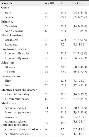
The sample comprised 80 patients with IBD, 70.0% of whom (n=56; 95% CI: 58.6-79.5) had a diagnosis of UC and 30.0% (n=24; 95% CI: 20.5-41.4) had a diagnosis of CD. Mean age was 40.3±11 years, with a range age between 19 to 59 years and frequency peaks of IBD at 26 and 42 years. A total of 2.5% (n=2; 95% CI: 0.7-8.7) were in disease activ-ity (one with diagnosis of UC and the other with CD) and 97.5% (n=78; 95% CI: 91.3-99.3) were in remission at the time of the interview. The female gender (66.2%), non-Cau-casian ethnicity (77.5%), residents of urban areas (92.5%), individuals with more than 8 years of schooling (70.0%), those from a low economic class (87.5%) and those with a monthly household ≥1 minimum salary (75.0%), using ami-nosalicylates (51.3%) predominated in the sample. Table 1
displays the demographic, socioeconomic andclinical data
on the population studied.
TABLE 1. Demographic, socioeconomic and clinic characterization of
outpatients with IBD, Recife, 2013
Variable n = 80 % 95% CI
Genre
Male 27 33.8 (24.3-44.6) Female 53 66.2 (55.4-75.6) Ethnicity
Caucasian 18 22.5 (14.7-32.8) Non-Caucasian 62 77.5 (67.2-85.2) Place of residence
Urban area 74 92.5 (83.8-96.9) Rural area 6 7.5 (3.1-16.2) Employment status
Economically active 42 52.5 (41.7-63.0) Economically inactive 38 47.5 (36.9-58.3) Schooling
≤8 years 24 30.0 (20.5-41.4) >8 years 56 70.0 (58.6-79.5) Economic class
High 10 12.5 (6.5-22.2) Low 70 87.5 (77.8-93.5) Monthly household income*
<1 minimum salary 20 25.0 (16.3-36.2) ≥1 minimum salary 60 75.0 (63.8-83.7) Medications
Aminosalicylates 41 51.3 (40.5-61.9) Immunosuppressants 17 21.3 (13.7-31.4) Corticoids 1 1.2 (0.2-6.7) Aminosalicylates+
Immunosuppressants 12 15.0
(8.8-24.4)
Aminosalicylates+ Corticoids 6 7.5 (3.5-15.4) All medications cited 3 3.7 (1.3-10.4)
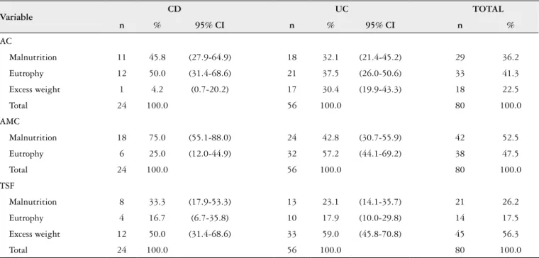
In the intra-group analysis of BMI, 1.8% (n=1; 95% CI: 0.3-9.4) of the patients with UC were malnourished, differing signiicantly from the patients in the other categories: 42.8%
(n=24; 95% CI:30.7-55.9) classiied as eutrophic and 55.4%
as overweight (n=31; 95% CI: 42.4-67.6). Non-signiicant differences in the frequency of BMI categories were found
among the patients with CD: 16.7% (n=4; 95% CI:6.7-35.8)
were malnourished, 50.0% (n=12; 95% CI:31.4-68.6) were
eutrophic and 33.3% were classiied as overweight (n=8; 95% CI:17.9-53.3) (data not presented in table). In the intergroup comparison, mean BMI was signiicantly higher among the
patients with UC (P=0.02) (Table 2). Only 4.2% (n=1; 95%
CI: 0.7-20.2) of the patients with CD and 5.4% (n=3; 95% CI: 1.8-14.6) of those with UC exhibited severe weight loss in the previous 6 months. The remaining portions of the
sample reported either maintaining their usual weight or gaining weight in the same period.
Based on the AC and AMC measures, the patients with CD had greater frequencies of malnutrition. TSF was used as a measure of subcutaneous fat and revealed a greater fre-quency of excess weight among the patients with UC (59.0%) (Table 3). In the intergroup comparison, the AC, AMC and TSF measures showed greater nutritional deicits among the patients with CD (P<0.05) (Table 3). In contrast, the
mea-sure of the AMT thickness classiied 100.0% (n=24; 95% CI: 86.2-100.0) of the patients with CD and 98.2% (n=54; 95%
CI:90.5-99.7) of those with UC as nutritionally adequate
(data not presented in table). No statistical difference was seen when comparing the nutritional status of the patients regarding the use of corticoids (Table 4).
TABLE .2. Anthropometric indicators among patients with CD and UC, Recife, 2013
Variable CD (n = 24) UC (n = 56) P-value*
BMI (Kg/m2) 23.5 ± 4.4 26.4 ± 5.2 0.020*
AC (% adequacy)I 90.1 ± 11.6 99.2 ± 15.3 0.011*
AMC (%adequacy)I 86.5 ± 7.6 93.3 ± 14.6 0.008*
TSF (%adequacy)I 108.0 ± 36.4 135.5 ± 61.0 0.044*
AMT (D) (mm) 21.2 ± 2.95 21.9 ± 3.8 0.407
WC (cm) 78.2 ± 8.5 85.9 ± 11.9 0.006*
W:H ratio (cm) 0.49 ± 0.07 0.53 ± 0.07 0.052
CD: Crohn’s disease; UC: ulcerative colitis; BMI: body mass index; AC: arm circumference; AMC: arm muscle circumference; TSF: triceps skinfold; AMT (D): adductor muscle of thumb on
dominant hand; WC: waist circumference; W:H: waist/height ratio. *Signiicant difference (P<0.05; Student’s t-test).
I % adequacy: the 50th percentile of the measures were used for the evaluation of the adequacy of AC, AMC and TSF, with malnutrition recorded in cases of patients with less than 90% adequacy.
TABLE 3. Frequency distribution of nutritional diagnoses according to anthropometric variables among outpatients with IBD, Recife, 2013
Variable
CD UC TOTAL
n % 95% CI n % 95% CI n %
AC
Malnutrition 11 45.8 (27.9-64.9) 18 32.1 (21.4-45.2) 29 36.2
Eutrophy 12 50.0 (31.4-68.6) 21 37.5 (26.0-50.6) 33 41.3
Excess weight 1 4.2 (0.7-20.2) 17 30.4 (19.9-43.3) 18 22.5
Total 24 100.0 56 100.0 80 100.0
AMC
Malnutrition 18 75.0 (55.1-88.0) 24 42.8 (30.7-55.9) 42 52.5
Eutrophy 6 25.0 (12.0-44.9) 32 57.2 (44.1-69.2) 38 47.5
Total 24 100.0 56 100.0 80 100.0
TSF
Malnutrition 8 33.3 (17.9-53.3) 13 23.1 (14.1-35.7) 21 26.2
Eutrophy 4 16.7 (6.7-35.8) 10 17.9 (10.0-29.8) 14 17.5
Excess weight 12 50.0 (31.4-68.6) 33 59.0 (45.8-70.8) 45 56.3
Total 24 100.0 56 100.0 80 100.0
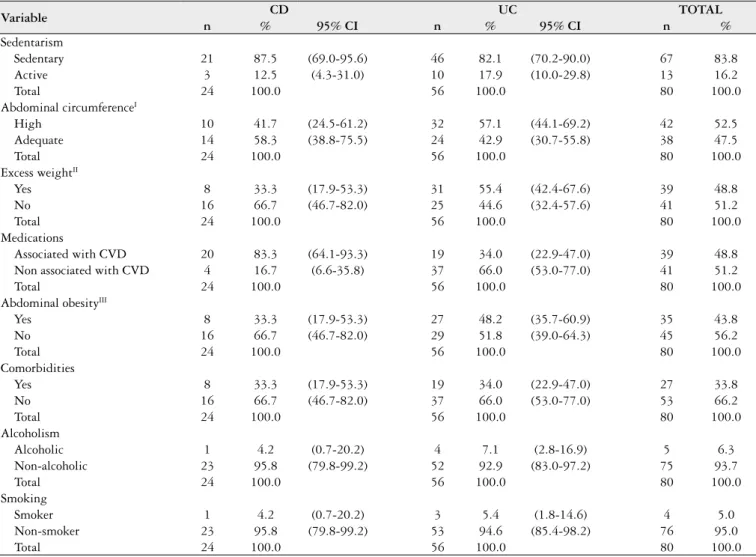
Table 5 displays the frequency distribution of risk factors for CVD. WC, BMI and W:H ratio were above the ideal ranges in 52.5% (95% CI:41.02-63.79), 48.8% (95% CI: 37.41-60.19) and 43.8% (95% CI:32.68-55.30) of the overall sample, respectively, with signiicant differences in the proportions between the patients with CD and those with UC.
Regarding lifestyle, the majority of the overall sample was classiied as non-alcoholic (95.8% of patients with CD and 92.9% of those with UC) and non-smoker (95.8% of patients with CD and 94.6% of those with UC). The major-ity of patients with CD (87.5%) and those with UC (82.1%) reported not performing physical activities and were therefore classiied as sedentary. With regard to comorbidities, the only signiicant difference was found in the patients with UC, with a greater frequency of the absence of comorbidities (66.0%). A signiicant difference between groups was found regarding the use of medications with and without effects associated with cardiovascular risk, as the majority of patients with CD (83.3%) used medications with associated risk, whereas 34.0% of those with UC made use of such medications.
TABLE 4. Anthropometric indicators among IBD patients with and
without use of corticosteroids, Recife, 2013
Variable
With corticosteroids
n=10
Without corticosteroids
n=70
P-value*
BMI (Kg/m2) 24.3 ± 4.25 25.7 ± 5.3 0.427
AC (% adequacy)I 98.4 ± 20.6 96.2 ± 13.9 0.745
AMC (%adequacy)I 93.4 ± 22.4 90.9 ± 11.5 0.736
TSF (%adequacy)I 123.2 ± 48.9 127.8 ± 57.3 0.810
AMT (D) (mm) 21.1 ± 2.73 21.8 ± 3.7 0.603
WC (cm) 80.5 ± 7.9 84.0 ± 11.9 0.372
W:H ratio (cm) 0.49 ± 0.07 0.52 ± 0.07 0.451
IBD: inlammatory bowel disease; BMI; body mass index; AC: arm circumference; AMC: arm muscle circumference; TSF: triceps skinfold; AMT (D): adductor muscle of thumb on dominant
hand; WC: waist circumference; W:H: waist/height ratio. *Student’s t-test.
I % adequacy: the 50th percentile of the measures were used for the evaluation of the adequacy of
AC, AMC and TSF, with malnutrition recorded in cases of patients with less than 90% adequacy.
TABLE 5. Frequency of risk factors for CVD among patients with CD and UC, Recife, 2013
Variable CD UC TOTAL
n % 95% CI n % 95% CI n %
Sedentarism
Sedentary 21 87.5 (69.0-95.6) 46 82.1 (70.2-90.0) 67 83.8 Active 3 12.5 (4.3-31.0) 10 17.9 (10.0-29.8) 13 16.2
Total 24 100.0 56 100.0 80 100.0
Abdominal circumferenceI
High 10 41.7 (24.5-61.2) 32 57.1 (44.1-69.2) 42 52.5 Adequate 14 58.3 (38.8-75.5) 24 42.9 (30.7-55.8) 38 47.5
Total 24 100.0 56 100.0 80 100.0
Excess weightII
Yes 8 33.3 (17.9-53.3) 31 55.4 (42.4-67.6) 39 48.8 No 16 66.7 (46.7-82.0) 25 44.6 (32.4-57.6) 41 51.2
Total 24 100.0 56 100.0 80 100.0
Medications
Associated with CVD 20 83.3 (64.1-93.3) 19 34.0 (22.9-47.0) 39 48.8 Non associated with CVD 4 16.7 (6.6-35.8) 37 66.0 (53.0-77.0) 41 51.2
Total 24 100.0 56 100.0 80 100.0
Abdominal obesityIII
Yes 8 33.3 (17.9-53.3) 27 48.2 (35.7-60.9) 35 43.8 No 16 66.7 (46.7-82.0) 29 51.8 (39.0-64.3) 45 56.2
Total 24 100.0 56 100.0 80 100.0
Comorbidities
Yes 8 33.3 (17.9-53.3) 19 34.0 (22.9-47.0) 27 33.8 No 16 66.7 (46.7-82.0) 37 66.0 (53.0-77.0) 53 66.2
Total 24 100.0 56 100.0 80 100.0
Alcoholism
Alcoholic 1 4.2 (0.7-20.2) 4 7.1 (2.8-16.9) 5 6.3 Non-alcoholic 23 95.8 (79.8-99.2) 52 92.9 (83.0-97.2) 75 93.7
Total 24 100.0 56 100.0 80 100.0
Smoking
Smoker 1 4.2 (0.7-20.2) 3 5.4 (1.8-14.6) 4 5.0
Non-smoker 23 95.8 (79.8-99.2) 53 94.6 (85.4-98.2) 76 95.0
Total 24 100.0 56 100.0 80 100.0
CVD: cardiovascular disease; CD: Crohn’s disease; UC: ulcerative colitis; CI: conidence interval.
DISCUSSION
Few studies have addressed IBD and risk factors asso-ciated with the development of CVD. The most current investigations have been directed toward the identiication of metabolic syndrome among individuals with IBD, with prevalence rates ranging from 7.1 to 17.7% among patients with CD and 16.0 to 29.5% among those with UC(25, 34, 47). However, the prevalence of metabolic syndrome varies widely depending on the criteria employed and the WC is one of the variables used to deine this condition. Obesity, especially abdominal obesity, is associated with coronary disease and death due to a cardiovascular cause. In the present sample of patients with IBD those nutritional abnormalities were frequent.
Data from the 3rd Health and Nutrition Study(16)
con-ducted in the state of Pernambuco, Brazil, where the present study was performed, reveal that overweight and obesity af-fected 68.5% of the population in 2006. However, few studies have addressed the magnitude of these conditions among outpatients with IBD in the state. Thus, the present indings are important, revealing high frequencies of excess weight (55.4% of patients with UC and 33.3% of those with CD) and an excessively large WC (57.1% of patients with UC and 41.7% of those with CD). In a study involving a comparison of individuals with IBD (n=489) and the general population in the state of Tayside, Scotland, 52.0% of patients with CD and 61.5% of those with UC exhibited overweight and obe-sity, which was similar to the 61.0% prevalence rate found in the general population(40). In a prospective case–control study (n=200), comprising 100 CD outpatients and 100 age-, sex- and socioeconomically-matched healthy controls, the
overall prevalence of overweight/obese (BMI≥25 kg/m2) in
CD was 40% compared with 52% of controls(26).
A number of factors have been associated with weight gain in patients with IBD, such as disease remission, with periods of the absence of symptoms leading to a diet with a greater energy density(30). In a more recent study by Nic-Suibhne et al.(26), there was a signiicant inverse association between current BMI and disease activity (CDAI) and with white blood cell count which suggest relected ‘wellness’ and well controlled disease in this outpatient setting. Silva et al.(35) found no difference in anthropometric measurements and body composition of patients whose inlammatory disease was in active phase or in remission. In the present study, just two patients were in active phase, thus the nutritional status was not evaluated regarding the disease activity.
The lower catabolism during remission secondary to the reduction in inlammation and smaller doses of medica-tions(11) may also be involved in the onset of obesity. This is especially true for corticoids, which alter nutritional status due to the increase in appetite and subsequent weight gain(10). However, BMI was not signiicantly associated with the need for corticosteroids in people with stable CD(26). In the pres-ent study, anthropometric indicators were not signiicantly different among IBD patients with and without use of cor-ticosteroids, probably because of the restricted number of
patients using this medication and in active phase of IBD. Studying ifty ive patients with IBD, Silva et al.(35) found that patients who used glucocorticoids in the 6 months preceding the assessment showed body fat percentage of 23.4±8.2%, while those who did not use, the percentage was 30.7±11.3. Jahnsen et al.(18) studying a total of 60 patients with CD, 60 patients with UC and 60 healthy subjects found that corticosteroid therapy had a negative impact on lean body mass in CD, without difference in body fat percentage. In other study, Mingrone et al.(24) compared 12 patients (6 men and 6 women) with biopsy-proven ileal CD with 11 healthy volunteers (6 men and 5 women). Five patients took no medi-cation and in seven patients was administered 29+/-18mg prednisone/d. Fat-free mass was not signiicantly different among groups, whereas fat mass was lower in patients than in control subjects. Thus, there are no conclusive studies investigating the effect of drug treatment on the nutritional status of patients with IBD.
Although the relationship between IBD and excess weight has not yet been fully clariied, a review of the literature per-formed by Bilski et al.(3) suggests that the pro-inlammatory cytokines found in obesity are the same as those found in IBD, especially tumor necrosis factor-alpha, which causes significant changes in the intestinal microbiota, with a consequent increase in intestinal permeability and the exa-cerbation of the inlammatory process, leading to changes in mesenteric fat and the intestinal mucosa, which act as a trigger and aggravating factor of fat tissue inlammation. In line with this event, NicSuibhne et al.(26) found that higher BMI was signiicantly associated with elevated C-reactive protein (CRP). CRP showed a signiicant but weak correla-tion with adiposity as determined by TSF and arm fat area (AFA), respectively. CRP is a marker linked with obesity in the general population and in those diagnosed with CD. However, the association between higher BMI and higher CRP occurs against a background of lower disease activity, lower white blood cell count, older age and more sedentary lifestyles in an outpatient setting. These authors suggest that the increasing rates of overweight and obesity in CD may begin to blur the lines between inlammatory and traditional risk factors for comorbid diseases in CD.
This study reports a high frequency of low levels of phys-ical activity among adults with CD and UC in an outpatient setting. Studying patients with stable CD NicSuibhne et al.(26) conirmed that BMI was positively associated with sedentary lifestyle, which may also be a predisposing factor to excess weight, as chronic IBD results in a limited capacity regarding activities of daily living and physical exercise. Moreover, in-suficient physical activity further provokes the buildup of fat tissue, leading to a low degree of cardiovascular and muscle itness, a poorer quality of life and even premature death(9).
transition among patients with IBD(14, 27). However, the fact that obesity has been increasing in this population may not mean that protein depletion is diminished over the time in patients with IBD. Thus, both nutritional disorders appear to coexist. Further studies are needed to conirm this inding.
Among the investigations addressing malnutrition in patients with IBD, Santos et al.(33) performed a retrospective analysis of the nutritional history of 45 outpatients with IBD and found higher malnutrition rates when using the AMC measure (47.4% among patients with CD and 36% among those with UC) in comparison to other indicators, which is in agreement with the present indings. Other studies employing the AMC measure found similar frequencies of muscle mass depletion independently of the phase of IBD (active or in remission) in both patients with CD and those with UC(35, 43). Studying 102 patients with IBD, Rocha et al.(31) found muscle depletion, as measures by arm muscle area, among patients in the active phase, which was also maintained during remis-sion. Muscle depletion may be explained by the exacerbated increase and action of pro-inlammatory cytokines, which directly affect muscles by stimulating proteolysis, with a con-sequent reduction in muscle mass that may be potentiated when associated with the chronic use of corticoids(8). The enteric loss of proteins during periods of active inlammation in both UC and CD as well as inadequate dietary intake constitute other factors that merit consideration(11).
Although the thickness of the AMT is also used for the evaluation of muscle depletion, this variable did not detect malnutrition in the present sample, as evidenced by the high prevalence rates of adequate nutrition in contrast to the rates determined when using AMC and AC. Data in the literature on the use of AMT for patients with IBD are extremely scarce. However, Urbano(42) evaluated the nutritional status of 59 outpatients with UC and found an inverse correlation between the AMT and the inlammatory proile, suggesting that a higher degree of inlammation is associated with a lower amount of lean muscle mass when evaluated using the AMT. Thus, the control of inlammation among the outpa-tients in the present study may explain the greater thickness of the AMT in the overall sample.
In the comparison between groups, the patients with UC had higher mean BMI, AC, AMC, TSF and WC, which is in agreement with data reported in the literature(13, 14, 27, 33). These indings demonstrate the preservation of nutritional status among such patients in comparison to those with CD. It is likely that the impairment of the small intestine in CD leads to a greater incidence of malnutrition and speciic nutrient deiciencies in comparison to UC(13).
Considering the high frequencies of both overweight/ obesity and skeletal muscle depletion, future studies should evaluate the joint impact of nutritional therapy to control excess weight and physical activity to minimize muscle deple-tion in patients with IBD. Physical activity has been suggested to be a protective factor against the onset of IBD. Despite the lack of consensus in the literature, physical exercise has
been associated to a reduction in symptoms and obesity may be associated with an increase in disease activity(3). However, gaps in knowledge on these issues remain.
The cross-sectional design can be considered a limitation of the present study. However, this design was chosen to describe the variables and distribution patterns due to its low cost and rapid execution. Moreover, the existence of a group for comparison purposes could allow extrapolating the indings. The use of nondiseased controls would be most desirable, but the present case series was useful for describing the characteristics of a local sample of patients with IBD, furnishing important information on the most common risk factors for the development of CVD. As such studies are scarce, further investigations into this line of research are needed.
The demographic and socioeconomic indings were in agreement with data reported in the literature showing a greater frequency of IBD among women, a bimodal age distribution occurring between 20 and 40 years, as well as greater frequencies among residents of urban areas, eco-nomically active individuals and those from lower economic classes(10, 35, 39). In a review of the literature, Loftus(22) suggests that the predominance of IBD in the female sex is due to the role of hormonal factors in the development of the disease. While the bimodal age distribution is in agreement previously published data, it is not known whether these differences are real or due to differences in the criteria used for the diagnosis of the disease. In a meta-analysis, Soon et al.(38) report that urban environments may increase the risk of developing IBD. This disease is known to affect economically active individuals more, which can then force individuals to go on leave from labor activities, thereby directly affecting income and economic class(10).
In conclusion, concomitant nutritional disorders were found in patients with IBD, along with cardiovascular risk factors, such as excess weight, abdominal obesity and a sed-entary lifestyle. Thus, factors related to lifestyle seem to be an important object of study in the multidisciplinary care required by patients with IBD. This study strengthens the need of a detailed nutritional evaluation of such patients with the investigation of a large number of indicators to establish the diagnosis of nutritional disorders and determine effective prevention and treatment strategies.
Author contribution
REFERENCES
1. ABEP – Associação Brasileira de Empresas de Pesquisa – 2012. Dados com
base no Levantamento Sócio Econômico 2010 – IBOPE. Available from: http:// www.abep.org/.
2. Bertolote JM. Secretaria Nacional de Políticas sobre Drogas. Glossário de álcool e drogas. 2Ed: Brasília; 2010.
3. Bilski J, Mazur-Bialy AI, Wierdak M, Brzozowski T. The impact of physical
activity and nutrition on inflammatory bowel disease: the potential role of cross talk between adipose tissue and skeletal muscle. J physiol pharmacol. 2013;64(2):143-55.
4. Blackburn GL, Bistrian BR, Maini BS, Schlamm HT, Smith MF. Nutritional
and metabolic assessment of the hospitalized patient. JPEN. 1977;1(1):11-22. 5. Blackburn GL, Thornton PA. Nutritional assessment of the hospitalized patients.
Med Clin North America. 1979;63(5):1103-15.
6. Bragagnolo R, Caporossi FS, Dock-Nascimento DB, Nascimento JESA.
Ad-ductor pollicis muscle thickness is a fast and reliable technique for nutritional assessment in surgical patients. JPEN. 2009;33:181-243.
7. Brasil. Ministério da Saúde. Secretaria de Atenção à Saúde. Departamento de
Atenção Básica. Prevenção clínica de doenças cardiovasculares, cerebrovasculares e renais/ Ministério da Saúde, Secretaria de Atenção à Saúde, Departamento de Atenção Básica: Brasília, Ministério da Saúde; 2006.
8. Cabral VLR, Carvalho L, Miszputen SJ. [Importance of serum albumin values in
nutritional assessment and inlammatory activity in patients with Crohn’s disease]. Arq Gastroenterol. 2001;38(2):104-8.
9. Coelho CF, Burini RC. Physical activity to prevent and treat non-communicable chronic diseases and functional disability. Rev Nutr. 2009;22(6):937-46. 10. Elia PP, Fogaça HS, Barros RGGR, Zaltman C, Elia CSC. [Descriptive analysis
of the social, clinical, laboratorial and anthropometric proiles of inlammatory bowel disease inwards patients from the “Clementino Fraga Filho” University Hospital, Rio de Janeiro, RJ, Brazil].Arq Gastroenterol. 2007;44(4):332-9. 11. Flora APL, Dichi I. Aspectos atuais na terapia nutricional da doença inlamatória
intestinal. Rev Bras Nutr Clín. 2006;21(2):131-7.
12. Frisancho AR. Anthropometric standards for the assessment of growth and nutritional status. University of Michigan, 1990. p. 189.
13. Goh J, O’Morain CA. Review article: nutrition and adult inlammatory bowel disease. Aliment Pharmacol Ther. 2003;17(3):307-20.
14. Harper JW, Sinanan MN, Zisman TL. Increased body mass index is associated with earlier time to loss of response to inliximab in patients with inlammatory bowel disease. Inlamm Bowel Dis. 2013;19(10):2118-24.
15. Hartmann C, Eliakim R, Shamir R. Nutritional status and nutritional therapy in inlammatory bowel diseases. World J Gastroenterology. 2009;15(21):2570-78. 16. III Pesquisa Estadual de Saúde e Nutrição: Saúde, Nutrição, Alimentação,
Condições Socioeconômicas e Atenção à Saúde no Estado de Pernambuco. Recife. 2006. [cited 2014 April 25]. Available from: http://pesnpe2006.blogspot.com/.
Andrade MIS, Maio R, Dourado KF, Macêdo PFC, Barreto-Neto AC. Paradoxo excesso de peso – depleção muscular e fatores de risco cardiovascular em pacientes ambulatoriais com doenças inlamatórias intestinais. Arq Gastroenterol. 2015,52(1):37-45.
RESUMO – Contexto – Evidências sugerem um processo de transição nutricional nos pacientes com doenças inlamatórias intestinais. A obesidade, que era um distúrbio incomum nas doenças inlamatórias intestinais, cresce em paralelo com a prevalência da obesidade em toda a população, podendo aumentar o risco de doenças cardiovasculares nesta população. Objetivo – Avaliar o estado nutricional e a ocorrência de fatores de risco cardiovas-cular em pacientes com doenças inlamatórias intestinais. Métodos – Estudo transversal, série de casos, realizado em pacientes ambulatoriais com doenças inlamatórias intestinais, adultos e de ambos os sexos. Foram obtidas variáveis demográicas, socioeconômicas, clínicas, antropométricas e os fatores de risco cardiovascular: sedentarismo, excesso de peso, obesidade abdominal, medicamentos, comorbidades, alcoolismo e tabagismo. O nível de signiicância foi de 5,0%. Resultados – A amostra foi constituída por 80 pacientes com doenças inlamatórias intestinais, 56 (70,0%) com etocolite ulcerativa inespecíica e 24 (30,0%) com doença de Crohn, sendo a idade média 40,3±11 anos e 66,2% do sexo feminino. Elevadas frequências de excesso de peso (48,8%) e obesidade abdominal (52,5%) foram identiicadas, segundo o índice de massa corporal e a circunferência da cintura, respectivamente, em ambos os grupos, e de forma mais importante na retocolite ulcerativa inespecíica. A depleção de massa muscular ocorreu em 52,5% dos pacientes segundo a circunferência muscular do braço, sendo mais depletados aqueles com (P=0,008). Dentre os fatores de risco para doenças cardiovasculares, os mais frequentes foram o sedentarismo (83,8%), o perímetro abdominal elevado (52,5%) e o excesso de peso (48,8%). Conclusão – A avaliação antropométrica completa mostrou elevada frequência de déicit proteico somático ao mesmo tempo em que se encontraram elevadas frequências de excesso de peso e obesidade abdominal.
DESCRITORES – Doenças inlamatórias intestinais. Sobrepeso. Desnutrição. Fatores de risco. Doenças cardiovasculares.
17. Instituto Brasileiro de Geograia e Estatística. Censo Demográico 2000. [Internet]. [cited 2014 April 25]. Available from: http://www.ibge.gov.br/home/presidencia/ noticias/20122002censo.shtm
18. Jahnsen J, Falch JA, Mowinckel P, Aadland E. Body composition in patients with inlammatory bowel disease: a population-based study. Am J Gastroenterol. 2003;98(7):1556-62.
19. Kamimura MA, Baxman A, Sampaio LR, Cuppari L. Avaliação nutricional. Guia de nutrição: nutrição clínica no adulto. 2Ed. São Paulo: Manole, 2006. p. 89-128. 20. Lameu EB, Gerude MF, Campos AC, Luiz RR. The thickness of the adductor
pollicis muscle relects the muscle compartment and may be used as a new an-thropometric parameter for nutritional assessment. Curr Opin Clin Nutr Metab Care. 2004;7(3):293-301.
21. Lochs H, Dejong C, Hammarqvist F, Hebuterne X, Leon-Sanz M, Schütz T, et al. ESPEN Guidelines on Enteral Nutrition: Gastroenterology. Clin Nutr. 2006;25:260-74.
22. Loftus Jr. EV. Clinical epidemiology of inlammatory bowel disease: Incidence, prevalence, and environmental inluences. Gastroenterology. 2004;126(6):1504-17. 23. Lohman TG, Roche AF, Martorell R. Anthropometric standardization reference
manual. Champaign: Human Kinetics Books, 1991.
24. Mingrone G, Benedetti G, Capristo E, De Gaetano A, Greco AV, Tataranni PA, et al. Twenty-four-hour energy balance in Crohn disease patients: metabolic implications of steroid treatment. Am J Clin Nutr. 1998;67(1):118-23. 25. Nagahori M, Hyun S, Totsuka T, Okamoto R, Kuwahara E, Takebayashi T et al.
Prevalence of metabolic syndrome is comparable between inlammatory bowel disease patients and the general population. J Gastroenterol. 2010;45(10):1008-13. 26. NicSuibhne T, Raftery TC, McMahon O, Walsh C, O’Morain C, O’Sullivan
M. High prevalence of overweight and obesity in adults with Crohn’s disease: associations with disease and lifestyle factors. J Crohns Colitis. 2013; 7(7):241–8. 27. Nguyen GC, Steinhart AH. Nationwide patterns of hospitalizations to centers
with high volume of admissions for inlammatory bowel disease and their impact on mortality. Inlamm Bowel Dis. 2008; 14:1688-94.
28. Pitanga FJG, Lessa I. Razão cintura-estatura como discriminador do risco coronariano de adultos. Rev Assoc Méd Bras. 2006;52(3):157-61.
29. Rajendran N, Kumar D. Role of diet in the management of inlammatory bowel disease. World J Gastroenterology. 2010;16(12):1442-1448.
30. Ripoli J, Miszputen SJ, Ambrogini Jr O, Carvalho L. Nutritional follow-up of patients with ulcerative colitis during periods of intestinal inlammatory activity and remission. Arq. Gastroenterol. 2010;47(1):49-55.
31. Rocha R, Santana GO, Almeida N, Lyra AC. Analysis of fat and muscle mass in patients with inlammatory bowel disease during remission and active phase. Br J Nutr. 2009;101:676-9.
32. Román AL, Muñoz F. Comorbidity in inlammatory bowel disease. World J Gastroenterology. 2011;17(22):2723-33.
34. Sezikli M, Cetinkaya ZA, Güzelbulut F, Yetkin DO. Metabolic Syndrome in Inlammatory Bowel disease: A real relationship or just a coincidence. Euroasian J Hepato-Gastroenterol. 2012;2(2):79-83.
35. Silva AF, Schieferdecker MEM, Rocco CS, Amarante HMBS. Relação entre estado nutricional e atividade inlamatória em pacientes com doença inlamatória intestinal. ABCD Arq Bras Cir Dig. 2010;23(3):154-8.
36. Sociedade Brasileira de Cardiologia. IV Diretriz Brasileira sobre Dislipidemias e Prevenção da Aterosclerose: Departamento de Aterosclerose da Sociedade Brasileira de Cardiologia. Arq Bras Cardiol. 2007;88(Suppl 1):2-19.
37. Sociedade Brasileira de Cardiologia; Sociedade Brasileira de Hipertensão; So-ciedade Brasileira de Nefrologia. VI Diretrizes Brasileiras de Hipertensão. Arq Bras Cardiol. 2010;95(Suppl 1):I-III.
38. Soon IS, Molodecky NA, Rabi DM, Ghali WA, Barkema HW, Kaplan GG. The relationship between urban environment and the inlammatory bowel diseases: a systematic review and meta-analysis. BMC Gastroenterol. 2012;12:51-64.
39. Souza MM, Barbosa DA, Espinosa MM, Belasco AGS.Qualidade de vida
de pacientes portadores de doença inlamatória intestinal. Acta Paul Enferm. 2011;24(4):479-84.
40. Steed H, Walsh S, Reynolds N. A brief report of the epidemiology of obesity in the inlammatory bowel disease population of Tayside, Scotland. Obes Facts. 2009;2(6):370-2.
41. Ungar B, Kopylov U, Goitein D, Lahat A, Bardan E, Avidan B, et al. Severe and morbid obesity in Crohn’s disease patients: prevalence and disease associations. Digestion 2013;88(1):26–32.
42. Urbano, APS. Avaliação do estado nutricional, ingestão de antioxidantes e extensão da lesão intestinal em pacientes portadores de colite ulcerativa. [Disser-tation]. São Paulo: Faculdade de Medicina de Botucatu, Universidade Estadual Paulista; 2011.
43. Valentini L, Schaper L, Buning C, Hengstermann S, Koernicke T, Tillinger W, et al. Malnutrition and impaired muscle strength in patients with Crohn’s disease and ulcerative colitis in remission. Nutrition. 2008;24(7-8):694-702.
44. World Health Organization. Department of Mental Health and Substance Abuse. Global status report on alcohol and health. Geneva: World Health Organization. 2011.
45. World Health Organization. Obesity: preventing and managing the global epidemic of obesity: report of a WHO consultation on obesity. Geneva: World Health Organization. 1998.
46. World Health Organization. Tobacco country profiles. 2nd ed. Proceedings of the 12th World Conference on Tobacco or Health. Geneva: World Health Organization. 2003.
47. Yorulmaz E, Adali G, Yorulmaz H, Ulasoglu C, Tasan G, Tuncer I. Metabolic syndrome frequency in inlammatory bowel diseases. Saudi J Gastroenterol. 2011;17(6):376-82.