LIKELIHOOD OF RETROGRADE DOUBLE-J STENTING ACCORDING
TO URETERAL OBSTRUCTING PATHOLOGY
ALEXANDRE DANILOVIC, IOANNIS M. ANTONOPOULOS, JOSE L. MESQUITA,
ANTONIO M. LUCON
Division of Urology, General Hospital, University of Sao Paulo Medical School, USP, Sao Paulo, Brazil
ABSTRACT
Objectives: To evaluate the likelihood of retrograde double-J stenting in urgent ureteral drain-age according to obstructing pathology.
Materials and Methods: From July 2002 to January 2003, 43 consecutive patients with ure-teral obstruction who needed urgent decompression were evaluated at our institution, where we per-formed a total of 47 procedures. Emergency was defined as ureteral obstruction associated with infec-tion, obstructive acute renal failure, or refractory pain. Ureteral obstruction was defined as intrinsic and extrinsic based on etiology and evaluated by ultrasound. Patients submitted to previous double-J stenting were excluded. Failures in retrograde ureteral stenting were treated with percutaneous nephrostomy. Results were analyzed with Fisher’s exact test and regression analysis.
Results: Failure in retrograde ureteral stenting occurred in 9% (2/22) and 52% (13/25) of the attempts in patients with intrinsic and extrinsic obstruction respectively (p < 0.001). Failures in stenting extrinsic obstructions occurred due to lack of identification of the ureteral meatus in 77% and impos-sibility of catheter progression in 23% (p < 0.05). All attempts of retrograde catheter insertion failed in obstructions caused by prostate or bladder pathologies (6/6). Inability to identify the ureteral me-atus was the cause of all failures.
Conclusion: Retrograde double-J stenting has a low probability of success in extrinsic ure-teral obstruction caused by prostate or bladder disease. Such cases might be best managed with percu-taneous nephrostomy.
Key words:ureter; obstruction; drainage; stents Int Braz J Urol. 2005; 31: 431-6
INTRODUCTION
Ureteral obstruction often presents as uro-logical urgency demanding surgical treatment with urinary diversion (1-5). The first successful endo-scopic ureteral drainage using a silicone catheter was reported by Zimskind et al. in 1967 (6). During the last decade, double-J stenting has been widely used by urologists. Despite endourological technical ad-vances, retrograde double-J stenting may be
double-J stenting in urgent ureteral drainage and to define criteria for selection of decompression method in order to reduce cost and to avoid time loss.
MATERIALS AND METHODS
Between July 2002 and January 2003, 43 consecutive patients with ureteral obstruction and need of urgent decompression were evaluated at our institution, where we performed a total of 47 proce-dures. The need for urgent decompression was de-fined as ureteral obstruction associated with infec-tion, obstructive acute renal failure, or refractory pain. All patients were evaluated with x-ray (KUB) and ultrasound in order to diagnose obstructive ur-opathy (10). Non-enhanced spiral CT was performed when standard evaluation was not satisfactory. Pa-tients submitted to previous retrograde double-J stenting were excluded.
Ureteral obstruction was classified accordingly to etiology as intrinsic (inside the ureteral lumen) or extrinsic (outside the ureteral lumen) (1,9,11,12).
All procedures were performed under general anesthesia, with fluoroscopic C-arm guidance (13). Retrograde pyelography was performed previously to each procedure when it was possible to identify the ureteral meatus. This was done using an open-ended ureteral catheter. This catheter was then used to pass a 0.35 mm hydrophilic guide wire (13). A non-hydrophilic polyurethane double-J ureteral catheter of various sizes (4,7;6;7 Fr) was used according to surgeon’s preference (14,15).
The adequate positioning of the double-J stent was confirmed by fluoroscopy at the end of the procedure. Failures in retrograde ureteral stenting were immediately treated with percutaneous nephrostomy. The percutaneous nephrostomy kit used was 14F/4.6 mm. The success of percutaneous nephrostomy placement was confirmed with antegrade pyelography after the procedure.
Statistical analysis was performed with Fisher’s exact test and regression analysis, with p < 0.05 considered significant.
RESULTS
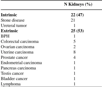
Intrinsic and extrinsic lesions were respon-sible for 47% and 53% of the obstructions respec-tively (Table-1).
Intrinsic (Table-2) and extrinsic (Table-3) groups were sex and age matched (Table-4).
The need for ureteral decompression differed between groups. The main indication for decompres-sion in the intrinsic group was pyelonephritis (77%) and in the extrinsic group it was acute renal failure (88%). The site of obstruction was preferentially dis-tal in extrinsic lesions, and proximal in intrinsic ones (84% vs. 41%, p < 0.001), and renal dilation was more pronounced in the extrinsic group (27% vs. 44%, p < 0.05).
The results show that retrograde ureteral stenting success was significantly lower in patients with extrinsic ureteral obstruction (Table-5).
Retrograde ureteral stenting failures in intrin-sic obstruction were caused by non-progression of the hydrophilic guide wire and by non-identification of the ureteral meatus (one case each). Failures in extrinsic obstruction were caused by non-progression of the hydrophilic guide wire in 3 patients (23%) and by non-identification of the ureteral meatus in 10 pa-tients (77%) (p < 0.05).
N Kidneys (%)
Intrinsic 22 (47) Stone disease 21
Ureteral tumor 1
Extrinsic 25 (53)
BPH 1
Colorectal carcinoma 5
Ovarian carcinoma 2
Uterine carcinoma 8
Prostate cancer 4
Endometrial carcinoma 1
Pancreas carcinoma 1
Testis cancer 1
Bladder cancer 1
Lymphoma 1
Table 1 – Specific causes of ureteral obstruction in 47
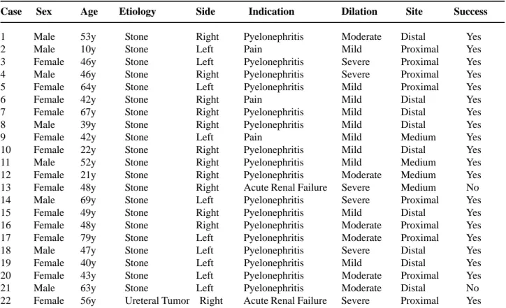
Table 2 – General data of the patients with intrinsic ureteral obstruction.
Case Sex Age Etiology Side Indication Dilation Site Success
1 Male 53y Stone Right Pyelonephritis Moderate Distal Yes
2 Male 10y Stone Left Pain Mild Proximal Yes
3 Female 46y Stone Left Pyelonephritis Severe Proximal Yes
4 Male 46y Stone Right Pyelonephritis Severe Proximal Yes
5 Female 64y Stone Left Pyelonephritis Mild Proximal Yes
6 Female 42y Stone Right Pain Mild Distal Yes
7 Female 67y Stone Right Pyelonephritis Mild Distal Yes
8 Male 39y Stone Right Pyelonephritis Mild Distal Yes
9 Female 42y Stone Left Pain Mild Medium Yes
10 Female 22y Stone Right Pyelonephritis Mild Distal Yes
11 Male 52y Stone Right Pyelonephritis Mild Medium Yes
12 Female 21y Stone Right Pyelonephritis Moderate Medium Yes
13 Female 48y Stone Right Acute Renal Failure Severe Medium No
14 Male 69y Stone Left Pyelonephritis Severe Proximal Yes
15 Female 49y Stone Right Pyelonephritis Mild Distal Yes
16 Female 48y Stone Right Pyelonephritis Moderate Proximal Yes
17 Female 79y Stone Left Pyelonephritis Moderate Proximal Yes
18 Male 47y Stone Left Pyelonephritis Severe Distal Yes
19 Female 40y Stone Left Pyelonephritis Mild Distal Yes
20 Female 43y Stone Left Pyelonephritis Moderate Proximal Yes
21 Male 63y Stone Left Pyelonephritis Moderate Distal No
22 Female 56y Ureteral Tumor Right Acute Renal Failure Severe Proximal Yes
All attempts of catheter insertion failed in obstructions caused by prostate or bladder patholo-gies (Table-6). Inability to identify the ureteral me-atus was the cause of all failures.
One retrograde double-J insertion became complicated with ureteral perforation distally to the extrinsic obstruction and was managed with percuta-neous nephrostomy. Follow-up was uneventful.
COMMENTS
The cornerstone for acute ureteral obstruc-tion treatment is ureteral decompression. The ideal method should be minimally invasive, fast, and inex-pensive. Currently, the most common methods in these situations are insertion of double-J catheter or place-ment of percutaneous nephrostomy. There is no con-sensus in the literature about which one is more ap-propriate, and usually the choice is left to the surgeon’s
Table 3 – General data of the patients with extrinsic ureteral obstruction.
Case Sex Age Etiology Indication Dilation Site Success
1 Male 84y BPH ARF Severe Distal No
2 Male 66y Prostate Tumor ARF Severe Distal No
3 Male 71y Prostate Tumor ARF Moderate Distal No
4 Male 56y Bladder Tumor ARF Severe Distal No
5 Female 30y Uterine Tumor ARF Moderate Distal No
6 Female 49y Lymphoma ARF Moderate Distal No
7 Male 75y Prostate Tumor ARF Severe Distal No
8 Male 75y Prostate Tumor ARF Moderate Distal No
9 Male 67y Colorectal Tumor ARF Moderate Distal No
10 Male 34y Testis Tumor ARF Severe Distal No
11 Female 78y Uterine Tumor ARF Severe Distal Yes
12 Female 78y Uterine Tumor ARF Severe Distal Yes
13 Female 41y Uterine Tumor ARF Moderate Distal Yes
14 Female 30y Uterine Tumor ARF Moderate Distal Yes
15 Female 57y Uterine Tumor ARF Severe Distal Yes
16 Female 57y Uterine Tumor ARF Moderate Distal Yes
17 Female 25y Ovarian Tumor ARF Mild Distal Yes
18 Female 52y Ovarian Tumor ARF Mild Distal Yes
19 Male 67y Colorectal Tumor ARF Moderate Distal Yes
20 Female 36y Uterine Tumor Pyelonephritis Severe Distal No
21 Female 46y Colorectal Tumor ARF Severe Distal No
22 Female 67y Pancreas Tumor ARF Mild Medium Yes
23 Female 64y Colorectal Tumor Pyelonephritis Moderate Medium Yes
24 Female 75y Endometrial Tumor Pyelonephritis Moderate Distal Yes
25 Female 35y Colorectal Tumor ARF Severe Distal No
ARF = acute renal failure, BPH = benign prostate hyperplasia
Table 5 – Success index of double-J insertion between
groups.
Intrinsic (%) Extrinsic (%) P value
Success 20 (81) 12 (48) < 0.001 Failure 2 (9) 13 (52)
of cases. Therefore, attempts of retrograde catheter insertion in patients with lower urinary tract condi-tions may be avoided, giving preference to percuta-neous nephrostomy.
Pearle and associates concluded that double-J catheter and percutaneous nephrostomy are equally good methods for ureteral decompression in
obstruc-Table 4 – Demographic data of intrinsic and extrinsic
obstruction cases.
Sex Intrinsic Extrinsic P value
Male 8 8 0.852
Female 14 13
Mean age (range) 47.5(10-79) 55 (30-84) 0.119
Table 6 – Success index of double-J insertion versus type
of disease causing extrinsic ureteral obstruction.
Prostate Other P value and Bladder (%) Tumors (%)
Success 0 12 (63.2) < 0.05
tive ureterolithiasis associated with infection (7). However, double-J catheters are prone to obstruct when used for long periods. Docimo & DeWolf re-ported a 30-day re-obstruction index up to 53% in extrinsic ureteral obstruction (9). Such problem may be adequately dealt with by simultaneous insertion of 2 double-J catheters in the obstructed ureteral unit (4,11,12).
The impact in quality of life caused by tem-porary urinary diversion was accessed by Joshi & colleagues and no functional or psychosocial dif-ference between double-J catheter and percutane-ous nephrostomy in ureteral decompression was found (5). Nevertheless, patients were followed for only 30 days. Possibly a longer follow-up may dis-close differences between both methods. Our im-pression is that an external prosthesis promotes pro-gressive loss of quality of life caused by more hos-pital visits due to nephrostomy displacement or in-fection.
The choice of ureteral drainage method should take cost into account. Both procedures are expensive as they are performed in the operating room under fluoroscopy. The double-J catheter used in the present study costs US$ 47 and the percutaneous nephrostomy kit costs US$ 88. As the double-J cath-eter ensures adequate urcath-eteral drainage, similar im-pact in quality of life and lower cost, it should be considered the preferential method for ureteral de-compression except for selected cases.
CONCLUSIONS
Retrograde double-J stenting has a low prob-ability of success in extrinsic ureteral obstruction caused by prostate or bladder disease. Such cases might be best managed with percutaneous nephrostomy.
REFERENCES
1. Yossepowitch O, Lifshitz DA, Dekel Y, Gross M, Keidar DM, Neuman M, et al.: Predicting the success of retrograde stenting for managing ureteral obstruc-tion. J Urol. 2001; 166: 1746-9.
2. Mokhmalji H, Braun PM, Portillo FJ, Siegsmund M, Alken P, Kohrmann KU: Percutaneous nephrostomy versus ureteral stents for diversion of hydronephrosis caused by stones: a prospective, randomized clinical trial. J Urol. 2000; 165: 1088-92.
3. Pappas P, Stravodimos KG, Mitropoulos D, Kontonopoulou C, Haramoglis S, Giannopoulou M, et al.: Role of percutaneous urinary diversion in ma-lignant and benign obstructive uropathy. J Endourol. 2000; 14: 401-5.
4. Watterson JD, Cadieux P, Denstedt JD: Ureteral stents: which, when, and why? AUA update series. 2002; vol. XXI, lesson 16, p. 122.
5. Joshi HB, Adams S, Obadeyi OO, Rao PN: Nephrostomy tube or “JJ” ureteric stent in ureteric obstruction: assessment of patient perspectives using quality-of-life survey and utility analysis. Eur Urol. 2001; 39: 695-701.
6. Zimskind PD, Kelter TR, Wilkerson SL: Clinical use of long-term indwelling silicone rubber ureteral splints inserted cystoscopically. J Urol. 1967; 97: 840-4.
7. Pearle MS, Pierce HL, Miller GL, Summa JA, Mutz JM, Petty BA, et al.: Optimal meted of urgent decom-pression of the collecting system for obstruction and infection due to ureteral calculi. J Urol. 1998; 160: 1260-4.
8. Park DS, Park JH, Lee YT: Percutaneous nephrostomy versus indwelling ureteral stents in patients with bilat-eral non-genitourinary malignant extrinsic obstruction. J Endourol. 2002; 16: 153-4.
9. Docimo SG, DeWolf WC: High failure rate of indwell-ing ureteral stents in patients with extrinsic obstruc-tion: experience at 2 institutions. J Urol. 1989; 142: 277-9.
10. Fernbach SK, Maizels M, Conway JJ: Ultrasound grad-ing of hydronephrosis: introduction to the system used by the Society for Fetal Urology. Pediatr Radiol. 1993; 23: 478-80.
11. Rotariu P, Yohannes P, Alexianu M, Rosner D, Lee BR, Lucan M, et al.: Management of malignant ex-trinsic compression of the ureter by simultaneous place-ment of two ipsilateral ureteral stents. J Endourol. 2001; 15: 979-83.
12. Liu JS, Hrebinko RL: The use of 2 ipsilateral ureteral stents for relief of ureteral obstruction from extrinsic compression. J Urol. 1998; 159: 179-81.
14. Candela JV, Bellman GC: Ureteral stents: impact of diameter and composition on patient symptoms. J Endourol. 1997; 11: 45-7.
15. Hübner WA, Plas EG, Stoller ML: The double-J ure-teral stent: in vivo and in vitro flow studies. J Urol. 1992; 148: 278-80.
Received: February 24, 2005 Accepted after revision: June 28, 2005
Correspondence address: Dr. Alexandre Danilovic Rua Alves Guimarães, 623 / 161 05419-001, São Paulo, SP, Brazil E-mail: [email protected]
EDITORIAL COMMENT
Double-J stenting has become an important endourological procedure in ureteral obstructive pa-thology. Successful stenting would reduce the mor-bidity of extrinsic ureteral obstruction. The authors reported a low success rate, especially in lower ure-teral obstruction due to bladder or prostate pathol-ogy. Deployment of metallic ureteral stents would be a solution for overcoming the obstruction in this situ-ation. Success would depend upon passing a guide wire. Failures in the retrograde approach can be over-come by antegrade stenting under ultrasound guided and fluoroscopic control. The upper tracts are usu-ally dilated and easy to puncture. The guide wire can be negotiated into the bladder by using an
angiogra-phy curved tip catheter. Once the guide wire is in the bladder it can be pulled outside the urethra by cys-toscopy. By pulling the guide wire in the opposite direction the curvatures can be straightened out, mak-ing it easy to dilate over which one can put either double-J stent or metallic stent.
In my experience, combining an antegrade and retrograde approach to ureteral obstruction suc-cess can be increased remarkably.
REFERENCE
1. Kulkarni R: Metallic ureteric stents: the current situ-ation. BJU Int. 2003; 92: 188-9.