www.rbo.org.br
0102-3616/$–see front matter © 2013 Sociedade Brasileira de Ortopedia e Traumatologia. Publicado pela Elsevier Editora Ltda. Todos os direitos reservados. Work performed in the Radiology and Orthopedics Services, Santa Casa da Misericórdia do Rio de Janeiro, Rio de Janeiro, RJ, Brazil *Corresponding author at: Av. Henrique Dodsworth 83/105, Copacabana, Rio de Janeiro, RJ. CEP: 22061-030
Email: [email protected] (R. Pires e Albuquerque).
Original Article
Comparative analysis between radiographic views for knee
osteoarthrosis (bipedal AP versus monopedal AP)
Rodrigo Pires e Albuquerque,
a,* Cristina Barbosa,b Dafne Melquíades,
bHilton Koch,
cJoão Maurício Barretto,
dAlexandre Albino and Waldeck Duarte Júnior
eaMSc and PhD in Medicine. Attending Physician in the Knee Group, Santa Casa de Misericórdia do Rio de Janeiro, Rio de Janeiro, RJ, Brazil
bPhysician in the Radiology Service, Santa Casa de Misericórdia do Rio de Janeiro, Rio de Janeiro, RJ, Brazil
cMSc and PhD in Medicine. Head of the Radiology Service, Santa Casa de Misericórdia do Rio de Janeiro, Rio de Janeiro, RJ, Brazil
dMSc and PhD in Medicine. Head of the Orthopedics Service, Santa Casa de Misericórdia do Rio de Janeiro, Rio de Janeiro, RJ, Brazil
ePhysician in the Orthopedics Service, Santa Casa de Misericórdia do Rio de Janeiro, Rio de Janeiro, RJ, Brazil
doi:
a b s t r a c t
Objective: A comparative analysis by applying the criteria of the original classification Ahlbäck in the anteroposterior (AP) bipedal knee in extension and anteroposterior (AP) monopodal knee in symptomatic knee arthrosis. With this analysis we intend to observe the agreement, any advantage or difference between the incidence and degree of joint involvement between the orthopedic surgeons and radiologists with the referring physician. Methods: From January 2012 to March 2012, was a prospective study of 60 symptomatic arthrosis knees (60 patients), clinically selected group of outpatient knee and radiographic proposals submitted to the search. Of the 60 patients, 39 were female and 21 male, mean age 64 years (ranging from 50 to 84 years). Of the 60 knees studied, 37 corresponded to the right side and 23 on the left side. Statistical analysis was performed by Kappa statistics, which evaluates the interobserver agreement for qualitative data. Results: According to the scale of Ahlbäck, there was a significant agreement (p < 0.0001) intra-observer in the classification of knee osteoarthritis among the five evaluators. There was a significant agreement (p < 0.0001) with inter-observer referring physician in the incidence of AP monopodal and AP bipedal for the four raters. Conclusion: The study found no difference between the incidence in the AP monopodal versus AP bipedal in osteoarthritis of the knee.
© 2013 Sociedade Brasileira de Ortopedia e Traumatologia. Published by Elsevier Editora Ltda. All rights reserved. A RT I C L E I N F O
Article history:
Received on April 27, 2012 Accepted on June 20, 2012
Palavras-chave: Estudo comparativo Osteoartrose do joelho Radiologia
r e s u m o
Objetivo: Fazer uma análise comparativa com a aplicação dos critérios da classificação original de Ahlbäck na incidência ântero-posterior (AP) bipodal do joelho em extensão e na incidência ântero-posterior (AP) monopodal do joelho, em joelhos artrósicos sintomáticos. Com esta análise pretendemos observar a concordância, diferença ou as vantagens eventuais entre as incidências e o grau de comprometimento articular entre os médicos ortopedistas e radiologistas com o médico de referência. Métodos: De janeiro de 2012 a março de 2012, foi feito um estudo prospectivo, de 60 joelhos artrósicos sintomáticos (60 pacientes), selecionados clinicamente no ambulatório do grupo de joelho e submetidos às incidências radiográficas propostas na pesquisa. Dos 60 pacientes, 39 eram do sexo feminino e 21 do masculino, com média de 64 anos (variando de 50 a 84). Dos 60 joelhos avaliados, 37 correspondiam ao lado direito e 23 ao esquerdo. A análise foi feita pela estatística de Kappa, que avalia a concordância interobservadores de dados de natureza qualitativa. Resultados: Segundo a escala de Ahlbäck, houve uma concordância significativa (p < 0,0001) intraobservador na classificação da osteoartrose do joelho entre os cinco avaliadores. Houve uma concordância significativa (p < 0,0001) interobservador com médico de referência na incidência em AP monopodal e AP bipodal para os quatro avaliadores. Conclusão: O estudo não observou diferença entre a incidência em AP bipodal versus o AP momopodal na osteoartrose do joelho.
© 2013 Sociedade Brasileira de Ortopedia e Traumatologia. Publicado pela Elsevier Editora Ltda. Todos os direitos reservados. Análise comparativa entre incidências radiográficas para a osteoartrose do joelho (AP bipodal versus AP monopodal)
Introduction
Physical examination and radiological examination are fundamental assessments for patients with knee osteoarthrosis. In radiological assessments on knee osteoarthrosis, the severity of joint impairment can be graded and the ligament instability or bone loss can be measured. The type of surgery and the implant needed can also be indicated. Even today, there is no consensus regarding standardization of radiological evaluations on patients with knee osteoarthrosis, in the worldwide literature.
The aim of the present study was to conduct a comparative analysis through applying the criteria of the original Ahlback classification1 in bipedal anteroposterior (AP) view of the
extended knee and in monopedal AP view of the extended knee, in symptomatic arthrotic knees. Through this analysis, we aimed to observe the concordance, possible advantages or differences between the views, and the degree of joint impairment between the orthopedic surgeons and radiologists and the reference physician.
Materials and methods
From January 2012 to March 2012, a prospective study was conducted on 60 symptomatic arthrotic knees (60 patients), which were selected clinically at the knee outpatient clinic of Santa Casa da Misericordia do Rio de Janeiro and were examined using the radiographic views proposed for this investigation.
The inclusion criteria for the patients was that they should be over the age of 50 years and present pain in the knee, together with a history and physical examination compatible
with osteoarthrosis, The patients needed to have never had previous knee surgery and be free from rheumatic pathological conditions. Among the 60 patients, 39 were female and 21 were male, with a mean age of 64 years (ranging from 50 to 84). We only evaluated the knee that was more painful. Thus, among the 60 knees evaluated, 37 were right and 23 were left knees.
The patients were taken to a preestablished radiological room and the Super 100R x-ray machine (Philips, Brazil) was used, with specifications of 50 kV and 31 mA. The patients were carefully positioned by the physician, aided by a radiology technician. The examination was assessed by the researchers with regard to image quality and was repeated if the technical quality was judged to be poor.
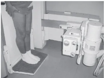
Two radiographic views were produced on each knee: View 1 (bipedal AP): an anteroposterior radiograph on the extended knee with bipedal weight-bearing. The tube-film distance was one meter, and the x-rays were centered on the lower pole of the patella (Fig. 1).
View 2 (monopedal AP): an anteroposterior radiograph on the extended knee with monopedal weight-bearing. The tube-film distance was one meter, and the x-rays were centered on the lower pole of the patella (Fig. 2).
The evaluation group was subdivided according to the degree of experience and was composed of five observers: two physicians who were residents in orthopedics (R3), two physicians who were residents in radiology (R3) and one physician who was a member of the Brazilian Society of Knee Surgery and had a doctorate. This last physician was taken to be the reference physician. The classifications obtained from view 1 (bipedal AP) and view 2 (monopedal AP) on each knee were compared to determine their concordance.
Measurements on the joint space were made manually by the five examiners. The assessment consisted of tracing out a vertical line from the extremity of the compartment evaluated (line A) and a second vertical line between the tibial spines (line B). The distance between the lines was measured (line C) and a third line was created at the midpoint between the two existing lines, parallel to them (line D). From line D, we ascertained the measurement of the joint space created between the convex surface of the femoral condyle and the upper margin of tibial plateau.
This was the location at which we graded the knee joint wear, using the Ahlback classification. This analysis was done using a single ruler graduated in millimeters that was supplied to the five examiners.2
In order to minimize the bias caused by interpretational difficulties or any possibility of forgetting the Ahlback classification, this was described on the response sheet handed out to each observer at the time of evaluating the radiographs, together with schematic drawings of the respective classifications. There was no limit to the amount of time taken for classifying the radiographs.
The data for the radiographic analyses were gathered blindly. In this, a physician colleague in the orthopedics and traumatology service who was the coordinator of the medical residence program gathered in the forms, typed up the interpretations from the five participants and sent the data to a statistician for review.
The analysis was done by means of kappa statistics,3 in
which the interobserver concordance regarding qualitative
Figure 4 - Monopedal AP radiograph with weight-bearing. Figure 3 - Bipedal AP radiograph with weight-bearing.
data was assessed. The hypothesis tested was whether the concordance represented by the letter p was zero, i.e. no interobserver concordance (Ho: p = 0 versus Ha: p ≠ 0).
If the hypothesis Ho was rejected, we would have evidence for believing that significant concordance existed between the observers. In the other hand, if we did not reject the hypothesis, there would then be no concordance between the evaluators. It is known that the values of kappa statistics can vary between negative values and 1,0, such that kappa equal to 1.0 expresses perfect concordance, while kappa close to zero expresses discordance, i.e. the concordance observed is no better than chance.
The criterion used for determining significance was the level of 1%, i.e. when the p value of the test was less than or equal to 0.01, significant concordance then existed.
Results
Table 1 presents the coefficients of concordance for kappa, their respective standard errors and descriptive levels (p values), and the percentage of the responses that were concordant in the intraobserver analysis.
In this study, it was observed that there was significant intraobserver concordance (p < 0.0001) in the classification of knee osteoarthrosis among the five evaluators.
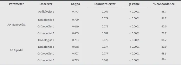
Although all the analyzes were significant, it was observed that radiologist 1 and the reference physician presented excellent intraobserver concordance (kappa ≥ 0.80). This showed that the monopedal AP technique was no significantly different from the bipedal AP technique. Table 2 presents the coefficients of concordance for kappa, their respective standard errors and descriptive levels (p values), and the percentage of the results that were concordant in the interobserver analysis in relation to the reference physician.
It was observed that there was significant interobserver concordance (p < 0.0001) with the reference physician, for both the monopedal AP and the bipedal AP technique, and for all four evaluators.
Although all the results were significant, it was observed that for the monopedal AP view, radiologists 1 and 2 presented higher concordance (kappa ≥ 0.70) than did orthopedists 1 and 2. For the bipedal AP view, the degree of concordance was equilibrated.
Parameter Observer Kappa Standard error p value % concordance
AP Monopedal
x Bipedal
Radiologist 1 0.819 0.066 < 0.0001 90.0
Radiologist 2 0.619 0.079 < 0.0001 76.7
Orthopedist 1 0.636 0.076 < 0.0001 73.3
Orthopedist 2 0.671 0.076 < 0.0001 78.3
Reference physician 0.800 0.066 < 0.0001 88.3
Source: Santa Casa, 2012.
Parameter Observer Kappa Standard error p value % concordance
AP Monopedal
Radiologist 1 0.773 0.069 < 0.0001 86.7
Radiologist 2 0.709 0.074 < 0.0001 81.7
Orthopedist 1 0.449 0.076 < 0.0001 60.0
Orthopedist 2 0.633 0.082 < 0.0001 76.7
AP Bipedal
Radiologist 1 0.754 0.075 < 0.0001 86.7
Radiologist 2 0.648 0.077 < 0.0001 80.0
Orthopedist 1 0.507 0.077 < 0.0001 68.3
Orthopedist 2 0.783 0.069 < 0.0001 86.7
Source: Santa Casa, 2012.
Table 1 - Intraobserver concordance for monopedal versus bipedal AP view.
Discussion
In our series of patients, females predominated over males. This is concordant with the concept that osteoarthrosis preferentially affects females.5
Galli et al.5 concluded that observers with difference
levels of experience gave rise to low levels of concordance when the Ahlback classification was used. Vilalta et al.6 found that experienced observers gave rise to individual variability thereby causing differences in the results and confusion in the literature. Gunther et al.7 observed that
especially in relation to evaluation of the knee joint space, the results from measurements were highly influenced by the evaluator’s experience. In a Brazilian study conducted by Albuquerque et al.,8 which compared three classifications
for knee osteoarthrosis, it was observed that the observer’s experience influenced the analysis on the reproducibility of the classification. Thus, we used observers with great experience in using these classifications, as well as not stipulating any limit on the time take to make responses, in an attempt to reproduce a more precise assessment.
In the worldwide literature, there is still no consensus regarding which classification to use in evaluating knee osteoarthrosis. Weidow et al.9 reported that the radiographic
classifications for the knee should be revised and improved by means of the examination technique used or the methods used. In orthopedic settings, the classification most used and best known is the Ahlback system.1 Rheumatologists prefer to use
the Kellgren and Lawrence classification,10 which emphasizes
the presence or absence of osteophytes and is performed in the supine position. We used the Ahlback classification because it is a system that evaluates the reduction of the joint space and is the best method for analyzing the progression of osteoarthrosis.11,12 Studies like that of Danielsson and
Hernborg13 demonstrated that osteophytes did not become
modified over the course of 16 years of evolution. On the other hand, Kijowski et al.14 concluded that knee osteoarthrosis
should be diagnosed by means of marginal osteophytes. The progression of the disease should be evaluated in terms of narrowing of the joint space, presence of subchondral sclerosis and presence of subchondral cysts.14 Felson et al.15 observed
that osteophytes present an association with poor alignment of the ipsilateral lower limb. Poor alignment is a potent risk factor for progression of the osteoarthrosis. We agree with the opinion of Danielsson and Hernborg,13 who validated use of
Ahlback assessments for knee arthrosis.
Alback’s original classification1 was modified in 1992 by Keyes et al.16 These authors included lateral-view
radiographs of the knee in the classification: these evaluate the integrity of the anterior cruciate ligament, by means of anterior subluxation of the tibia over the femur, along with wear of the posterior tibial plateau. Our investigation used Ahlback’s original classification, since our aim was to compare the bipedal AP view of the knee with the monopedal AP view.
Ravaud et al.2 found that rulers graduated in
mil-limeters produced reproducible measurements of the joint space in radiographic views of the knee.
Moreover, this method is simple and inexpensive. Our investi-gation agrees with the conclusions of Ravaud et al.2 and
reaf-firms that this is a simple method associated with low cost, which should always be borne in mind with public healthcare expenditure.
Vince et al.17 observed in a study in the United Kingdome
that there is still no consensus among British orthopedists regarding which view should be requested for assessing patients with knee osteoarthrosis, which demonstrates the importance of our investigation.
Bhatnagar et al.18 observed that 86% of British orthopedists
requested radiographs with weight-bearing, but that only 12% used a view with the knee flexed, which demonstrates the relevance of our investigation. The posteroanterior view with the knee flexed and bearing weight has been proven in several studies to be the best radiological examination for showing tibiofemoral arthrosis.19,20 We did not use this view because we
believe that it is likely to be difficult to reproduce and painful for patients with knee osteoarthrosis. However, some other authors did not observe any difficulty in producing radiographs with this view, with the knee flexed.21
Brandt et al.22 and Kijowski et al.23 conducted comparative
analyses on patients with osteoarthrosis, between the AP view with weight-bearing and the knee extended and the arthroscopic findings. They emphasized that in patients with osteoarthrosis, joint space evaluation and osteophytes are not appropriate parameters for analyzing this disease. They suggested that further research should be developed with the aim of discovering a complementary examination that would present higher accuracy. We believe that knee arthroscopy is an excellent therapeutic method, but it is an invasive procedure and should not be used as a diagnostic method.
The AP view with the knee extended is greatly used in clinical practice. However, the importance of applying weight needs to be emphasized.1,24 This aids in evaluating the
joint spaces, particularly in the central region of the knee, thereby differentiating whether there is ligament instability or whether this instability is associated with bone loss. Buckland-Wright25 considered that using the AP view with
weight-bearing for evaluating the degree of impairment of the joint space was obsolete. This author advocated that a view with the knee flexed should be used. Our study was composed of radiographic views of the knee in the bipedal and monopedal positions. Inoue et al.26 did not observe any
difference between bipedal AP and monopedal AP views of the knee for evaluating the alignment and measuring the joint space. Leach et al.27 reported that the monopedal AP view
could be used, although they preferred the bipedal AP view. In some of their patients, they observed that for the monopedal AP view to be produced, it was compensated with the toes touching on the side that was not evaluated. Boegard et al.28
conducted a comparative study on the PA view with the knee flexed, between bipedal and monopedal weight-bearing. According to these authors, the PA view with bipedal weight-bearing should be used, and the monopedal PA view with the knee flexed is indicated only when there is a need to analyze the lateral compartment. Specogna et al.29 demonstrated
evaluating patients with knee osteoarthrosis associated with varus deformity, and even recommended that bipedal weight-bearing should be used routinely for preoperative assessments. In our opinion, the view using monopedal weight-bearing is difficult to apply among elderly populations and is more associated with the risk of falling over (with fracturing), due to changes in balance and muscle strength.
Knee osteoarthrosis is a common and complex disease. There are many controversies regarding this topic: the radiographic analysis, the classification used, the method for measuring the joint space, use or nonuse of fluoroscopy, and the degree of angling of the knee. The present study suggests that this line of research on radiographic views of the knee remains unconcluded and open for further studies. Likewise, research is needed in order to create and develop a radiographic classification system for the knee in order to reach a consensus between the different medical specialties.
Conclusion
This study did not observe any difference between the bipedal and monopedal AP views in cases of knee osteoarthrosis.
Conflicts of interest
The authors declare no conflicts of interest.
R E F E R E N C E S
1. Ahlbäck S. Osteoarthrosis of the knee. A radiographic investigation. Acta Radiol Diagn (Stockh). 1968:Suppl 277:7-72. 2. Ravaud P, Chastang C, Auleley GR, Giraudeau B, Royant V,
Amor B, et al. Assessment of joint space width in patients with osteoarthritis of the knee: A comparison of 4 measuring instruments. J Rheumatol. 1996;23(10):1749-55.
3. Bartko JJ, Carpenter WT. On the methods and theory of reliability. J Nerv Ment Dis. 1976; 163(5):307-16.
4. Hernborg J, Nilsson BE. Age and sex incidence of osteophytes in the knee joint. Acta Orthop Scand. 1973;44(1):66-8.
5. Galli M, De Santis V, Tafuro L. Reliability of the Ahlbäck classification of knee osteoarthritis. Osteoarthritis Cartilage. 2003;11(8):580-4.
6. Vilalta C, Núñez M, Segur JM, Domingo A, Carbonell JA, Maculé F. Knee osteoarthritis: interpretation variability of radiological signs. Clin Rheumatol. 2004;23(6):501-4.
7. Günther KP, Sun Y. Reliability of radiographic assessment in hip and knee osteoarthritis. Osteoarthritis Cartilage. 1999;7(2):239-46. 8. Albuquerque RP, Giordano V, Sturm L, Azevedo V, Leão A, Amaral
NP. Análise da reprodutibilidade de três classificações para a osteoartrose do joelho. Rev Bras Ortop. 2008;43(8):329-35. 9. Weidow J, Cederlund CG, Ranstam J, Kärrholm J. Ahlbäck
grading of osteoarthritis of the knee: poor reproducibility and validity based on visual inspection of the joint. Acta Orthop. 2006;77(2):262-6.
10. Kellgren JH, Lawrence JS. Radiological assessment of osteo-arthrosis. Ann Rheum Dis. 1957;16(4):494-502.
11. Altman RD, Fries JF, Bloch DA, Carstens J, Cooke TD, Genant H, et al. Radiographic assessment of progression in osteoarthritis. Arthritis Rheum. 1987;30(11):1214-25.
12. Petersson IF, Boegård T, Saxne T, Silman AJ, Svensson B. Radiographic osteoarthritis of the knee classified by the Ahlbäck and Kellgren & Lawrence systems for the tibiofemoral joint in people aged 35-54 years with chronic knee pain. Ann Rheum Dis. 1997;56(8):493-6.
13. Danielsson L, Hernborg J. Clinical and roentgenologic study of knee joints with osteophytes. Clin Orthop Relat Res. 1970;69:302-12. 14. Kijowski R, Blankenbaker DG, Stanton PT, Fine JP, De
Smet AA. Radiographic findings of osteoarthritis versus arthroscopic findings of articular cartilage degeneration in the tibiofemoral joint. Radiology. 2006;239(3):818-24. 15. Felson DT, Gale DR, Elon Gale M, Niu J, Hunter DJ, Goggins
J, et al. Osteophytes and progression of knee osteoarthritis. Rheumatology (Oxford). 2005;44(1):100-4.
16. Keyes GW, Carr AJ, Miller RK, Goodfellow JW. The radiographic classification of medial gonarthrosis. Correlation with operation methods in 200 knees. Acta Orthop Scand. 1992;63(5):497-501.
17. Vince AS, Singhania AK, Glasgow MM. What knee X-rays do we need? A survey of orthopaedic surgeons in the United Kingdom. Knee. 2000;7(2):101-104.
18. Bhatnagar S, Carey-Smith R, Darrah C, Bhatnagar P, Glasgow MM. Evidence-based practice in the utilization of knee radiographs – A survey of all members of the British Orthopaedic Association. Int Orthop. 2006;30(5):409-11. 19. Rosenberg TD, Paulos LE, Parker RD, Coward DB, Scott SM.
The forty-five-degree posteroanterior flexion weight-bearing radiograph of the knee. J Bone Joint Surg Am. 1988;70(10):1479-83.
20. Mason RB, Horne JG. The posteroanterior 45 degrees flexion weight-bearing radiograph of the knee. J Arthroplasty. 1995;10(6):790-2.
21. Davies AP, Calder DA, Marshall T, Glasgow MM. Plain
radiography in the degenerate knee. A case for change. J Bone Joint Surg Br. 1999;81(4):632-5.
22. Brandt KD, Fife RS, Braunstein EM, Katz B. Radiographic grading of the severity of knee osteoarthritis: relation of the Kellgren and Lawrence grade to a grade based on joint space narrowing, and correlation with arthroscopic evidence of articular cartilage degeneration. Arthritis Rheum. 1991;34(11):1381-6.
23. Kijowski R, Blankenbaker D, Stanton P, Fine J, De Smet A. Arthroscopic validation of radiographic grading scales of osteoarthritis of the tibiofemoral joint. AJR Am J Roentgenol. 2006;187(3):794-9.
24. Walker PS, Hajek JV. The load-bearing area in the knee joint. J Biomech. 1972;5(6):581-9.
25. Buckland-Wright C. Which radiographic techniques should we use for research and clinical practice? Best Pract Res Clin Rheumatol. 2006;20(1):39-55.
26. Inoue S, Nagamine R, Miura H, Urabe K, Matsuda S, Sakaki K, et al. Anteroposterior weight-bearing radiography of the knee with both knees in semiflexion, using new equipment. J Orthop Sci. 2001;6(6):475-80.
27. Leach RE, Gregg T, Siber FJ. Weight-bearing radiography in osteoarthritis of the knee. Radiology. 1970;97(2):265-8. 28. Boegård T, Rudling O, Petersson IF, Sanfridsson J, Saxne T,
Svensson B, et al. Postero-anterior radiogram of the knee in weight-bearing and semiflexion. Comparison with MR imaging. Acta Radiol. 1997;38(6):1063-70.