07 artigo 457
ORIGINAL ARTICLE
1 – MSc in Surgical Clinical Medicine from the Federal University of Paraná (UFPR); Professor in the Specialization Course on Sports Traumatology and Arthroscopy,UFPR; Member of the Orthopedics and Traumatology Service, Hospital do Trabalhador, UFPR, Curitiba, PR, Brazil.
2 – PhD in Mechanical Engineering; Professor in the Academic Department of Mechanics, Federal Technological University of Paraná (UTFPR), Curitiba, PR, Brazil. 3 – MSc in Surgical Clinical Medicine from UFPR; Professor and Coordinator of the Specialization Course on Sports Traumatology,UFPR; Member of the Orthopedics and
Traumatology Service, UFPR, Curitiba, PR, Brazil.
4 – PhD in Surgical Clinical Medicine from UFPR; Professor in the Specialization Course on Sports Traumatology and Arthroscopy,UFPR; Member of the Orthopedics and Traumatology Service,UFPR, Curitiba, PR, Brazil.
5 – Physician and Professor in the Specialization Course on Sports Traumatology and Arthroscopy,UFPR; Member of the Orthopedics and Traumatology Service,UFPR, Curitiba, PR, Brazil.
6 – Physician andProfessor in the Specialization Course on Sports Traumatology and Arthroscopy,UFPR, Curitiba, PR, Brazil. 7 – PhD in Surgical Clinical Medicine. Adjunct Professor in the Department of Surgery, UFPR, Curitiba, PR, Brazil. Work performed inthe Orthopedics and Traumatology Service,Federal University of Paraná (SOT-UFPR).
Correspondence: Mauro Batista Albano, Rua Padre Dehon 1119 ap 410, V. Hauer, Curitiba, PR. Tel. (41) 3287- 4221 e-mail: [email protected] Received for publication: 11/24/2010, accepted for publication: 10/25/2011
bIOMECHANICAL STUDy OF TRANSCORTICAL OR
TRANSTRAbECULAR bONE FIXATION OF PATELLAR TENDON GRAFT
WITH bIOAbSORbAbLE PINS IN ACL RECONSTRUCTION IN SHEEP
Mauro Batista Albano1, Paulo César Borges2, Mario Massatomo Namba3, João Luiz Vieira da Silva4, Francisco de Assis Pereira Filho5, Edmar Stieven Filho6, Jorge Eduardo Fouto Matias7
The authors declare that there was no conflict of interest in conducting this work
This article is available online in Portuguese and English at the websites: www.rbo.org.br and www.scielo.br/rbort ABSTRACT
Objective: To determine the initial resistance of fixation using the Rigid Fix® system, and compare it with tradi-tional fixation methods using metal interference screws; and to evaluate the resistance of the fixation with the rigid fix system when the rotational position of the bone block is altered in the interior of the femoral tunnel. Methods: forty ovine knee specimens (stifle joints) were submitted to anterior cruciate ligament reconstruction (ACL) using a bone-tendon-bone graft. In twenty specimens, the Rigid Fix method was used; this group was subdivided into two groups: ten knees the pins transfixed only the spongious area of the bone block, and ten for fixation passing through the layer of cortical bone. In the twenty remaining speci-mens, the graft was fixed with 9mm metal interference
screws. Results: comparison of the RIGIDFIX® method
with the metal interference screw fixation method did not show any statistically significant differences in terms of maximum load and rigidity; also, there were no statisti-cally significant differences when the rotational position of the bone block was altered inside the femoral tunnel. For these evaluations, a level of significance of p < 0.017 was considered. Conclusion: fixation of the bone-tendon-bone graft with 2 bioabsorbable pines, regardless of the rotational position inside the femoral tunnel, gave a com-parable fixation in terms of initial resistance to the metal interference screw, in this experimental model.
Keywords - Anterior Cruciate Ligament; Bone-Patellar Tendon-Bone Graft; Knee; Sheep
INTRODUCTION
In reconstructing the anterior cruciate ligament, graft fixation is an extremely important factor. The fixation method has to be rigid and resistant to trac-tion forces so that postoperative rehabilitatrac-tion using current principles can be applied(¹).
Autografts are preferred because of their low com-plication rate. Among these, the ones most used in an-terior cruciate ligament reconstruction surgery are the
flexor tendons (semitendinosus and gracilis), bone-tendon-bone removed from the patella, the central third of the patellar tendon and the anterior tuberosi-ty of the tibia. Bone-tendon-bone grafts enable rigid fixation inside the tunnels. The fixation technique using interference screws is the one most used for this graft and is considered to be the gold standard for fixation(2-8).
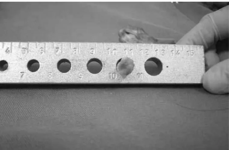
Figure 1 – Photograph of graft with bone block of 10 mm in diameter. reconstructions have been described. The commonest
of these are lesions to the graft at the bone-tendon transition at the time of femoral fixation, rupture of the posterior cortical bone and screw divergence in relation to the tunnel direction(9-14).
This has led to development of new fixation meth-ods with different materials, especially using implants made of bioabsorbable material.
Recently, a system using bioabsorbable pins of 2.7 mm in diameter by 42 mm in length for bone-tendon-bone fixation and 3.3 mm for quadruple semitendino-sus-gracilis grafts was developed. Among the advan-tages of the system the following have been reported: greater graft-bone block contact in the tunnel (100%); potentially less risk of rupture of the posterior cortical bone, and the possibility of graft fixation even if such situations occur; possibility of performing magnetic resonance imaging after the operation without signal interference; and, in cases of revision, the femoral tunnel will be closed and there will not be any need to remove the metal implant(15).
This method using special guides that position two implants parallel to each other and perpendicular to the orientation of the femoral tunnel and consequently to the graft.
Because this is a recent method, there are only a few studies demonstrating the resistance and rigidity of this fixation system.
The aims of the present study were to determine the initial fixation resistance of the system using transverse bioabsorbable pins and to compare this with the traditional fixation method for bone-tendon-bone grafts using 9 mm metal interference screws, and to evaluate the fixation resistance of this system when the rotational positioning of the bone block in-side the femoral tunnel is modified.
MATERIAL AND METHODS
Forty knee (stifle joint) specimens (18 right and 22 left) from skeletally immature sheep of ages ranging from 12 to 18 months were acquired from a sheep meat trading company. After the animals had been slaughtered for human consumption, the knees were separated during the deboning process, with preserva-tion of the femur and the proximal third of the tibia, together with the knee joint and extensor apparatus. The specimens were wrapped and preserved at -20°C until the date of the surgery. At the time of the pro-cedure, the specimens were defrosted in groups of
10, at room temperature, before performing the sur-gery. From all the knees, a patellar tendon graft of 1 cm in width with two bone blocks was harvested. The blocks removed from the patella were 2.8 mm in length by 10 mm in diameter (Figure 1).
The tibial block was of conical shape, with a width of 2 cm and a depth of 2 cm in the proximal portion, by 3 cm in length. An opening was made in the most proximal portion of the patellar bone block, with the purpose of positioning a no. 5 suturing thread for graft traction. The femur of each specimen was isolated to make a tunnel of 10 mm in diameter, positioned in the intercondylar space and oriented at one o’clock in the left knees and at 11 o’clock in the right knees, and preserving posterior cortical bone of 1 to 2 mm in thickness.
For the tunnels in 20 specimens, the RigidFix® guide was adapted. This consists of two nails joined together in a U shape. One of the guide nails was positioned inside the femoral tunnel and the other was external, with a device to adapt to two jackets, through which two openings were made such that they intercepted the tunnel perpendicularly, halfway along its diameter in the coronal plane (Figures 2A and 2B).
The jackets were left at the entrances to the ope-nings in the lateral cortical bone of the femur, to serve as guides for drilling and inserting two bioabsorbable pins of diameter 2.7 mm, through the bone blocks of the proximal end of the grafts.
45
Figure 2 – Photograph demonstrating the adaptation of the Ri-gidfix® guide: (A) front view; (B) side view.
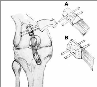
Figure 3 – Illustration demonstrating the two bone-block fixation methods using transverse pins.
A
B
GRAFT WITH BIOABSORBABLE PINS IN ACL RECONSTRUCTION IN SHEEP
length, inserted by means of Kirschner guidewires. The screws were positioned on the spongy bone, parallel to the orientation of the bone block inside the tunnel (Figure 4).
The specimens were identified regarding the fixa-tion method using adhesive tape on the proximal por-tion of the femur and then were wrapped for refreezing at -20ºC until 24 hours before performing the tests, when they were defrosted at room temperature.
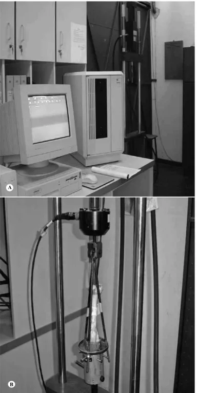
The tests were performed at the Federal Technologi-cal University of Paraná (UTFPr), on a MTS 810 univer-sal hydraulic machine. The specimens were adapted to the machine such that traction was performed longitudi-nally at a velocity of 50 mm/min (Figures 5A and 5B).
allow the fixation pins to transfix the bone plate, and this was used in ten specimens. The other method al-lowed the pins to transfix only the spongy part of the bone block, which was used in another 10 specimens (Figure 3).
The next step was to drill the graft inside the tun-nel, aided by the guide jackets and using special drills of diameter 2.7 mm, and to insert the pins using an impacting device and hammer. The specimens were duly identified by means of adhesive tape applied to the proximal portion of the femur, and then were wrapped and refrozen at -20ºC.
Another 20 knees were operated, taking the same care regarding defrosting, graft harvesting and cons-truction of the femoral tunnel, as described above. The graft was placed under traction inside the tunnel in the same manner, but the graft fixation was done by using interference screws made of cannulated steel,
Figures 5A and 5B – Photographs showing the composition of the MTS 810 hydraulic machine.
A
B
Figure 6 – Graph comparing the maximum load values between the different fixation groups: interference screw, fixation to cortical bone and fixation to spongy bone.
NEWTONS
MAXIMUM FORCE
Interference Cortical fixation
Spongy fixation
Min-Max
25%-75% Median STATISTICAL ANALYSIS
To assess the hypothesis that the variables pre-sented normal distribution, the Shapiro-Wilks test was used. For situations in which the maximum force did not meet the condition of normal distribution, the groups were compared in pairs by means of the non-parametric Mann-Whitney test.
For the rigidity variable, which met the condition
of normal distribution, the groups were compared in pairs by means of Student’s t test for independent samples, taking into consideration the homogeneity of the variance. The significance level used was p < 0.05, and this was corrected using the Bonferroni mul-tiple comparison test (values of p < 0.017 indicated statistical significance).
RESULTS
The mean traction resistance obtained in a single cy-cle in the groups of transverse bioabsorbable pins did not present statistically significant differences between each other or in relation to the control group with interference screws. Table 1 and Figure 6 show the mean values of the different groups and their variations, and Table 2 shows the p values of the comparisons made using the nonparametric Mann-Whitney method, taking p < 0.017. The mean rigidity results also did not show any sta-tistically significant differences between the different fixation methods, taking p < 0.017. The mean rigidity is presented in Table 3 and the p values for comparisons between the different groups are in Table 4.
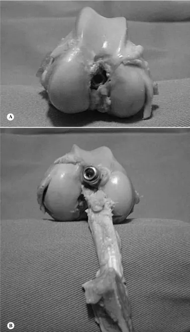
The types of failure and the place of occurrence in the specimens fixed with interference screws are shown in Table 5 and Figures 7A and 7B.
Table 1 – Comparison of mean maximum loads between the meth-ods of fixation using a metal interference screw and fixation using the RigidFix® technique to transfix the cortical and spongy bone.
Group N Mean Median Minimum Maximum Standard deviation
Interference 20 653.7 677 112 963 180.79 Pins/
cortical bone 10 607.5 598.5 278 940 198.7 Pins/
47
Figures 7A and 7B – Photographs of specimens showing lo-cations and most frequent types of failure found in the fixation method with interference screws.
A
B GRAFT WITH BIOABSORBABLE PINS IN ACL RECONSTRUCTION IN SHEEP
Table 5 – Types and locations of failure in the fixation method using interference screws.
Types Number of
oc-currences Percentage
Proximal detachment 10 50%
Distal detachment 2 10%
Loosening of screw and/or slippage
of bone block in femoral tunnel 8 40%
Total 20 100%
Tables 6 and 7 show the places of occurrence of fail-ures in the groups fixed with transverse pins.
Figures 8A and 8B show the types of failure found in the groups in which bioabsorbable transverse pins were used.
Tabela 7 – Tipos e local de falha no método de fixação com RigidFix®, fixação na esponjosa.
Tipos Número de
ocor-rências Porcentagem
Destacamento na
tot Proximal 1 10%
Destacamento na tot Distal 2 20%
Fratura do bloco ósseo l 5 50%
Falha do implante no
túnel femoral 1 10%
Rompimento do tendão
no 1/3 médio 1 10%
Total 10 100%
Table 2 -P values of comparisons made in the different groups.
Groups under comparison p* value
Interference vs. fixed in the rigid part 0.35
Interference vs. fixed in the spongy part 0.267
Fixed in the rigid part vs. fixed in the spongy part 0.123
Table 3 – Comparison of mean rigidity between the fixation meth-od using a metal interference screw and the fixation methmeth-od using pins transfixing to the cortical and spongy bone.
Group n Mean Median Minimum Maximum Standard deviation
Interference
screw 20 76.6 72.5 53 106 15.13
Pin to cortical
bone 10 65.8 64.5 38 103 22.19
Pin to spongy
bone 10 74.4 78 44 104 18.61
Table 4 – Comparison of p values for assessing rigidity between the fixation method using a metal interference screw and the fixation method using transverse pins to the cortical bone in one groups and spongy bone in the other group.
Groups under comparison p* value
Interference vs fixed in rigid part 0.127
Interference vs fixed in spongy part 0.73
Fixed in rigid part vs fixed in spongy part 0.36
Table 6 – Types and locations of failure in the fixation method using bioabsorbable pins fixed to the cortical bone.
Types Number of occurrences Percentage
Proximal detachment 0
Distal detachment 2 20%
Fracture of bone block 6 60%
Failure of implant in femoral
tunnel 2 20%
Figures 8A and 8B – Photograph showing types of failure in the method with bioabsorbable pins.
A
B
DISCUSSION
In the literature, there are few studies evaluating this method experimentally(16-18) and none that pre-sent or discard the possibility of fixation of a bone--tendon-bone graft by means of the spongy portion of the bone block. Based on this point, we decided to evaluate the method that uses 2.7 mm bioabsorbable pins, with two different types of fixation relating to graft rotation inside the tunnel, and to compare these two types with the method that uses 9 mm metal in-terference screws.
The initial hypothesis in subdividing the group in which the bioabsorbable pins were used was that fixation of implants by means of the cortical face of the bone block of the graft inside the femoral tunnel would be more resistant to traction forces than would
passage of the pins solely into the spongy bone. In doing the tests, no statistically significant difference was found, taking p < 0.017 between the two subgroups.
Passage of the 2.7 mm pin through the cortical bone caused fragility in this more resistant portion of the graft, and therefore the resistance depended al-most exclusively on the resistance of the two columns parallel to the more distal hole of the graft. This is the commonest site for fractures in this type of fixation (60% in this experiment).
In the group fixed with pins in the spongy bone, the tendon structure continued to be in line with the bone plate and therefore the traction forces were transmit-ted through the cortical bone. The more resistant bone plate remained intact, without holes for the pins to pass through, thus allowing transmission of the trac-tion forces to the proximal pin. The resistance of the fixation depended on the density of the spongy bone and its adherence to the cortical bone, and the resis-tance to traction may have come from the sum of the two pins, thereby partially compensating for the lower resistance of this portion of the graft.
In the present study, blocks of 10 mm in diameter were used, taking into consideration the work per-formed by Zantop et al(17), who used the same experi-mental model and observed that the diameter of the bone block was an important factor in the resistance of the fixation when using transfixing pins. These authors recommended against using blocks smaller than 9 mm in diameter.
The absence of soft tissues in the experimental model made it easier to check the positioning of the transverse holes created for passing the bioabsorbable pins into the femoral tunnel. This did not add any technical difference in carrying out the experimen-tal surgery, given that in humans, this observation method is frequently used with the aid of a camera placed by means of the tibial tunnel(19).
Placement of the specimen at an angle in order to align the direction of traction produced by the ma-chine with the orientation of the tunnel leads to the be-lief that, both in the case of the screw and in the case of the bioabsorbable implant, the resistance could be greater given that the axis of the forces acting on the knee under normal conditions of movement act tan-gentially to the joint line(8).
Sheep were used as the experimental model be-cause of the similarity of the anatomical properties and because they have been accepted as an experi-mental model in evaluating reconstructions of the anterior cruciate ligament(20-27).
49 GRAFT WITH BIOABSORBABLE PINS IN ACL RECONSTRUCTION IN SHEEP
to any modification of the technique for inserting bio-absorbable implants for fixation of bone-tendon-bone grafts. There will certainly be a need to conduct fur-ther tests on cyclical traction, in order to demonstrate whether this technical variant would be sufficiently secure in cases of necessity.
CONCLUSION
Comparison of the method using 2.7 mm bio-absorbable pins for fixation of a bone-tendon-bone graft transversely inside the femoral tunnel with the fixation method using an interference screw did not shown any statistically significant difference with
regard to maximum load and rigidity for the initial fixation in a single traction cycle.
With this experimental model, similar resistance was obtained for bone block fixations in the femoral tunnel using bioabsorbable pins transfixed solely in spongy bone and using pins transfixing the cortical bone plate.
Acknowledgement
The authors express their gratitude to Dr Carlos Hen-rique Ramos, who drew the illustration for Figure 3, demonstrating the two types of bone block fixation using transverse pins.
REFERENCES
1. Shelbourne KD, Nitz P. Accelerated rehabilitation after anterior cruciate ligament reconstruction. Am J Sports Med. 1990;18(3):292-9.
2. Brand J Jr, Weiler A, Caborn DN, Brown CH Jr, Johnson DL. Graft fixation in cruciate ligament reconstruction. Am J Sports Med. 2000;28(5):761-74.
3. Snyder-Mackler L, Delitto A, Bailey SL, Stralka SW. Strength of the quadríceps femoris muscle and functional recovery after reconstruction of the anterior cruci-ate ligament. A prospective, randomized clinical trial of electrical stimulation. J Bone Joint Surg Am. 1995;77(8):1166-73.
4. Arnoczky SP, Tarvin GB, Marshall JL. Anterior cruciate ligament replacement using patellar tendon. An evaluation of graft revascularization in the dog. J Bone Joint Surg Am. 1982;64(2):217-24.
5. Noyes FR, Butler DL, Grood ES, Zernicke RF, Hefzy MS. Biomechanical analy-sis of human ligament grafts used in knee-ligament repairs and reconstructions. J Bone Joint Surg Am. 1984;66(3):344-52.
6. Kurosaka M, Yoshiya S, Andrish JT. A biomechanical comparison of different surgical techniques of graft fixation in anterior cruciate ligament reconstruction. Am J Sports Med. 1987;15(3):225-9.
7. Abate JA, Fadale PD, Hulstyn MJ, Walsh WR. Initial fixation strength of poly-lactic acid interference screws in anterior cruciate ligament reconstruction. Arthroscopy. 1998;14(3):278-84.
8. Fu FH, Bennett CH, Lattermann C, Ma CB. Current trends in anterior cruciate ligament reconstruction. Part 1: Biology and biomechanics of reconstruction. Am J Sports Med. 1999 ;27(6):821-30.
9. Brown CH Jr, Carson EW. Revision anterior cruciate ligament surgery. Clin Sports Med. 1999;18(1):109-71.
10. Kousa P, Järvinen TL, Kannus P, Ahvenjärvi P, Kaikkonen A, Järvinen M. A bioabsorbable plug in bone-tendon-bone reconstruction of the anterior cruciate ligament: Introduction of a novel fixation technique. Arthroscopy. 2001;17(2):144-50.
11. Matthews LS, Soffer SR. Pitfalls in the use of interference screws for anterior cruciate ligament reconstruction: brief report. Arthroscopy. 1989;5(3):225-6.
12. Allum R. Complications of arthroscopic reconstruction of the anterior cruciate ligament. J Bone Joint Surg Br. 2003 Jan;85(1):12-6.
13. Azar FM, Arthur ST. Complications of anterior cruciate ligament reconstruction. Techn Knee Surg. 2004;3:238–50.
14. Pierz K, Baltz M, Fulkerson J. The effect of Kurosaka screw divergence on the holding strength of bone-tendon-bone grafts. Am J Sports Med. 1995;23(3):332-5.
15. Johnson & Johnson Gateway. RIGIDFIX®: ACL Cross Pin System. Disponível em: www.jnjgateway.com
16. Weimann A, Zantop T, Rümmler M, Hassenpflug J, Petersen W. Primary stability
of bone-patellar tendon-bone graft fixation with biodegradable pins. Arthroscopy. 2003;19(10):1097-102.
17. Zantop T, Ruemmler M, Welbers B, Langer M, Weimann A, Petersen W. Cyclic loading comparison between biodegradable interference screw fixation and biodegradable double cross-pin fixation of human bone-patellar tendon-bone grafts. Arthroscopy. 2005;21(8):934-41.
18. Milano G, Mulas PD, Ziranu F, Deriu L, Fabbriciani C. Comparison of femoral fixation methods for anterior cruciate ligament reconstruction with patellar ten-don graft: a mechanical analysis in porcine knees. Knee Surg Sports Traumatol Arthrosc. 2007;15(6):733-8.
19. Chandratreya AP, Aldridge MJ. Top tips for RIGIDfix femoral fixation. Arthros-copy. 2004;20(6):e59-61.
20. Zantop T, Weimann A, Wolle K, Musahl V, Langer M, Petersen W. Initial and 6 weeks postoperative structural properties of soft tissue anterior cruciate liga-ment reconstructions with cross-pin or interference screw fixation: an in vivo study in sheep. Arthroscopy. 2007;23(1):14-20.
21. Jackson DW, Grood ES, Arnoczky SP, Butler DL, Simon TM. Cruciate recon-struction using freeze dried anterior cruciate ligament allograft and a ligament augmentation device (LAD). An experimental study in a goat model. Am J Sports Med. 1987;15(6):528-38.
22. Schindhelm K, Rogers GJ, Milthorpe BK, Hall PJ, Howlett CR, Sekel R, et al. Autograft and Leeds-Keio reconstructions of the ovine anterior cruciate liga-ment. Clin Orthop Relat Res. 1991;(267):278-93.
23. Drez DJ Jr, DeLee J, Holden JP, Arnoczky S, Noyes FR, Roberts TS. An-terior cruciate ligament reconstruction using bone-patellar tendon-bone al-lografts. A biological and biomechanical evaluation in goats. Am J Sports Med. 1991;19(3):256-63.
24. Jackson DW, Grood ES, Goldstein JD, Rosen MA, Kurzweil PR, Cummings JF, et al. comparison of patellar tendon autograft and allograft used for an-terior cruciate ligament reconstruction in the goat model. Am J Sports Med. 1993;21(2):176-85.
25. Walton M. Absorbable and metal interference screws: comparison of graft security during healing. Arthroscopy. 1999;15(8):818-26.
26. Boszotta H, Anderl W. A primary stability with tibial press-fit fixation of pa-tellar ligament graft: a experimental study in ovine knees. Arthroscopy. 2001;17(9):963-970.