SOCIEDADE BRASILEIRA DE ORTOPEDIA E TRAUMATOLOGIA
w w w . r b o . o r g . b r
Original
Article
Conventional
plate
and
screws
in
medial
opening-wedge
high
tibial
osteotomy:
are
they
sufficiently
stable?
A
retrospective
study
夽
Rodrigo
Salim
∗,
Fabricio
Fogagnolo,
Mauricio
Martins
Perina,
Ugo
Messas
Rubio,
Mauricio
Kfuri
Junior
UniversidadedeSãoPaulo,FaculdadedeMedicina,HospitaldasClínicas,RibeirãoPreto,SP,Brazil
a
r
t
i
c
l
e
i
n
f
o
Articlehistory: Received27June2016 Accepted9September2016 Availableonline3January2017
Keywords:
Retrospectivestudy Osteotomy Knee Osteoarthritis
a
b
s
t
r
a
c
t
Objective:Opening-wedgeosteotomyoftheproximaltibiaisawidelyperformedprocedure fortreatingmedialgonarthrosisinactivepatientsandinthepresenceofvarusmalalignment ofthelowerlimb.Thefixationmethodiscontroversial,andtheuseofconventionalimplants hasbeenabandonedinfavorofimplantswithmoremodernlockingscrews.Theaimofthe presentclinicalstudywastoassessthemaintenanceofthecorrectionachievedincases whereinfixationwasperformedusingconventionalimplants.
Methods:Thisretrospectivestudyincluded51patientswhounderwentopening-wedgehigh tibialosteotomywhereinfixationwasperformedusingconventionalimplants(4.5-mmDCP plateandnon-lockingscrews).Radiologicalfindingsregardingpatellarheight,tibialslope, andvaruscorrectionpostoperativelyandafterconsolidationwereanalyzedtoassessthe maintenanceofthecorrectionachievedbyosteotomy.
Results:Themeanlossofcorrectionangle,calculatedbythedifferencebetweenthe cor-rection anglein theimmediatepostoperative period andthatafter consolidation,was 0.92◦±0.9◦.In addition, changes in patellar height determinedby the Blackburne–Peel
method andinthesagittal slopeofthe tibialplateauwerenot significant orclinically relevant.
Conclusions: Theuseofconventionalplatesandscrewsisviableinthefixationof opening-wedgehightibialosteotomybecausetheyprovideenoughstabilitytomaintaintheachieved correctionuntilconsolidation,withoutsignificantchanges.
©2016SociedadeBrasileiradeOrtopediaeTraumatologia.PublishedbyElsevierEditora Ltda.ThisisanopenaccessarticleundertheCCBY-NC-NDlicense(http:// creativecommons.org/licenses/by-nc-nd/4.0/).
夽
WorkperformedintheUniversidadedeSãoPaulo,FaculdadedeMedicina,HospitaldasClínicas,DepartamentodeBiomecânica, MedicinaeReabilitac¸ãodoAparelhoLocomotor,RibeirãoPreto,SP,Brazil.
∗ Correspondingauthor.
E-mail:[email protected](R.Salim). http://dx.doi.org/10.1016/j.rboe.2016.09.007
O
sistema
convencional
de
placa
e
parafusos
na
osteotomia
tibial
alta
com
cunha
de
abertura
medial
é
suficientemente
estável?
Um
estudo
retrospectivo
Palavras-chave: Estudoretrospectivo Osteotomia Joelho Osteoartrite
r
e
s
u
m
o
Objetivo:Aosteotomiacomcunhadeaberturadatíbiaproximaléumprocedimento ampla-menterealizadoparaotratamentodagonartrosemedialempacientesativosenapresenc¸a demaualinhamentoemvarodomembroinferior.Ométododefixac¸ãoécontroversoeo usodeimplantesconvencionaisfoisubstituídopelousodeimplantescomparafusosde bloqueiomaismodernos.Oobjetivodopresenteestudoclínicofoiavaliaramanutenc¸ãoda correc¸ãorealizadanoscasosemqueafixac¸ãofoirealizadacomimplantesconvencionais. Métodos: Esteestudoretrospectivoincluiu51pacientessubmetidosaosteotomiatibialalta comcunhadeaberturaemqueafixac¸ãofoirealizadautilizandoimplantesconvencionais (placadeDCPde4,5mmeparafusosnãobloqueados).Osachadosradiológicosreferentes àalturadapatela,àinclinac¸ãotibialeàcorrec¸ãodovaronopós-operatórioimediatoe apósconsolidac¸ãoforamanalisadosparaavaliaramanutenc¸ãoda correc¸ãoobtidapela osteotomia.
Resultados: Aperdamédiadeângulodecorrec¸ão,calculadapeladiferenc¸aentreoângulode correc¸ãonopós-operatórioimediatoeapósaconsolidac¸ão,foide0,92◦±0,9◦.Alémdisso,
alterac¸õesnaalturapatelar, avaliadaspelométodo deBlackburne-Peel,enainclinac¸ão sagitaldoplatôtibialnãoforamsignificativasouclinicamenterelevantes.
Conclusão: Ousodeplacaseparafusosconvencionaiséumaalternativaviávelnafixac¸ão daosteotomiatibialaltacomcunhadeabertura,poisproporcionamestabilidadesuficiente paramanteracorrec¸ãoobtidaatéaconsolidac¸ão,semalterac¸õessignificativas.
©2016SociedadeBrasileiradeOrtopediaeTraumatologia.PublicadoporElsevier EditoraLtda.Este ´eumartigoOpenAccesssobumalicenc¸aCCBY-NC-ND(http:// creativecommons.org/licenses/by-nc-nd/4.0/).
Introduction
Proximaltibialosteotomyisawidelyperformedsurgical pro-cedureforthetreatmentofunicompartimentalkneearthrosis associatedwiththemalalignmentofthemechanicalaxisof thelimb(varus), particularlyinrelatively youngand active patients.Theprocedureallowsarthroplastytobedelayedfor morethan10yearsinapproximately80%ofpatients.1,2Medial openingwedgehightibialosteotomyhasbecomeincreasingly popularinrecentyearsbecauseitisaneasytechniquethat allowsfineadjustmentstothedesiredcorrectionduringthe surgicalprocedure3anddoesnotrequireasurgicalapproach to the fibula or superior tibiofibular articulation. However, this typeofprocedure resultsina smallbonecontact sur-faceatthe osteotomysitethatisoftenlimitedtotheapex ofosteotomy,whichincreasesthechanceoffixationfailure andlossofcorrection.4Forthisreason,severalauthorshave stressedtheimportanceofpreservingtheintegrityofthe cor-tex oppositethe baseofthe osteotomywedgeas ameans ofpreventingsecondarydeformities,andthereare descrip-tionsofsurgicalstrategiesforthispurpose.5Biomechanical andclinicalstudieshaveemphasizedtheimportanceofthe implantusedinthefixationofopeningwedgeosteotomies,in additiontothegeometryoftheosteotomy,andnovelimplants havebeendevelopedwiththeaimofincreasingstability.6,7 Someimplantshavewedgesorblocksofvaryingsizesthatare placedsoastosupportthecortexoftheopeningwedge,and othersuselockingscrewsthatcreateangularstabilityrelative
totheplates.8Indevelopingcountries,itissometimes diffi-culttoobtainthemostmodernandexpensiveimplantsfor thefixationofosteotomies,andconventionalimplantsthat theoreticallywouldnotbethefirstchoiceareusedasan alter-native.Thisretrospectivestudyreportsaseriesofcasesofhigh tibialosteotomyperformedusingthemedialopeningwedge technique,whereinthefixationswereperformedusing con-ventionalDCP(Synthes,Paoli,USA)platesandinvestigatesthe efficacyoftheseimplantsinthemaintenanceoftheachieved correctionuntilunionoftheosteotomysite.
Methods
patientswas48.8years (range,18–62years). Allprocedures wereperformedbyoneofthehospital’sseniorsurgeonsor undertheirsupervision.ThestudywasapprovedbytheEthics Committee in Medical Research. Radiographic assessment wasperformedpreoperativelyandineveryfollow-upvisitat 4,8,16and52weekspostoperatively,bymeansofastanding anteroposterior(AP)weightbearingviewandaregularlateral view.Fulllengthpanoramicviewswereobtainedatthe ini-tialassessmentpreoperativelyandatthe52-weeks-mark.For anteroposteriorradiographs,thepatientsstoodwiththe patel-laecenteredoverthefemoralcondylesandfeetstraightahead toattainatrueanteroposteriorimageandtocontrolforeffects offoot rotationonmeasuresoflower extremityalignment. TheX-raybeamwascenteredontheknee atadistanceof 2.5metersandbeamexposurewasdeterminedbasedoneach patient’slegmass.Kneearthrosiswasclassifiedaccordingto themodifiedAhlbäckclassification.9
Surgicaltechnique
Thepatientsunderwent ligamentexaminationunder anes-thesia and were placed in the supine position on the radiolucenttable,withapneumatictourniquetonthethigh. Allpatientshadundergonediagnosticarthroscopytoassess thestateofthelateralcompartmentoftheknee and treat-mentofunstablechondralormeniscallesions.Anteromedial incisions (ofapproximately 8cm) were madelengthwise in thekneeskin,andtheproximallimitwaslocated1–2cm dis-tallyfromthejointline.Thecruralfasciaandpesanserinus tendonswereelevatedandretractedposteriorly.Themedial collateral ligament was completely released from its tibial insertioninallcases.AHohmann-typeretractor,whichwas posteriorly placedto thetibia, protectedthe neurovascular structures.Thedesignandcutoftheosteotomywasfollowed accordingtothetechniquedescribedbyStaubli.8Underimage intensification,thefirstosteotomyinthecoronalplanewas
performedposteriorly totheanteriortibialtuberosityinan obliquemanner.Itstarted1cmproximallytothepatellar liga-mentinsertionandextendeddistallyandposteriorlyuntilthe secondaxialcomponentoftheosteotomywasmet.Ina pro-fileview,thetwolinesofosteotomyformedanobtuseangle. Guidewireswereobliquelyinsertedtoguidethemedial open-ingwedgeaxialtibialosteotomy,thecutofwhichstartedatthe medialcortexofthetibia4cmfromthejointlineandextended totheprojectionoftheheadofthefibulainthe anteroposte-riorview.Thiscutwasdesignedtoendatapointlocated2cm fromthejointlineand1cmfromthelateralcortexofthetibia, whichwaspreserved(incompleteosteotomy).Theosteotomy wasperformedwithanoscillatingsawandbroadosteotomes, andthewedgewasperformedtoachievethedesired correc-tion.Abladeretractorwasposteriorlyplacedinthemedial cortexofthetibiaforthispurpose.Theaxiswascorrected withtheaidofthecauterycord,extendedfromthecenterof thefemoralheadtothecenteroftheankletoreproducethe mechanicalaxisofthelimb.TheFujisawapointwasusedasa referenceforthecorrectionlimit,thuseliminatingtheneedof preoperativeplanningoftheamountofangularcorrection.8,10 Thefixationofthe osteotomywassubsequently performed usinga4.5-mmDCPplatewithfourorfiveholes.Spongious screws(6.5mm)andcorticalscrews(4.5mm)wereusedinthe metaphysisanddiaphysis,respectively.Thefixationwas sup-plementedinallcaseswitha6.5-mmspongiousandpartially threadedscrewthatcrossedtheosteotomy,insertedthrough thelateralcortexproximallytotheapexoftheosteotomyand extending tothe medialplateau, as described byPaccola.5
Fig. 1shows the radiographs of a patient who underwent surgery with the described technique. After fixation, bone grafting withautologousbone extracted from thepatient’s iliaccrestwasusuallyperformedincasesinwhichthe open-ing ofthe medialcortex in the osteotomy site was larger than1cm(arbitrarilydefinedininstitution’sroutine)oratthe discretionoftheseniorsurgeon.Intheremainingpatients, thegapwasfilledwithabsorbablegelatinsponge(Gelfoam®,
Fig.2–Radiographsinanteroposterior(AP)andprofile(P)viewsofanopeningwedgeosteotomydemonstratingthe methodsusedtomakemeasurements.
Pfizer),andthefragmentsofspongiousboneremovedfromthe osteotomyitselfwereplacedintheprojectionofthecortex, aroundthegelatinsponge.Intraoperativeradiographicimages wereobtainedinallcasesattheendofprocedure.
Physiotherapywasstartedonthefirstdayafterthe proce-dure,withexercisestoimprovetherangeofmovementsand isometricexercisestostrengthentheglutealandquadriceps musclesandforactivemobilizationoftheknee.Weight bear-ingwaspartiallylimitedforeightweeksandwasallowedto increaseaftertheverificationofconsolidationinradiographs obtainedat4,8,16,and52weekspostoperatively.No immo-bilizationororthosiswasrequired.
Radiologicalassessment
To assess the maintenance of the correction achieved by osteotomy,theanglesoftheproximalpartofthetibiawere measured according to the method described by Poignard etal.11Thevaluesrecordedintheradiographsimmediately aftertheprocedureswerecomparedwiththoserecordedin theradiographsobtainedafterconsolidation,andthe differ-enceswere calculated (Fig. 2). Similarly,thepatellar height indexusingtheBlackburne-Peelmethodandthesagittalslope ofthejointline(tibialslope)weremeasured.Thedifferences betweentheseanglesallowedustoconfirmwhethertherehad beenalossofthecorrectionachievedbythesurgical proce-dure.Unionoftheosteotomysiteprogressesovertimefrom lateraltomedialanditwasjudgedaccordingtothepresence oftrabecularbonecrossingtheinitialgapduringfollow-upAP radiographicassessments.
Statisticalanalysis
Thepairedt-testwasusedtocomparepatellarheight,tibial slope, and varus correction postoperatively and after con-solidationbecause itconsiders groupedresponses, andthe assumptionofindependencebetweentheobservationswas
notadequate.StatisticalanalysiswasperformedusingSAS® 9.2software.
Results
Ofthe54patientswhounderwentsurgeryduringthisperiod, threewerelosttothe2-yearfollow-upandwerethusexcluded. Therefore,thefinalsampleincluded51patientswho under-wentopeningwedgeosteotomytocorrectvarusdeformityin theproximalendofthetibia, withfixationusinga4.5-mm largefragmentDCPplate.Autogenousbonegraftingharvested fromiliaccrestwasdoneonlyinninecases.Casedistribution according tothedegreeofarthrosis(Ahlbäck classification) is shown inTable 1. Inthis series of cases,three patients exhibitedfixationfailurewithlooseningofimplantsandloss ofcorrection.Theyreceivedfixationwithfixed-angleplates andprogressedwithadequateconsolidationandcorrection. Onepatienthadahematomainthesurgicalsitethatrequired surgicaldrainageandthatsubsequentlyresolved.At1year postoperatively,allosteotomieswereconsolidated.Nocases ofinfection,thromboembolicevents,orneurovascular com-plicationswerereported.
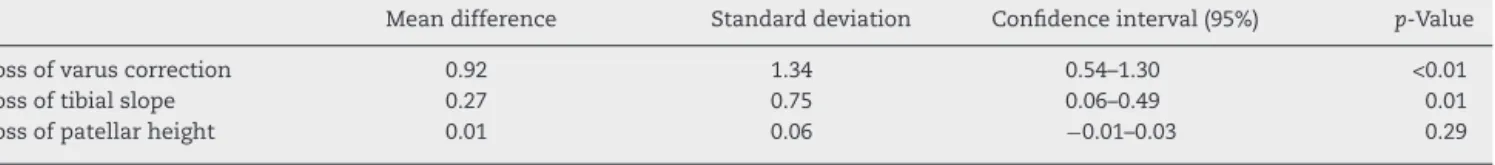
Table2summarizesthefindingsofthepresentstudy.There wasnoclinicallysignificantdifferenceintheassessed param-etersbetweenthepostoperativeradiographsandradiographs obtainedaftertheconsolidationoftheosteotomies.Changes in patellar height determined using the Blackburne-Peel
Table1–Distributionofpatientsaccordingtomodified Ahlbäckclassification.
Ahlbäck n
1 21
2 12
3 12
Table2–Differencesinthethreeassessedparametersbetweenradiographsobtainedimmediatelyaftertheprocedures andthoseobtainedafterosteotomyconsolidation.
Meandifference Standarddeviation Confidenceinterval(95%) p-Value
Lossofvaruscorrection 0.92 1.34 0.54–1.30 <0.01
Lossoftibialslope 0.27 0.75 0.06–0.49 0.01
Lossofpatellarheight 0.01 0.06 −0.01–0.03 0.29
method,tibialslope,andtibialcorrectioninthe anteroposte-riorplanewerenotsignificantandwerewithinthevariability expectedfor this typeofdimension. Thedifference inthe proximaltibialanglewasstatisticallysignificant.Themean correctionlossangle,whichwascalculatedbythedifference betweenthecorrectionangleintheimmediatepostoperative periodandafterconsolidation,was0.92◦±0.9◦.
Discussion
Themaincontributionofthepresentstudy wasto demon-strate that conventional and less expensive implants may provideadequatestabilityforacommonlyperformed proce-dureinactivepatientswithkneeosteoarthritis.Althoughthe numberofosteotomiesforthetreatmentofmedial gonarthro-sishasdecreasedovertheyearsinfavorofarthroplasties,this procedure isstillwidelyperformed. It improvessymptoms and functional capacityand allowsa delay inarthroplasty in a large number of patients.12 Several factors are asso-ciatedwiththe successorfailure ofosteotomies;however, correctionmaintenanceisundoubtedly importantfor long-termoutcomes.13 Thetypeoffixationandselectedimplant have a decisive role in the stability of the fixation. More-over, the small surface ofbone contact in opening wedge osteotomyleadstoahigherincidenceofcomplications.4,14 Nelissenetal.14studiedfixationusingshortplateswith retrac-torsandreportedahigherrateofcomplicationsincasesin whichmoreextensivecorrectionswereperformed.Withthe adventofimplantswithlockingscrews,whichprovideangular stabilitytotheplate,thepopularityofthistypeoffixation sig-nificantlyincreased.8,15,16Biomechanicalstudiesconfirmthat implantswithlockingscrewsenhancethestabilityofthe fix-ationofopeningwedgeosteotomies.15,17However,thehigher costoftheseimplantslimitstheirroutineuseineconomically lessdevelopedcountries.Moreover,importantchangesmay occurinthesagittalplaneandpatellarheight,dependingon theosteotomytechniqueused.18Changesinthesagittalslope ofthetibialplateauandpotentialreductioninpatellarheight afteranopeningwedgeosteotomyintheproximalendofthe tibiamayleadtosignificantbiomechanicalchangesthatcan compromiselong-termoutcomes.19
Themainfindingsofthepresent studyshowedthatthe osteotomytechniqueandfixationmethodwithconventional platesandscrews(whichareintheorylessstable)usedinour hospitalprovidedenoughstabilitytomaintainthecorrection achieveduntilconsolidationbecausethedifferencesobserved intheassessedparameterswerenotsignificant.Therewereno clinicallyrelevantchangesinthefrontalplane,sagittalplane, orpatellarheightwiththefixationmethod.Theuseofalateral spongiousscrewcrossingtheosteotomymayhaveprovided morestabilitytothefixation.Arecentbiomechanicalstudy
conductedatourhospitalshowedthatthe additionofthis screw,eveninthepresenceofagapinthelateralcortex,makes the resistancetothe fixationgapcomparabletothatofan intactlateralcortex.20
Althoughthestudyhadlimitations,suchasthe method-ologicallimitationinherenttoastudyofcasesandtheabsence of a comparison with other methods of fixation, the dif-ferences observed between the measured angles were not clinicallyorstatisticallysignificant,whichallowedusto con-cludethattheconventionalplatesandscrewsusedwiththe described techniqueprovidedenoughstabilityforthistype offixation. Theseimplants canstill beusedinthe clinical routine,withtheadvantageofbeinganaffordablesolution.
Conclusions
Theuseofconventionalplatesandscrewsisviableinthe fix-ation ofopeningwedgehightibialosteotomybecausethey provideenoughstabilitytomaintainthecorrectionachieved untilconsolidation,withoutsignificantchanges.
Conflicts
of
interest
Theauthorsdeclarenoconflictsofinterest.
r
e
f
e
r
e
n
c
e
s
1.HuiC,SalmonL,KokA,WilliamsH,HockersN,TempelW, etal.Long-termsurvivalofhightibialosteotomyformedial compartmentosteoarthritisoftheknee.AmJSportsMed. 2011;39(1):64–70.
2.EfeT,AhmedG,HeyseT,BoudriotU,TimmesfeldN, Fuchs-WinkelmannS,etal.Closing-wedgehightibial osteotomy:survivalandriskfactoranalysisatlong-term followup.BMCMusculoskeletalDisord.2011;12(1):46. 3.McNamaraI,BirminghamTB,FowlerPJ,GiffinJR.Hightibial
osteotomy:evolutionofresearchandclinicalapplications–a Canadianexperience.KneeSurgSportsTraumatolArhrosc. 2013;21(1):23–31.
4.DuivenvoordenT,BrouwerR,BaanA,BosP,ReijmanM, Bierma-ZeinstraS,etal.Comparisonofclosing-wedgeand opening-wedgehightibialosteotomyformedial
compartmentosteoarthritisoftheknee:arandomized controlledtrialwithasix-yearfollow-up.JBoneJointSurg Am.2014;96(17):1425–32.
5.PaccolaCA,FogagnoloF.Open-wedgehightibialosteotomy:a technicaltricktoavoidlossofreductionostheopposite cortex.KneeSurgSportsTraumatolArthrosc.
2005;13(1):19–22.
7. AgneskirchnerJD,FreilingD,HurschlerC,LobenhofferP. Primarystabilityoffourdifferentimplantsforopeningwedge hightibialosteotomy.KneeSurgSportsTraumatolArthrosc. 2006;14:291–300.
8. StaubliA,JacobH.Evolutionofopen-wedgehigh-tibial osteotomy:experiencewithaspecialangularstabledevice forinternalfixationwithoutinterpositionmaterial.Int Orthop.2010;34(2):167–72.
9. AhlbäckS.Osteoarthrosisoftheknee.Aradiographic investigation.ActaRadiolDiagn(Stockh).1968;277Suppl:7–72. 10.FujisawaY,MasuharaK,ShiomiS.Theeffectofhightibial
osteotomyonosteoarthritisoftheknee.Anarthroscopicstudy of54kneejoints.OrthopClinNorthAm.1979;10(30):585–608. 11.PoignardA,LachanietteC,AmzallagJ,HernigouP.Revisiting
hightibialosteotomy:fiftyyearsofexperiencewiththe opening-wedgetechnique.JBoneJointSurgAm.2010;92 Suppl2:187–95.
12.W-DahlA,RobertssonO,LohmanderLS.Hightibial
osteotomyinSweden,1998-2007:apopulation-basedstudyof theuseandrateofrevisiontokneearthroplasty.ActaOrthop. 2012;83(3):244–8.
13.El-AzabHM,MorgensternM,AhrensP,SchusterT,ImhoffAB, LorenzSG.Limbalignmentafteropen-wedgehightibial osteotomyanditseffectontheclinicaloutcome. Orthopedics.2011;34(10):e622–8.
14.NelissenEM,vanLangelaanEJ,NelissenRG.Stabilityof medialopeningwedgehightibialosteotomy:afailure analysis.IntOrthop.2010;34(2):217–23.
15.GomollAH.Hightibialosteotomyforthetreatmentof unicompartmentalkneeosteoarthritis:areviewofthe literature,indications,andtechnique.PhysSportsmed. 2011;39(3):45–54.
16.JungWH,ChunCW,LeeJH,HaJH,KimJH,JeongJH. Comparativestudyofmedialopening-wedgehightibial osteotomyusing2differentimplants.Arthroscopy. 2013;29(6):1063–71.
17.RajaIzahamRMA,AbdulKadirMR,AbdulRashidAH,Hossain MG,KamarulT.FiniteelementanalysisofPudduandTomofix platefixationforopenwedgehightibialosteotomy.Injury. 2012;43(6):898–902.
18.AmisA.Biomechanicsofhightibialosteotomy.KneeSurg SportsTraumatolArthrosc.2013;21(1):197–205.
19.d’EntremontAG,McCormackRG,HorlickSGD,StoneTB, ManzaryMM,WilsonDR.Effectofopening-wedgehightibial osteotomyonthethree-dimensionalkinematicsoftheknee. BoneJointJ.2014;96-B:1214–21.