INTRODUCTION
Retinal pigment epithelium (RPE) tear is a rare devastating com-plication of age-related macular degeneration (AMD). An RPE tear develops when the pigment epithelium detaches from the neurosen-sorial layer with its basement membrane and retracts(1). RPE tears may develop spontaneously in eyes with AMD or after photocoagulation and photodynamic therapy. There are also cases reported to occur after nd:YAG laser capsulotomy and cataract surgery(2-4). After anti-vas cular
endothelial growth factor (VEGF) therapies became widely used for cho roidal neovascularization, the incidence of RPE tears has increased recently(5-7). Some authors have stated that the contraction of choroi-dal neovascularization lying beneath the RPE after anti-VEGF injection causes this complication(8).
RPE tears are diagnosed by clinical examination, luorescein angio-graphy, optical coherence tomography (OCT), and fundus autoluores-cence imaging of the macula. RPE tears have a characteristic
appea-Anatomical and visual outcomes of ranibizumab injections in retinal pigment
epithelium tears
Resultados anatômicos e visuais de injeções de ranibizumab em roturas do epitélio pigmentado da retina
MuhaMMet KazıM erol1, ozdeMır ozdeMır2, denız turgut Coban1, basaK bostanCı Ceran1, esın sogutlu sarı3
Submitted for publication: November 3, 2014 Accepted for publication: April 6, 2015
1 Ophthalmology Department, Antalya Education and Research Hospital, Antalya, Turkey. 2 Ophthalmology Department, Zekai Tahir Burak Women’s Health Education and Research Hospital,
Ankara, Turkey.
3 Ophthalmology Department, Faculty of Medicine, Balıkesir University, Balıkesir, Turkey.
Funding: No specific financial support was provided for this study.
Disclosure of potential conflicts of interest: None of the authors have any potential conflict of interest to disclose.
Corresponding author: Ozdemir Ozdemir. Zekai Tahir Burak Kadın Sağlığı Eğitim ve Araştırma Hastanesi, Göz Hastalıkları Polikliniği, Talatpaşa Bulvarı, Altındağ, Ankara - 06100, Turkey
E-mail: [email protected]
Approved by the research ethics committee of Zekai Tahir Burak Women’s Health Education and
Research Hospital. Project number: Karar No: 29-24/09/2014
ABSTRACT
Purpose: To report the anatomical and visual results in patients diagnosed as ha-ving retinal pigment epithelium (RPE) tears after receiha-ving ranibizumab injections. Methods: Eyes diagnosed as having RPE tears with a minimum 6-month follow-up were retrospectively evaluated. Each eye was treated with at least three doses of ranibizumab at monthly intervals. Best-corrected visual acuity (BCVA), anterior segment findings, intraocular pressure, and fundus examination results were evaluated during control visits. Color fundus photography, fundus fluorescein angiographies, fundus autofluorescence, and spectral domain optical coherence tomography (SD-OCT) images were obtained. The height of pigment epithelial detachment (PED) was measured by SD-OCT.
Results: Twelve eyes with RPE tears were studied. Nine eyes (75%) developed RPE tears during ranibizumab injections for choroidal neovascularization (eight eyes with vascularized PED and one eye with choroidal osteoma), and tears occurred in three eyes before any injections. The median number of ranibizumab injections after diagnosis of RPE tears was 3 (min 2, max 5). In the most recent follow-up visit, there was no statistically significant correlation between the grade of RPE and logMAR of BCVA (p>0.05, r=0.112). Eight of twelve eyes had PED, and seven of these had irregular PED contours before injection therapy. The mean PED height was 447 ± 122 µm.
Conclusions: In this series, RPE tears developed mostly after intravitreal anti-VEGF injections for vascularized PED. Increased vertical height and irregular contours of the PEDs can be risk factors for the formation of RPE tears. The continuation of anti-VEGF therapy after tear formation is beneficial for vision improvement in eyes with RPE tears.
Keywords: Macular degeneration; Retinal detachment; Retinal pigment epithe-lium; Intravitreal injections; Antibodies, monoclonal, humanized; Tomography, optical coherence; Vascular endothelial growth factor; Fluorescein angiography
RESUMO
Objetivo: Apresentar os resultados anatômicos e visuais de injeções de ranibizumab em pacientes que foram diagnosticados com roturas do epitélio pigmentado da retina (RPE). Métodos: Olhos com um mínimo de seis meses de acompanhamento após diagnóstico de roturas do RPE foram avaliados retrospectivamente. Cada olho foi tratado com, pelo menos, três doses de ranibizumab em intervalos mensais. Acuidade visual com a melhor correção (BCVA), achados do segmento anterior, pressão intraocular e exames de fundo de olho foram avaliados nas visitas de controle. Retinografia colorida, angiografias fluoresceínicas, autofluorescência de polo posterior e tomografia de coerência óptica imagens de domínio espectral (SD-OCT) foram obtidos. A altura do descolamento do epitélio pigmentado (PED) foi medida com SD-OCT.
Resultados: Doze olhos com roturas do epitélio pigmentado da retina foram incluí-dos no estudo. Nove olhos (75%) desenvolveram roturas do epitélio pigmentado da retina durante as injeções ranibizumab para neovascularização de coroide (oito olhos com descolamento do epitélio pigmentado vascularizado e um olho com osteoma de coroide), a rotura ocorreu em três olhos antes de quaisquer injeções. A mediana do número de injeções de ranibizumab após o diagnóstico da rotura do RPE foi de 3 (mínimo 2, máximo 5). Na visita de acompanhamento mais recente, não houve cor-relação estatisticamente significante entre o grau de RPE e logMAR de BCVA (p>0,05, r=0,112). Oito dos doze olhos tinham descolamento do epitélio pigmentado, desses, 7 olhos tinham PEDs com contornos irregulares antes da injeção. A altura média do PED foi 447 ± 122 µm.
Conclusões: Nesta série, as roturas de epitélio pigmentado da retina aconteceram principalmente após a injeção intravítrea anti-VEGF para descolamento do epitélio pigmentado vascularizado. O aumento da altura vertical e contornos irregulares dos PEDs podem ser considerados fatores de risco para a formação da rotura de epitélio pigmentado da retina.
rance on luorescein angiography. During luorescein angiography, the bare area is hyperluorescent in the early phase and leakage does not occur, unlike choroidal neovascularization. The scrolled region of the RPE is particularly dark and blocks the underlying luorescence. On occasion, the scrolled area of the RPE has been termed “doubly hypoluorescent.” OCT scans through the retracted RPE show a very intense hyperrelectivity. A deep hyperrelectivity under the line corres-ponding to the RPE is evident in the area of the bare choroid. Fundus autoluorescence shows patchy or hazy hyperluorescence(1-8).
The incidence of RPE tear formation following anti-VEGF treat-ment has been reported to range between 1.8% and 27% in recent studies(9). Diferent treatment protocols and follow-up periods may explain the wide incidence range and diiculties in diagnosing RPE tears. In previous case studies, the spontaneous healing of RPE tears has been demonstrated by using time-domain OCT(10,11). In a study conducted by Caramoy et al., the use of anti-VEGFs was proposed to slow down the scarring process, prevent photoreceptor damage, and give RPE a chance to heal(12).
In this study, we evaluated the anatomical and visual results in patients diagnosed as having RPE tears after receiving ranibizumab injections as anti-VEGF treatment.
METHODS
The charts of 12 eyes of 12 choroidal neovascularization patients with at least 6 months follow-up after being diagnosed with RPE tears were retrospectively evaluated. This study was conducted in accor-dance with the ethical principles of the Declaration of Helsinki. All patients signed an informed consent form before undergoing any treatment.
Full ophthalmic examination, including best-corrected visual acuity (BCVA), anterior segment, and fundus and intraocular pressure were evaluated at the initial visit. Spectral domain (SD)-OCT was taken by using a Cirrus HD-OCT (Carl Zeiss Meditec Inc., Germany). The height of pigment epithelial detachment (PED) was evaluated by using SD-OCT, as previously described by Chan et al.(5). Fundus photos, fun-dus autoluorescent images, and funfun-dus luorescein angiographies of the eyes were obtained by using a Visucam NM/FA fundus camera (Carl Zeiss, Dublin, California) in 45° mode.
In our clinic, patients diagnosed with choroidal neovasculariza-tion receive ranibizumab (Lucentis, Genentech Inc., San Francisco, CA, USA) injections (0.5 mg/0.5 ml) at monthly intervals for the irst 3-month period. After the third injection, eyes with more than a 50-μm increase in central foveal thickness and/or one or more lines of visual
acuity loss on a Snellen chart receive repeated injections. This treat-ment protocol was not changed for patients who developed RPE tears during ranibizumab therapy.
RPE tears were graded as described by Sarraf et al.(13). The grading was performed on the basis of the greatest length of a defect in the vector direction of the tear and foveal involvement by using luores-cein angiographic analysis: Grade 1 tears (diameter <200 µm), Grade 2 tears (diameter between 200 µm and 1 disc diameter), Grade 3 tears (diameter >1-disc diameter), and Grade 4 tears (Grade 3 tears that in volved the foveal center).
S
TATISTICALANALYSISStatistical analysis was performed by using a computer program (SPSS 18.0; SPSS Inc., Chicago, IL, USA). Results are reported as the mean ± standard deviation (SD), median, minimum (min), maximum (max), frequency, or percentage. BCVA results were converted to logMAR for statistical evaluation. The Wilcoxon signed rank t-test was used for comparisons. The correlations between the grade of the RPE tear and BCVAs in the irst and last follow-ups were assessed by using Spearman’s rank correlation coeicient. A p value of <0.05 and an r value of >0.5 were considered to indicate statistical signiicance.
RESULTS
Twelve eyes of 12 patients were diagnosed as having RPE tears. The demographic properties of the patients are shown in table 1. Seven (58%) of the 12 patients were females. The mean age of the patients was 68.5 ± 14.5 years. Nine eyes (75%) developed RPE tears during ranibizumab injections. The median number of ranibizumab injections before RPE tears for nine eyes was 2 (min 1, max 3). Eight of the nine eyes with RPE tears had choroidal neovascularization with vascularized PED secondary to AMD, and the other one had choroidal neovascularization secondary to choroidal osteoma. The other three eyes with RPE tears were referred to our clinic from another hospital. Therefore, it was unknown whether PED existed before RPE tear formation. The patient histories of these patients showed that there were developments of choroidal neovascularization after cataract surgery (in one eye 15 days later and in two eyes 1 year later).
The mean follow-up time after the diagnosis of RPE tears was 12.1 ± 4.9 months. The median ranibizumab injection after the diagnosis of RPE tears was 3 (min 2, max 5). In all of the patients, ranibizumab was used as an anti-VEGF treatment agent. In the last follow-up visit, the BCVAs of the patients (logMAR 0.60 ± 0.52) were better than
Table 1. Characteristics, best-corrected visual acuity, grade of RPE tears, and numbers of ranibizumab injections of the patients
Patient no Age Sex
First BCVA (logMAR)
Last BCVA (logMAR)
Grade of RPE tear
Number of ranibizumab injections before RPE tear
Number of ranibizumab injections after RPE tear
Mean PED height (micron)
01 72 M 1.0 0.1 1 0 4 No PED
02 76 F 0.2 0.1 1 2 3 344
03 57 M 0.3 0.1 2 2 3 387
04 71 F 0.7 0.5 3 1 3 402
05 75 F 0.7 0.7 2 2 2 498
06 70 F 1.8 1.3 3 1 4 689
07 75 M 0.7 0.4 4 0 4 No PED
08 75 F 0.7 1.3 4 0 5 No PED
09 74 M 0.4 0.4 3 2 3 230
10 29 F 1.0 0.7 2 2 2 No PED
11 68 F 1.0 0.7 3 2 5 455
12 81 M 2.0 0.9 2 3 3 578
those at the irst follow-up visit (logMAR 0.85 ± 0.45); however, the diference was not statistically signiicant (p=0.12). The median grade of the RPE tears was 2.5 (range, 1-4). In the last follow-up visit, there was a positive correlation between the RPE grade and logMAR BCVA, but the correlation was not statistically signiicant (p>0.05, r=0.112). In ive of the nine eyes that developed RPE tears during ranibi-zumab therapy, subretinal hemorrhages were evident. Five of these eyes developed RPE tears after the second injection, and two eyes hemorrhaged after the irst dose. After tear development, hemorrha-ge was observed in only two eyes.
During the follow-up period, the RPEs (one Grade 1 and one Grade 2) in two eyes reattached. In these eyes, reattachments of RPE tears were observed by using SD-OCT and fundus autoluorescence imaging. Initially, the RPE tear areas were hypoautoluorescent and the tear borders were hyperautoluorescent. However, in the last visit, it was observed in two eyes that the hypoautoluorescence initially observed in the tear area had changed to hyperautoluorescence, and in one of the eyes, hyperautoluorescent spots were still evident in the hypoautoluorescent area. Eight of the twelve eyes with RPE tears had PED. The mean PED height of the eight eyes was 447 ± 122 µm initially. The PEDs of seven of these eyes had irregular contours.
R
EPRESENTATIVECASEEXAMPLESCase 3: A 57-year-old man was admitted to our clinic with vision loss in the left eye. The BCVA in the left eye was 1.0 logMAR. On the basis of the OCT and fundus luorescein angiography indings, he was diagnosed with AMD, and an intravitreal ranibizumab treatment protocol was started. When his BCVA improved to 0.3 logMAR after the second dose, a Grade 2 RPE tear was observed in the inferior macula. Two doses of ranibizumab were administered after tear for-mation. Six months later, his BCVA improved to 0.1 logMAR. In fundus autolorescence imaging, there was hyperautoluorescent spotting in the denuded RPE area, and subretinal and intraretinal hyper-relective spots were seen in the OCT (Figure 1).
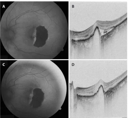
Case 9: A 74-year-old male was with vision loss in both eyes was examined. His BCVA indicated the ability to count ingers at 1 m in the right eye and to count ingers at 2 m in the left eye. Fundus examina-tion in the right eye revealed two disc-sized subretinal hemorrhages on the macula. He was diagnosed with AMD. After the second dose of ranibizumab, the subretinal hemorrhage was resorbed, BCVA increa-sed to 0.4 logMAR (Figure 2 A), but OCT revealed an RPE tear (Figure 2 B). Three additional ranibizumab injections were administered after tear formation, and his BCVA remained stable at 0.4 logMAR (Figures 2 C and 2 D).
DISCUSSION
We observed that nine of the 12 eyes developed RPE tears during the treatment of choroidal neovascularization with ranibizumab in-jections. We continued to inject intravitreal ranibizumab after the de-velopment of RPE tears. After all injections, two of the 12 eyes appeared to be stable, nine of the 12 eyes showed improvement, and only one eye showed worsening of visual acuity at the end of the follow-up periods. At the last follow-up visits, however, the visual acuity had decreased in the patients with a high degree of tears, but the decrea-se was not statistically signiicant. According to our experience, con-tinuation of anti-VEGF injections for patients who developed RPE tears during injection therapy appears to be necessary. In our study population, the heights of the PEDs were >400 microns, and PEDs in seven of the eight eyes had irregular contours.
The pathogenesis of RPE tears is not fully understood. Contrac-tion of the choroidal neovascularizaContrac-tion under the weak RPE, in crea sed luid transport secondary to exudative AMD under the RPE, globe de-formation, vitreous syneresis, vitreo-macular traction due to vitreous incarceration in the injection hole, and detachment of the tight jun ctions between RPE cells are some of the mechanisms that have been considered(14,15).
RPE tears usually develop from the temporal edge of the PED. The separated part of the RPE folds parallel to the PED and reveals the
Figure 1. A) Fundus autoluorescent imaging of the left eye in case 3. The retinal pigment epithelium (RPE) tear in the inferior macula is hypoautoluorescent, and the denuded RPE area shows spotted hyperautoluorescence. B) In luorescein angiography, the RPE tear in the inferior macula is hyper-luorescent, and the denuded RPE area is hypoluorescent. C) The spectral domain optical coherence tomography (SD-OCT) section passes through the RPE tear and pigment epithelial detachment (PED). D) Fundus autolorescence imaging 3 months after the RPE tear formation.
A
C
B
bare Bruch’s membrane for a few days(2,8,12). After RPE tear formation, the PED usually becomes lat spontaneously. Reattachment of the free RPE edges to the Bruch membrane at a diferent location has recently been reported(2). There is a possibility of improvement in patients with lower grade RPE tears(12).
Overexpression of new tissue from the tear border provides soli-dity to the RPE layer. The intraretinal hyper-relective spots in SD-OCT and the corresponding hyperluorescent spots in fundus autolores-cence may be secondary to RPE migration(12). The existence of the hyperautoluorescent spots may be similar to the ones in central se rous retinopathy(16). The reason for this is the corrupted pump me-chanism in the RPE denuded region and phagocytosis in the outer segment. The hyperrelective spots in fundus autolorescence ima-ging may represent the subretinal or intraretinal outer photoreceptor seg ments that are phagocytized by the macrophages because the photoreceptor cells can live up to 325 days in the RPE denuded area(17). Moreover, it has been demonstrated that the precipitates in the su bretinal outer segment layer acted as hyperrelective spots in OCT ophthalmoscopy of central serous retinopathy(18,19).
Previous studies have reported repopulation and reattachment of RPE cells after tear formation, but these studies did not demons-trate healing of the hypoautoluorescence in the tear region(2,8). Pece et al. used OCT to show that reattachment of the margins of an RPE tear were healed by tissue remodeling and described how the disease can recur(20). In our study, reattachment of the RPE tear was demonstra-ted by using SD-OCT and fundus autoluorescence imaging only in 2 eyes. In addition, the hypoautoluorescent area in the tear region healed partially in one eye after 6 months. In another eye, hyperau-toluorescent spots had appeared in the hypoauhyperau-toluorescent area. Similarly, Caramoy et al. reported a case in which healing in the hy-poautoluorescent area was observed after 2.5 years. In that study using SD-OCT, 7 (19.4%) of 36 eyes showed patchy or hazy hyperluo-rescent areas in fundus autoluorescence imaging, and the majority of the eyes (83.3%) showed hyperrelective dots that possibly
repre-sented hard exudates and intraretinal RPE migration. The authors stated that fundus autoluorescence imaging showed a considerable amount of RPE proliferation, repopulation, and migration(12).
In a recent study that included 1298 AMD patients, the pooled rate of RPE tears was 1.8% in the 0.5-mg ranibizumab group, 3.0% in the 0.3-mg ranibizumab group, and 1.6% in the control group. Better visual acuity results were achieved with ranibizumab treatment ver-sus control treatment in patients with RPE tears. Moreover, the po-tential beneit of continued ranibizumab therapy was suggested for patients with RPE tears secondary to neovascular AMD(21). Garg et al. reported that 15 eyes from 15 patients developed an RPE tear, which gave an incidence of 1.6% in 920 eyes with exudative AMD treated with intravitreal bevacizumab. Six of the 15 eyes were continued to be injected with bevacizumab/ranibizumab after tear development, and four of these six eyes showed visual improvement(22). The risk for RPE tear after bevacizumab injection in eyes with PED seems to be moderate. Weinberger et al. investigated RPE tears after intravitreal bevacizumab in 31 eyes with PED and observed RPE tears in four eyes without vision loss. The authors concluded that continuation of anti-VEGF injections for patients who develop RPE tears during injection therapy seems to be necessary(23).
In a few studies, certain factors, such as large PED diameter, ver tical height, and subretinal luid, have been associated with an increase in RPE tear rate(5,24). The contour of the PED is an important factor for predicting RPE tear formation risk. Moroz et al. retrospectively evalua-ted 24 consecutive patients with choroidal neovascular membrane associated with PED, and reported the development of RPE tears after the irst injection in six patients. They described two typical patterns in the eyes, which developed tears. One pattern was multifocal wrinkles and waves with RPE elevations, and the second was step-like in terruptions of the continuity of the RPE line(25). Knowing these risk factors is important for identifying eyes that are likely to develop this complication. The anatomical SD-OCT characteristics of a PED leading to RPE tear after anti-VEGF therapy has recently been
descri-Figure 2. A) The initial fundus autoluorescent imaging of the RPE tear with two disc-sized hypoauto-luorescent areas. B) The initial spectral domain optical coherence tomography (SD-OCT) with retinal pigment epithelium (RPE) tear. C) After three ranibizumab injections, hyperautoluorescent spots were observed in the hypoautoluorescent area, and D) the height of the pigment epithelial detachment (PED) was decreased.
A
C
B
bed by Nagiel et al. and has been prospectively described by Sarraf et al.(26,27). Eyes with vascularized PEDs secondary to AMD have a risk for RPE tear following intravitreal anti-VEGF injection. Those authors explained that the contraction of neovascular tissue adhering to the undersurface of the RPE and rapid involution may cause a substantial contractile force that tears this already-strained tissue layer(26). A base-line PED height >550 µm, presence of a Grade 1 tear, and positive ring sign are high-risk factors for the subsequent development of an RPE tear(27). In our study, the mean PED height of eyes with RPE tears was 447 µm. In addition, before the development of tear formation, irre-gularity in the PED area was detected in seven of eight eyes with PED. One of the patients in our study had choroidal neovascularization secondary to choroidal osteoma. Choroidal osteomas may decalcify and cause degeneration in the underlying retina, including the RPE. In this patient, the RPE tear occurred between the decalciied and calciied regions, which can be explained by the fragility of the area caused by degeneration of the RPE(28). The RPE tear mechanism may be diferent for choroidal neovascularization secondary to choroidal osteoma. Sen et al. demonstrated that multiple anti-VEGF injections caused Bruch membrane rupture in angioid streaks in which the Bruch membrane was calciied and brittle, as in choroidal osteoma(29). To our knowledge, our case is the irst choroidal osteoma case to show the development of an RPE tear after ranibizumab therapy.
The limitations of this study included the limited sample size, lack of a comparison with a control group, lack of standard etiological and anatomical classiications, short follow-up period, and retrospective design. On the other hand, this was a single-center study and the same technicians performed the fundus autolorescence imaging and SD-OCT, which may have helped to standardize the input. For distin-guishing risk factors and mechanisms for RPE tear formation either spontaneously or after VEGF therapy, randomized studies in larger pa tient series are needed.
CONCLUSION
In our series, RPE tears developed mostly after intravitreal anti-VEGF injections administered to treat vascularized PED. The study results demonstrate that SD-OCT, along with fundus autolorescence ima-ging, provided valuable information about the healing process of RPE tears. The continuation of anti-VEGF therapy after tear formation is beneicial for vision improvement in eyes with RPE tears. The PED height and irregularity in the edges of PED areas can be an early in-dicator of RPE tear formation.
REFERENCES
1. Ronan SM, Yoganathan P, Chien F, Corcóstegui IA, Blumenkranz MS, Deramo VA, et al. Retinal pigment epithelial tears after intravitreal injection of bevacizumab (Avastin) for neovascular age-related macular degeneration. Retina. 2007;27(5):535-40. 2. Moreira CA Jr, Arana LA, Zago RJ. Long-term results of repeated anti-vascular
endo-the lial growth factor endo-therapy in eyes with retinal pigment epiendo-thelial tears. Retina. 2013; 33(2):277-81.
3. Barkmeier AJ, Carvounis PE. Retinal pigment epithelial tears and the management of exudative age-related macular degeneration. Semin Ophthalmol. 2011;26(3):94-103. 4. Pece A, Introini U, Bottoni F, Brancato R. Acute retinal pigment epithelial tear after
pho-todynamic therapy. Retina. 2001;21(6):661-5.
5. Chan CK, Meyer CH, Gross JG, Abraham P, Nuthi AS, Kokame GT, et al. Retinal pigment
epithelial tears after intravitreal bevacizumab injection for neovascular age-related ma cular degeneration. Retina. 2007;27(5):541-51.
6. Bakri SJ, Kitzmann AS. Retinal pigment epithelial tear after intravitreal ranibizumab. Am J Ophthalmol. 2007;143(3):505-7.
7. Erol MK, Ozdemir O, Coban DT, Ceran BB, Bulut M. Ranibizumab treatment for choroidal neovascularization secondary to causes other than age-related macular degeneration with good baseline visual acuity. Semin Ophthalmol. 2014;29(2):108-13.
8. Chang LK, Sarraf D. Tears of the retina pigment epithelium: an old problem in a new era. Retina. 2007;27(5):523-34.
9. Smith BT, Kraus CL, Apte RS. Retinal pigment epithelial tears in ranibizumab-treated eyes. Retina. 2009;29(3):335-9.
10. Pece A, Vitale L, Milani P, Pierro L. Spontaneous reattachment of the margins of a macu-lar retinal pigment epithelium tear: optical coherence tomography documentation of a case. Ophthalmologica. 2010;224(3):159-61.
11. Peiretti E, Iranmanesh R, Lee JJ, Klancnik JM Jr, Sorenson JA, Yannuzzi LA. Repopula-tion of the retinal pigment epithelium after pigment epithelial rip. Retina. 2006;26(9): 1097-9.
12. Caramoy A, Fauser S, Kirchov B. Fundus autoluorescence and spectral domain optical coherence tomography indings suggesting tissue remodeling in retinal pigment epi-thelium tear. Br J Ophthalmol. 2012;96(9):1211-6.
13. Sarraf D, Reddy S, Chiang A, Yu F, Jain A. A new grading system for retinal pigment epithelial tears. Retina. 2010;30(7):1039-45.
14. Gass JD. Pathogenesis of tears of the retinal pigment epithelium. Br J Ophthalmol. 1984; 68(8):513-9.
15. Singh RP, Sears JE. Retinal pigment epithelial tear after pegaptanib injection for exuda-tive age-related macular degeneration. Am J Ophthalmol. 2006;142(1):160-2. 16. Erol MK, Özdemir Ö, Çoban DT, Karaçor A, Bulut M, Söğütlü Sarı E. Fundus
autoluores-cence in acute and chronic central serous chorioretinopathy. Turk J Ophthalmol. 2013; 43:94-8.
17. Caramoy A, Kirchhof B, Fauser S. Retinal pigment epithelium tears secondary to age-re-lated macular degeneration: a simultaneous confocal scanning laser ophthalmos copy and spectral-domain optical coherence tomography study. Arch Ophthalmol. 2011;129(5): 575-9.
18. Kon Y, Iida T, Maruko I, Saito M. The optical coherence tomography-ophthalmoscope for examination of central serous chorioretinopathy with precipitates. Retina. 2008; 28(6):864-9.
19. Ozdemir O, Erol MK. Morphologic changes and visual outcomes in resolved central serous chorioretinopathy treated with ranibizumab. Cutan Ocul Toxicol. 2014;33(2):122-6. 20. Pece A, Vitale L, Milani P, Pierro L. Spontaneous reattachment of margins of a macular
pigment epithelium tear: optical coherence tomography documentation of a case. Oph thalmologica. 2010;224(3):159-61.
21. Cunningham ET Jr, Feiner L, Chung C, Tuomi L, Ehrlich JS. Incidence of retinal pigment epithelial tears after intravitreal ranibizumab injection for neovascular age-related macular degeneration. Ophthalmology. 2011;118(12):2447-52.
22. Garg S, Brod R, Kim D, Lane RG, Maguire J, Fischer D. Retinal pigment epithelial tears after intravitreal bevacizumab injection for exudative age-related macular degene-ration. Clin Experiment Ophthalmol. 2008;36(3):252-6.
23. Weinberger AWA, Thiel M, Mohammadi B, Theofylaktopoulos I, Thumann G, Walter P. Retinal pigment epithelium tears after intravitreal bevacizumab in pigment epithe-lium detachment. Am J Ophthalmol. 2007;144(2):294-6.
24. Chiang A, Chang LK, Yu F, Sarraf D. Predictors of anti-VEGF associated retinal pigment epithelial tears using FA and OCT analysis. Retina. 2008;28(9):1265-9.
25. Moroz I, Moisseiev J, Alhalel A. Optical coherence tomography predictors of retinal pig-ment epithelial tear following intravitreal bevacizumab injection. Ophthalmic Surg Lasers Imaging. 2009;40(6):570-5.
26. Nagiel A, Freund KB, Spaide RF, Munch IC, Larsen M, Sarraf D. Mechanism of retinal pigment epithelium tear formation following intravitreal anti-vascular endothelial growth factor therapy revealed by spectral-domain optical coherence tomography. Am J Ophthalmol. 2013;156(5):981-8.
27. Sarraf D, Chan C, Rahimy E, Abraham P. Prospective evaluation of the incidence and risk factors for the development of RPE tears after high- and low-dose ranibizumab therapy. Retina. 2013;33(8):1551-7.
28. Gass JD, Guerry RK, Jack RL, Harris G. Choroidal osteoma. Arch Ophthalmol. 1978;96(3): 428-35.