341
Rev Bras Oftalmol. 2013; 72 (5): 341-3
R
ELATODEC
ASOThe authors declare no conflicts of interest
Recebido para publicação em 15/2/2012 - Aceito para publicação em 4/8/2012
Kabuki Syndrome: a case report with
severe ocular abnormalities
Síndrome de Kabuki: relato de caso com
alterações oculares severas
Flavio Mac Cord Medina
1, Pamela Rodriguez
1, Renata Tavares de Souza Cabral
1, Marcia Brazuna de Castro
1,
Juan Clinton Llerena Junior
2, Ricardo Miguel Japiassú
11 Department of Ophthalmology, Hospital Universitário Pedro Ernesto, Universidade do Estado do Rio de Janeiro (UERJ) – Rio de Janeiro (RJ), Brazil; 2 Instituto Fernandes Figueira, Fundação Oswaldo Cruz – Rio de Janeiro (RJ), Brazil;
Study carried out at Retina and Vitreous Service – Departament of Ophthalmology, Hospital Universitário Pedro Ernesto, Universidade do Estado do Rio de Janeiro (UERJ) – Rio de Janeiro (RJ), Brazil.
A
BSTRACTKabuki syndrome is a rare congenital anomaly, characterized by five fundamental features, the “Pentad of Niikawa”: dysmorphic facies, skeletal anomalies, dermatoglyphic abnormalities, mild to moderate mental retardation and postnatal growth deficiency. Patients present characteristic external ocular features, nonetheless they may also present significant ocular abnormalities. We report a case of a brazilian child diagnosed with Kabuki syndrome, addressing the clinical features observed, with emphasis on the ocular manifestations. This case highlights the existence of this syndrome and all of its complexity. The identification of preventable causes of loss of vision underlines the value of detailed ophthalmologic examination of Kabuki syndrome patients.
Keywords: Congenital disorders; Coloboma; Glaucoma; Strabismus; Case reports
R
ESUMOA síndrome de Kabuki é uma anomalia congênita rara, caracterizada por cinco aspectos fundamentais a “Pêntade de Niikawa”: face dismórfica, anomalias esqueléticas, alterações dermatoglíficas, retardo mental leve a moderado e retardo de crescimento pós-natal. Os pacientes apresentam aspectos oculares externos característicos. Além disso, também podem apresentar anormalidades oculares significativas. Reportamos um caso de uma criança brasileira diagnosticada com síndrome de Kabuki, relatando os aspec-tos clínicos observados, com ênfase nas manifestações oculares. Esse caso chama a atenção para a existência dessa síndrome e toda sua complexidade. A identificação de causas evitáveis de perda de visão reforça a importância do exame oftalmológico detalhado de pacientes com síndrome de Kabuki.
342 Medina FMC, Rodriguez P, Cabral RTS, Castro MB, Llerena Junior JC, Japiassú RM
Rev Bras Oftalmol. 2013; 72 (5): 341-3
I
NTRODUCTIONT
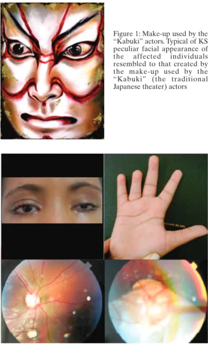
he diagnosis of Kabuki syndrome (KS, OMIM 147920) is based on the presence of five fundamen-tal features (“Pentad of Niikawa”): dysmorphic facies, skeletal anomalies, dermatoglyphic abnormalities, mild to moderate men-tal retardation and postnamen-tal growth deficiency(1). Characteristicexternal ocular features typical of KS include long eyelid cleft, hypertelorism, bowed eyebrows with scarce hair and sparse la-teral third, long and curved eyelashes and eversion of the outer part of inferior eyelid. These features give the patients a pecu-liar mask-like facies. The designation of KS was labeled because the peculiar facial appearance of the affected individuals resembled to that created by the make-up used by the “Kabuki” (the traditional japanese theater) actors (Figure 1).
Initially, the majority of patients reported with this condition were japanese, and its occurrence in Japan was estimated to be one in 32,000(2). Because the characteristic facial features are
clearly identifiable regardless of ethnic background, non-japanese patients with KS have also been recently reported(3). Few cases
have been previously reported in Brazil, with emphasis in dental findings(4-6).
Patients with KS are frequently referred for ophthalmo-logical evaluation because of the characteristic external ocu-lar features. However, patients may also present significant ocular abnormalities. Kluijt et al. reported six of their own cases and reviewed 200 cases of KS, 144 of them had significant ocular abnormalities in addition to the characteristics external ocular features typical of KS(7). Some
of the reported ocular features such as ptosis, cataracts, refractive errors, strabismus and retinal coloboma may cause severe visual impairment. Prepapillary gliosis and tortuous retinal vessels have also been described(8), as well as non
ocu-lar findings as celiac disease, diaphragmatic defects(9) and
congenital heart, ear, and renal defects(10). Phenotypic overlap
can lead to the diagnosis of charge syndrome, especially since the typical facial features of Kabuki syndrome may not be apparent in early infancy.
Mental handicap of these patients could be worsened by poor vision, which could be prevented in some cases by adequate treatment and follow-up at an early age.
The purpose of the paper is to increase the awareness amongstpediatricians of KS together with its eyefindings and the importance of ophthalmologic evaluation including thorough posterior segment examination.
C
ASER
EPORTA 15-year-old brazilian girl diagnosed with KS by the geneticist was referred to ophthalmological evaluation. She is the second child for a non-consanguineous healthy couple, with no similar cases within the family.
She presented moderate learning and coordination difficulties and general developmental delay. Physical exam revealed short stature, shortness of the fingers and dermatoglyphic abnormalities (figure 2). Radiographs showed a T11 butterfly vertebra and bilateral shortening of the fourth metacarpal bones.
Clinical ocular evaluation showed long eyelid cleft, hypertelorism, bowed eyebrows with scarce hair and sparse la-teral third, long and curved eyelashes and eversion of the outer
part of inferior eyelid (figure 2).
Best corrected visual acuity was 20/60 in right eye and counting finger in left eye. Static retinoscopy showed myopic astigmatism (- 3,50 esf - 2,00 cyl at 155o) in the right eye and was
impossible in the left eye because of scissors reflex.
The patient was unable to fix and follow with the left eye. She had left exotropia and Hirschberg of 45°. There was a limitation of extraocular movement towards adduction on left eye. Anterior segment biomicroscopy showed bilateral blue sclera, left microcornea and microphthalmia. Fundus examination revealed, on right eye, a small inferior coloboma and large optic disc excavation and, on left eye, a large inferior chorioretinal coloboma defect (figure 2).
The patient was started on treatment for glaucoma and received myopic correction.
Figure 1: Make-up used by the “Kabuki” actors. Typical of KS peculiar facial appearance of the affected individuals resembled to that created by the make-up used by the “Kabuki” (the traditional Japanese theater) actors
343
Rev Bras Oftalmol. 2013; 72 (5): 341-3
D
ISCUSSIONThere are few reports of the ocular manifestations of KS in the literature. External ocular features are usually emphasized. Significant ocular abnormalities may be present and this case highlights the existence of this syndrome and all of its complexity. Our patient presented all five cardinal manifestations of KS and all external features characteristics of Kabuki make-up. Nevertheless, our patient presented bilateral severe ocular abnormalities and low vision. Right eye presented advanced glaucoma, a small retinal coloboma defect and moderate myopia. Left eye findings included microphthalmia, microcornea, upper eyelid congetinal ptosis, exotropia, and large chorioretinal coloboma (table 1).
Few patients present as many ocular manifestations as our patient. One patient has been previously described with bilate-ral large chorioretinal coloboma, but no apparent visual loss(11).
Mental handicap of these patients can be worsened by poor vision, which can be prevented in some cases by adequate treatment and follow-up at an early age. Refractive errors and strabismus must be promptly treated with glasses and occlusion therapy. Ptosis mandates surgical correction at an early age when it causes visual axis obstruction. Periorbital dysmorphism in KS can be submitted to a surgical correction, with simultaneous repair of the medial and outer canthus to obtain an adequate fissure length(12).
Because of the extent of retinal abnormalities and age of first ophthalmological evaluation, left eye was not considered for correction of ametropy and occlusion therapy. Best corrected visual acuity in right eye may be underestimated because of development delay.
Although KS is rare, more children can be diagnosed as pediatricians gain awareness of this syndrome. The identification of preventable causes of loss of vision underlines the value of detailed ophthalmologic examination of KS patients.
R
EFERENCES1. Niikawa N, Matsuura N, Fukushima Y, Ohsawa T, Kajii T. Kabuki make-up syndrome: a syndrome of mental retardation, unusual facies, large and protruding ears, and postnatal growth deficiency. J Pediatr. 1981;99(4):565-9.
2. Niikawa N, Kuroki Y, Kajii T, Matsuura N, Ishikiriyama S, Tonoki H, et al. Kabuki make-up (Niikawa-Kuroki) syndrome: a study of 62 pa-tients. Am J Med Genet. 1988;31(3):565-89.
3. Wessels MW, Brooks AS, Hoogeboom J, Niermeijer MF, Willems PJ. Kabuki syndrome: a review study of three hundred patients. Clin Dysmorphol. 2002;11(2):95-102.
4. Gabrieli APT, Rovaris FV, Bisol LE, Borges L, Michelin MM, Lovatto L. Síndrome da maquiagem de kabuki. Acta Ortop Bras. 2002;10(3):57-61.
5. dos Santos BM, Ribeiro RR, Stuani AS, de Paula e Silva FW, de Queiroz AM. Kabuki make-up (Niikawa-Kuroki) syndrome: dental and cran-iofacial findings in a Brazilian child. Braz Dent J. 2006;17(3):249-54. 6. Rocha CT, Peixoto IT, Fernandes PM, Torres CP, de Queiroz AM. Dental findings in Kabuki make-up syndrome: a case report. Spec Care Dentist. 2008;28(2):53-7.
7. Kluijt I, van Dorp DB, Kwee ML, Toutain A, Keppler-Noreuil K, Warburg M, et al. Kabuki syndrome - report of six cases and review of the literature with emphasis on ocular features. Ophthalmic Genet. 2000;21(1):51-61.
8. Chuah JL, Chuah JK, Brown R. New fundus findings in a case of Ka-buki syndrome. Eye (Lond). 2009;23(6):1483-5.
9. Geneviève D, Amiel J, Viot G, Le Merrer M, Sanlaville D, Urtizberea A, et al. Atypical findings in Kabuki syndrome: report of 8 patients in a series of 20 and review of the literature. Am J Med Genet A. 2004;129A(1):64-8.
10. Ming JE, Russell KL, Bason L, McDonald-McGinn DM, Zackai EH. Coloboma and other ophthalmologic anomalies in Kabuki syndrome: distinction from charge association. Am J Med Genet A. 2003;123A(3):249-52.
11. Chaudhry IA, Shamsi FA, Alkuraya HS, Al-Sharif A. Ocular manifes-tations in Kabuki syndrome: the first report from Saudi Arabia. Int Ophthalmol. 2008;28(2):131-4.
12. Sakurai H, Nozaki M, Takeuchi M, Soejima K, Kajimoto M, Hori S. Periorbital correction in Kabuki syndrome. Plast Reconstr Surg. 2003;111(4):1461-4.
Author corresponding
Flávio Mac Cord Medina Clínica São Vicente
Rua João Borges, 204 – Gávea
Zip code 22451-100 – Rio de Janeiro (RJ), Brazil Phone: (21) 2529-4432;
E-mail: [email protected]
“Pentad of Niikawa” Kabuki make-up Vision threatening disorders
Dysmorphic facies + Long eyelid cleft + Strabismus +
Skeletal anomalies + Hypertelorism + Nystagmus
-Dermatoglyphic abnormalities + Bowed eyebrows with scarce hair + Eyelid ptosis + and sparse lateral third
Mild to moderate mental retardation + Long and curved eyelashes + Retinal findings + Postnatal growth deficiency + Eversion of the outer part of + Refractive errors +
inferior eyelid
Glaucoma +
Cataract
-Corneal findings +
Table 1
Characteristics of Kabuki syndrome
“Pentad of Niikawa” fundamental features, Kabuki make-up characteristics and vision threatening disorders compared to findings previously described in the literature.