R
ESUMOA
BSTRACTPrognóstico visual de ‘crosslinking’ para ceratocone com
base em tomografia de córnea pré-operatória
Visual prognosis of crosslinking for kearatoconus
based on preoperative corneal tomography
Bernardo Lopes¹, Isaac Ramos¹, Tobias Koller², Theo Seiler²,Renato Ambrósio Jr¹.
1 Rio de Janeiro Corneal Tomography and Biomechanics Study Group, Rio de Janeiro/RJ, Brazil. 2 Institute for Refractive and Ophthalmic Surgery, Germany.
Study conducted at the Renato Ambrósio Eye Institute, Rio de Janeiro/RJ, Brazil.
Conflicts of interest: Renato Ambrósio Jr. é consultor da Oculus Optikgeräte GmbH (Wetzlar, Germany)
Received for publication 06/06/2013 - Accepted for publication em 09/04/2014
Objetivo: Verificar índices tomográficos do pré-operatório de pacientes com ceratocone submetidos à crosslinking corneano (CXL)
como fatores preditivos para a melhora na acuidade visual corrigidas (AVc) após um ano. Métodos: Estudo retrospectivo que incluiu
63 olhos de 53 pacientes com ceratocone progressivo submetidos à CXL segundo o protocolo de Dresden: deseptelização corneana, riboflavina 0,1% por 30 minutos e luz ultra-violeta A (UVA) a uma irradiância de 3mW/cm² por 30 minutos. Foram avaliados exames de tomografia corneana com sistema de Scheimpflug rotacional (Pentacam, Oculus) antes do CXL e a acuidade visual
corrigida antes e após a cirurgia. A análise estatística foi feita com o teste de Kolmorov-Smirnov, teste t de student e curvas de
característica operador-receptor (ROC). Resultados: Houve diferença estatisticamente significante (p<0,05) entre os pacientes
que obtiveram melhora de AVc em um ano e os que não experimentaram melhora na AVc nesse período nos índices tomográficos pré-operatórios relacionados com espessura e volume corneano. Entre os pacientes que obtiveram melhora na AVc todos possuíam volume corneano em 6,0mm maior que 14,55mm³ e 97,2% deles possuíam volume corneano em 6,5mm maior que 17,76mm³. Assim como, 94,29% desses pacientes apresentavam paquimetria média em 4,0mm maior que 487 µm e 82,86% paquimetria no ponto mais
fino maior de 421 µm. Conclusão: Pacientes com ceratocone menos avançado (volume e espessura da córnea maiores) no período
pré-operatório obtiveram mais chances de ter melhora da AVc um ano após CXL. Estudos prospectivos envolvendo outras variá-veis relacionadas com a aberrometria total e o estudo biomecânico da córnea são relevantes para se aumentar a capacidade prognóstica do resultado após CXL.
Descritores: Ceratocone; Tomografia da córnea; Prognóstico; Acuidade visual
Purpose: To verify pre-operative tomographic indices as predictive parameters for the improvement in best corrected visual acuity (BCVA) in one year after corneal collagen crosslinking (CXL) procedure for keratoconus.Methods: Retrospective study that included 63 eyes of 53 patients with progressive keratoconus submitted to CXL following the Dresden’s protocol: topical anesthesia, 9.0 mm of epithelial abrasion, riboflavin 0.1% drops for 30 minutes and ultraviolet-light A (UVA) with an irradiance of 3mW/cm² for 30 minutes. Corneal tomography taken by Scheimplug rotational system (Pentacam, Oculus) before CXL was evaluated along with pre and 1 year post-operative BCVA. Statistical analysis was accomplished with the Kolmogorov-Smirnov test, student’s t-test and Receiver Operating Characteristic (ROC) curve.Results: There were statistically significant differences (p<0.05) between patients who improved BCVA in one year after CXL and those who did not experienced improvement in BCVA in the same period of time in the pre-operative tomographic indices related to corneal volume and thickness. Among those who had its BCVA improved all of them had a corneal volume in 6.0mm grater then 14.55mm³ and 97.2% of them had corneal volume in 6.5mm grater then 17.76mm³. So with 94.29% of them hadthickness in 4.0mm grater then 487µm and 82.86% had thickness on the thinnest point grater then 421µm. Conclusion: Patients with less advanced keratoconus (grater corneal volume and thickness) in the pre-operative had more chances to improve its BCVA in one year after CXL. Prospective studies involving others variables related to total aberrometry and corneal biomechanics are relevant to increase the prognostic capability of CXL result.
I
NTRODUCTIONK
eratoconus is a progressive, usually bilateral cornealectasia whose treatment has changed in recent years. Until the past decade, treatments such as hard contact lenses, intrastromal corneal ring segment implantation, and
corneal transplantation aimed to improve visual performance (1,2).
In the late 1990s and early 21st century(3), the University of
Dresden, Germany, began to study a new conservative treatment for keratoconus in human corneas based on the formation of covalent bonds in corneal collagen, called cross-linking (CXL). Its primary goal is to prevent the progression of ectasia by
increasing the biomechanical stiffness of the cornea(4,5).
In its original protocol(3), CXL was induced by ultraviolet
A radiation and riboflavin (vitamin B2), a water-soluble photosensitiser that penetrates the stroma in the absence of epithelium. Several studies have demonstrated its safety, stability, and ability to improve corneal topography and tomography
indices, as well as corrected visual acuity (CVA)(7-11). CXL has
also been suggested as an adjunctive therapy to rigid contact
lenses and intrastromal corneal ring segment implantation(12).
However, complications related to the procedure, although infrequent, have been observed; they include scars and corneal opacities, persistent epithelial defect and, more rarely, bacterial keratitis(13,14).
Because CXL is not an innocuous treatment, prognostic factors able to predict the stability or improvement of patients submitted to the procedure are very important to refine its indication criteria. The present study aimed to identify preoperative corneal tomography indices that may predict whether the CVA of patients subjected to the procedure is likely to improve in one year.
M
ETHODSData from patients with progressive keratoconus submitted to CXL were analysed retrospectively. Inclusion criteria were progressive ectasia followed up for at least six months and characterised by an increase in maximum keratometry (Kmax) by 1.00 D (equivalent to 3 standard deviations [SD] of the device repeatability) [15] and Kmax <65.00 D. Exclusion criteria were preoperative superficial corneal opacities, as Scheimpflug images are unreliable in these cases; corneal pachymetry <400 µm even after instillation of hypotonic riboflavin; pregnancy; and other eye diseases, especially endothelial disease, history of recurrent erosion, and connective tissue disorders.
CXL was performed after topical anaesthesia with oxybuprocaine alternated with tetracaine eye drops every 3 minutes for 15 minutes. Corneal abrasion with a diameter of 9 mm was then performed. Riboflavin 0.1%, prepared immediately before treatment by diluting 0.5% aqueous riboflavin into a 20% dextran T-500 solution, was instilled every 3 minutes for 30 minutes. Central corneal ultrasonic pachymetry was then performed. In eyes where corneal thickness after epithelial removal was less than 400 µm, 0.1% hypotonic riboflavin without dextran was added until the thickness reached 400 µm; this was carefully monitored in all cases prior to the procedure. The presence of riboflavin in the anterior chamber was observed by slit lamp examination with a cobalt blue filter. The eye was then irradiated for 30 minutes using UVA light with an irradiance of
3 mW/cm² (UV-X, Peschke Meditrade). During irradiation, riboflavin 0.1% was administered every 3 minutes and oxybuprocaine was administered as needed. At the end of the procedure, a 0.3% ofloxacin solution and bandage contact lenses were applied. The antibiotic was maintained for two days; after epithelial healing, patients used topical fluorometholone twice a day for one week.
CVA, tested with optotypes on a logarithmic scale, was assessed preoperatively and one year after treatment. Pre- and postoperative corneal tomography data were obtained with a rotating Scheimpflug system (Pentacam 70700™, Oculus).
Statistical analysis was done using BioEstatTM 5.3 software.
The Kolmogorov-Smirnov test was used to assess whether the
study population was normally distributed. Student’s t-test was
used to compare patients with and without improvement in CVA. ROC curves were calculated to determine the best cut-off values for parameters with statistically-significant differences. p-values <0.05 were considered statistically significant.
R
ESULTSOf the 53 patients (63 eyes), 21 were female and 32 were male. Mean age was 29.4 ± 9.4 years (range: 15 to 56 years). After one year, CVA had improved in 35 eyes (55.6%), remained stable in 12 eyes (19%), and worsened in 16 eyes (25.4%).
Table 1 presents the differences in CVA, indices of central keratometry, and corneal thickness and volume before and one year after CXL. Statistically-significant differences were observed, with CVA improvement, flattening of the flattest corneal meridian (K1), reduction of corneal volume between the 2 mm and 4.5 mm diameters, and corneal thinning at the thinnest point and at the 2 mm diameter.
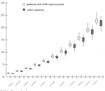
Patients whose CVA improved in one year had a statistically higher (p<0.05) preoperative corneal volume at the 2.0 to 7.0 mm diameters, as shown by 11 tomographic indexes (Table 2, Figure 1). Among the indices that performed better (largest area under the ROC curve), corneal volumes at 6.0 and 6.5 mm presented a sensitivity of 100% and 97.14% at the 14.55 and 17.76 mm³ cut-off points, respectively. In addition to the good sensitivity, only 12% of patients with these characteristics presented a decrease in CVA. The corneal volume at 7 mm had a specificity of 60.7% at the 21.43 mm³ cut-off point (Table 3, Figure 1).
Patients whose CVA improved in one year (p<0.05) also showed better preoperative corneal thickness values in five tomographic indexes: pachymetry at the thinnest point and at the 2, 4, 6, and 8 mm diameters (Table 2, Figure 2). Of these, pachymetry at 4 mm and at the thinnest point had a sensitivity of 94.29% and 82.86% at the 487 µm and 421 µm cut-off points, respectively (Table 3). For these pachymetry values at 4 mm and at the thinnest point, 12.8% and 13.6% of patients, respectively, presented a decrease in CVA.
D
ISCUSSIONTable 2
Preoperative tomographic indexes
Patients with CVA improvements Other patients
Mean Range Mean Range P-value
C.Vol.7,0mm: 22,87 ± 1,20 21,05 – 26,14 21,64± 1,65 18,14 – 24,71 0,0021
C.Vol.6,5mm: 19,16 ± 1,01 17,71 – 21,91 18,13 ± 1,41 15,05 – 20,77 0,0024
C.Vol.6,0mm: 15,82 ± 0,85 14,62 – 18,11 14,98 ± 1,18 12,37 – 17,21 0,0029
C.Vol.5,5mm: 13,05 ± 0,72 11,98 – 14,94 12,36 ± 0,98 10,22 – 14,21 0,0034
C.Vol.5,0mm: 10,45 ± 0,6 9,51 – 11,95 9,9 ± 0,79 8,23 – 11,39 0,0044
C.Vol.4,5mm: 8,37 ± 0,5 7,55 – 9,56 7,94 ± 0,64 6,64 – 9,12 0,0058
C.Vol.4,0mm: 6,41 ± 0,4 5,72 – 7,30 6,09 ± 0,49 5,13 – 6,98 0,0090
C.Vol.3,5mm: 4,89 ± 0,32 4,3 – 5,56 4,66 ± 0,38 3,94 – 5,32 0,0131
C.Vol.3,0mm: 3,47 ± 0,24 2,99 – 3,92 3,31 ± 0,28 2,82 – 3,79 0,0220
C.Vol.2,5mm: 2,38 ± 0,18 2,01 – 2,67 2,27 ± 0,20 1,94 – 2,61 0,0318
C.Vol.2,0mm: 1,49 ± 0,12 1,24 – 1,67 1,43 ± 0,13 1,23 – 1,65 0,0499
Pach Mín.: 453,23 ± 44,25 317 – 520 431,43 ± 40,32 359 – 502 0,0491
Pach 2mm: 475,49 ± 38,45 365 – 539 451,61 ± 38,75 392 – 516 0,0197
Pach 4mm: 528,23 ± 31,40 459 – 604 –2,43 ± 37,41 445 – 574 0,0059
Pach 6mm: 590,69 ± 29,90 535 – 661 567,04 ± 35,25 503 – 638 0,0075
Pach 8mm: 663,97 ± 31,25 609 – 739 639,82 ± 35,04 565 – 716 0,0070
C. Vol.: corneal volume; Min. Pach.: pachymetry at the thinnest point; Pach.: pachymetry
Pre CXL Post CXL
Mean ± SD Range Mean ± SD Range P-value
CVA (LogMAR) 0,40 ± 0,31 -0,08 – 1,3 0,31 ± 0,29 -0,15 – 1,3 0,0010
K1 46,11 ± 4,48 37,5 – 61,2 45,52 ± 3,94 37,4 – 56,3 0,0421
K2 50,48 ± 5,22 42,3 – 66 49,9 ± 5,13 42,4 – 66,9 0,1613
Astig 4,37 ± 2.35 0,1 – 10,4 4,37 ± 2,51 0,7 –13,4 0,9898
C.Vol. D 2,0mm 1,46 ± 0,13 1,23 – 1,67 1,43 ± 0,13 1,09 – 1,69 0,0063
C.Vol. D 2,5mm 2,33 ± 0,19 1,94 – 2,67 2,28 ± 0,2 1,79 – 2,69 0,0079
C.Vol. D 3,0mm 3,4 ± 0,27 2,82 – 3,92 3,33 ± 0,27 2,7 – 3,93 0,0108
C.Vol. D 3,5mm 4,79 ± 0,37 3,94 – 5,56 4,7 ± 0,36 3,92 – 5,53 0,0167
C.Vol. D 4,0mm 6,27 ± 0,47 5,13 – 7,3 6,17 ± 0,44 5,18 – 7,23 0,0266
C.Vol. D 4,5mm 8,18 ± 0,6 6,64 – 9,56 8,06 ± 0,54 6,83 – 9,42 0,0491
C.Vol. D 5,0mm 10,2 ± 0,74 8,23 – 11,95 10,08 ± 0,65 8,61 – 11,74 0,0824
C.Vol. D 5,5mm 12,74 ± 0,91 10,22 – 14,94 12,62 ± 0,78 10,86 – 14,65 0,1548
C.Vol. D 6,0mm 15,45 ± 1,09 12,37 – 18,11 15,34 ± 0,92 13,27 – 17,75 0,2548
C.Vol. D 6,5mm 18,7 ± 1,31 15,05 – 21,91 18,61 ± 1,1 16,16 – 21,48 0,4061
C.Vol. D 7,0mm 22,33 ± 1,54 18,14 – 26,14 22,25 ± 1,29 19,3 – 25,66 0,5819
Min Pach 443,67 ± 43,9 317 – 520 426,9 ± 50,36 290 – 515 0,0006
Pach 2mm 464,87 ± 40,37 365 – 539 452,54 ± 41,69 349 – 539 0,0030
Pach 4mm 516,76 ± 36,53 445 – 604 512,92 ± 31,1 454 – 592 0,2264
Pach 6mm 580,17 ± 34,45 503 – 661 581,94 ± 31,43 518 – 656 0,5264
Pach 8mm 653,24 ± 35,11 565 – 739 657,7 ± 38,45 533 – 732 0,1330
Table 1
Tomographic indices before and after CXL
AUROC 95% CI Cut-off point Sensitivity (%) Specificity (%)
C.Vol.7,0mm: 0,718 0,591 60,824 >21,43 85,71 60,71
C.Vol.6,5mm: 0,721 0,594 60,827 >17,76 97,14 50,00
C.Vol.6,0mm: 0,715 0,588 60,822 >14,55 100,00 46,43
C.Vol.5,5mm: 0,715 0,587 60,821 >12,12 94,2 50,00
C.Vol.5,0mm: 0,710 0,582 60,817 > 9,75 88,57 57,14
C.Vol.4,5mm: 0,696 0,567 60,806 > 7,69 94,29 46,43
C.Vol.4,0mm: 0,691 0,562 60,801 > 5,87 91,43 46,43
C.Vol.3,5mm: 0,680 0,551 60,792 > 4,63 77,14 57,14
C.Vol.3,0mm: 0,675 0,545 60,788 > 3,3 77,14 57,14
C.Vol.2,5mm: 0,670 0,540 60,784 > 2,22 80,00 50,00
C.Vol.2,0mm: 0,656 0,525 60,771 > 1,4 77,14 50,00
Pach Mín.: 0,659 0,528 60,773 > 421 82,86 46,43
Pach 2mm: 0,687 0,558 60,798 > 452 77,14 57,14
Pach 4mm: 0,700 0,571 60,809 > 487 94,29 50,00
Pach 6mm: 0,690 0,561 60,800 > 561 88,57 53,57
Pach 8mm: 0,688 0,559 60,799 > 636 80,00 57,14
C. Vol.: corneal volume; Min. Pach.: pachymetry at the thinnest point; Pach.: pachymetry; AUROC: area under the ROC curve; 95% CI: 95% confidence interval
Table 3
Receiver operating characteristic (ROC) curve analysis
Figure 1. Box-plot presenting the comparison of corneal volume between patients with or without CVA improvement.
C. Vol.: Corneal volume
Figure 2. Box-plot comparing corneal thickness among patients with or without CVA improvement.
Min. Pach.: pachymetry at the thinnest point; Pach.: pachymetry
an objective measure visual function widely used in the ophthalmic practice.
In the present sample, flattening at K1 and reduced corneal volume and thickness were observed. These results are in agreement with the literature and may indicate compression of corneal collagen fibres and increased corneal resistance.
CVA improved in 55.6% of patients submitted to CXL —
slightly less than the 65% observed by Wollensak et al.(3) The
CVA of patients with greater corneal volume and thickness was more likely to improve in one year.
Our results presented high sensitivity, suggesting that the
less advanced the ectasia, the greater the chance of CVA improvement and the lesser the chance of CVA worsening one year after CXL. Conversely, patients with more advanced ectasia are unlikely to achieve CVA improvement in one year. These results can help refine the indications for the procedure, so as to avoid its indication to patients who are unlikely to improve.
Prospective studies involving other variables related to total aberrometry and biomechanical studies of the cornea are important to improve our ability to accurately predict the outcomes of CXL. Further studies involving other means of
Correesponding author:
Rua Conde de Bonfim, 211/712 Tijuca, Rio de Janeiro/RJ, Brazil CEP: 20520-050
assessment of brain neuroplasticity, may help clarify how visual quality improves in these patients and how this affects their daily activities (17,18).
In the present study, patients with less advanced keratoconus (higher volume and corneal thickness) preoperatively were more likely to achieve CVA improvement one year after CXL. Prospective studies involving other variables related to total aberrometry and biomechanical studies of the cornea would help improve our ability to predict the outcomes of CXL.
R
EFERENCES1. Rabinowitz YS. Keratoconus. SurvOphthalmol. 1998;42(4):297-319. Review.
2. Siganos D, Ferrara P, Chatzinikolas K, Bessis N, Papastergiou G. Ferrara intrastromal corneal rings for the correction of keratoconus. J Cata-ract RefCata-ract Surg. 2002;28(11):1947-51.
3. Wollensak G, Spoerl E, Seiler T. Riboflavin/ultraviolet-a-induced col-lagen crosslinking for the treatment of keratoconus. Am J Ophthalmol. 2003;135(5):620-7.
4. Spoerl E, Huhle M, Seiler T. Induction of cross-links in corneal tissue. Exp Eye Res. 1998;66(1):97-103.
5. Wollensak G, Spoerl E, Seiler T. Stress-strain measurements of human and porcine corneas after riboflavin-ultraviolet-A-induced cross-link-ing. J Cataract Refract Surg. 2003;29(9):1780-5.
6. Renesto Ada C, Sartori M, Campos M. [Cross-linking and intrastromal corneal ring segment]. Arq Bras Oftalmol. 2011;74(1):67-74. Review. Portuguese.
7. Spoerl E, Mrochen M, Sliney D, Trokel S, Seiler T. Safety of UVA-ribo-flavin cross-linking of the cornea. Cornea. 2007;26(4):385-9. Review. 8. Vinciguerra P, Albè E, Trazza S, Rosetta P, Vinciguerra R, Seiler T, et
al. Refractive, topographic, tomographic, and aberrometric analysis of keratoconic eyes undergoing corneal cross-linking. Ophthalmol-ogy. 2009;116(3):369-78.
9. Koller T, Pajic B, Vinciguerra P, Seiler T. Flattening of the cornea after collagen crosslinking for keratoconus. J Cataract Refract Surg. 2011;37(8):1488-92.
10. Greenstein SA, Shah VP, Fry KL, Hersh PS. Corneal thickness changes after corneal collagen crosslinking for keratoconus and corneal ecta-sia: one-year results. J Cataract Refract Surg. 2011;37(4):691-700. 11. Raiskup-Wolf F, Hoyer A, Spoerl E, Pillunat LE. Collagen crosslinking
with riboflavin and ultraviolet-A light in keratoconus: long-term re-sults. J Cataract Refract Surg. 2008;34(5):796-801.
12. AlmodinE,Arschinoff S, Almodin J, Ferrara P. [Addictive treatment of keratoconus with collagen crosslinking after Ferrara ring implant]. Rev BrasOftalmol.2009;68(3):138-45. Portuguese.
13. Koller T, Mrochen M, Seiler T. Complication and failure rates after corneal crosslinking. J Cataract Refract Surg. 2009;35(8):1358-62. 14. Pérez-Santonja JJ, Artola A, Javaloy J, Alió JL, Abad JL. Microbial
keratitis after corneal collagen crosslinking. J Cataract Refract Surg. 2009;35(6):1138-40.
15. Koller T, Iseli HP, Hafezi F, Vinciguerra P, Seiler T. Scheimpflug imag-ing of corneas after collagen cross-linkimag-ing. Cornea. 2009;28(5):510-5. 16. Ginsburg AP. Contrast sensitivity: determining the visual quality and function of cataract, intraocular lenses and refractive surgery. CurrOpinOphthalmol. 2006;17(1):19-26. Review.
17. Braddick O, Atkinson J. Development of human visual function. Vi-sion Res. 2011;51(13):1588-609. Review.