8 4 Arq Bras Oftalmol. 2014;77(2):84-7
Original Article
Changes in corneal sensitivity following cross-linking for progressive early-stage keratoconus
Alterações da sensibilidade corneana após cross-linking para ceratocone progressivo em estágio inicial
Anelisede Medeiros lAgo1, Belquiz r. do AMArAl nAssArAllA2,lArissA rossAnA souzA stivAl1, João Jorge nAssArAllA Junior3,4
Submitted for publication: June 20, 2013 Accepted for publication: October 25, 2013
Study conducted at Goiania Eye Institute, Goiânia, GO, Brazil. 1 Goiania Eye Institute, Goiânia, GO, Brazil.
2 Department of Cornea and Refractive Surgery, Goiania Eye Institute, Goiânia, GO, Brazil. 3 Department of Retina & Vitreous, Goiania Eye Institute, Goiânia, GO, Brazil. 4 Faculty of Health Sciences, University of Brasília, Brasília, DF, Brazil.
Funding: No specific financial support was available for this study.
Disclosure of potential conflicts of interest: None of the authors have any potential conflicts of interest to disclose.
Corresponding author: Anelise de Medeiros Lago. Rua Soledade, 1.268 Apto. 405 Edifício Re -si dencial Petrópolis - Esteio (RS) - 793260-150 - Brazil - E-mail: [email protected]
ClinicalTrials.govID: NCT01743443
Research Ethics Committee: Goiânia Eye Institute, Goiânia, Goiás, Brazil.
INTRODUCTION
Keratoconus is a corneal ectatic disease where the cornea assu-mes a conical shape because of thinning, inducing irregular astigma-tism and leading to marked impairment of vision. Keratoconus typi-cally initiates at puberty and may progress, depending on individual characteristics, until the third or fourth decade of life; alternatively, it may commence later and arrest at any age. The disease is associated with several conditions, particularly those that encourage eye rubbing. This theory is supported by the fact that patients with keratoconus frequently have itchy eyes and ocular irritation. An autosomal domi-nant inheritance is reported in approximately 10% patients(1).
Keratoconus occurs in virtually every ethnic group. There is no gen der predominance, and it almost always presents bilaterally, alth ough the clinical features are frequently asymmetrical. The rate of progression varies between individuals and also between the two eyes. The severity at the stop of progression is also highly variable and can range from mild irregular astigmatism to severe thinning, scarring, and protrusion(1,2).
ABSTRACT
Purpose: To evaluate changes in corneal sensitivity following corneal cross-linking (CXL) in patients with progressive earlier stage keratoconus.
Methods: Thirty-eight eyes of 19 patients (11 women, 8 men) were included in a prospective, nonrandomized clinical study. The mean patient age was 22 years (range, 18-26 years). Inclusion criteria were early stage bilateral progressive kera-toconus, a transparent cornea, and a thickness of ≥440 µm in the thinnest area of the cornea. Using the Cochet-Bonnet esthesiometer, central corneal sensitivity was measured before surgery, 7 days after surgery, and once a month thereafter until recovery of baseline preoperative levels. Central corneal sensitivity >40 mm was considered normal.
Results: Corneal sensitivity gradually returned to preoperative levels in all trea-ted eyes. The mean central corneal sensitivity was 52.2, 24.0, 38.2, 42.5, 50.0, and 52.5 mm before surgery, 7 days after surgery, and at 1, 2, 3, and 4 months after surgery, respectively. Normal levels of corneal sensation, but not baseline (preo-perative) levels, were observed 2 months after surgery. The preoperative levels were obser ved 3 months after surgery.
Conclusions: Our results suggest that central corneal sensitivity can be decrea sed for as long as 3 months after CXL for progressive earlier stage keratoconus.
Keywords: Keratoconus/therapy; Collagen/radiation effects; Riboflavin/therapeu-tic use; Ultraviolet therapy; Cross-linking reagents; Corneal sensitivity
RESUMO
Objetivo: Avaliar as alterações da sensibilidade corneana após cross-linking (CXL) da córnea em pacientes com ceratocone progressivo em estágio inicial.
Métodos: Trinta e oito olhos de 19 pacientes (11 mulheres, 8 homens) foram incluídos em um estudo clínico prospectivo, não randomizado. A média de idade dos pacientes era de 22 anos (variação, 18-26 anos). Os critérios de inclusão foram ceratocone progressivo bilateral em estágio inicial, córnea transparente e espessura da córnea ≥440 µm usando o estesiômetro de Cochet-Bonnet, mediu-se a sensibilidade da córnea no pré-operatório, após 7 dias, e uma vez por mês até a recuperação dos níveis pré-operatórios. Foram considerados normais, valores de sensibilidade corneana superiores a 40 mm. Resultados: A sensibilidade da córnea retornou gradualmente aos níveis pré-ope-ratórios em todos os olhos tratados. A média de sensibilidade corneana central foi de 52,2, 24,0, 38,2, 42,5, 50,0 e 52,5 mm, antes da cirurgia, aos 7 dias, e em 1, 2, 3 e 4 meses após a cirurgia, respectivamente. Níveis normais de sensibilidade, mas não os níveis pré-operatórios basais, foram observados dois meses após a cirurgia. Níveis pré-operatórios foram observados três meses após a cirurgia.
Conclusão: Nossos resultados sugerem que após CXL para ceratocone progressivo em estágio inicial, a sensibilidade corneana permanece diminuída por até 3 meses.
Descritores: Ceratocone/terapia; Colágeno/efeitos de radiação; Riboflavina/uso terapêutico; Terapia ultravioleta; Reagentes para ligações cruzadas; Sensibilidade da córnea
Several possible alternatives to manage keratoconic corneas have been reported in the literature, including gas-permeable contact lenses, intracorneal ring segment implantation, and corneal transplantation. However, these options have been limited to the treatment of the consequences of progressive corneal weakening, without any effect on the cause of the disease(2).
Corneal collagen cross-linking (CXL) has been established as an effective surgical treatment for increasing the biomechanical stability of the cornea to prevent the progression of corneal ectasia that occurs in keratoconus(3-5) or after laser refractive surgery(6,7). This
te chni que uses a combination of riboflavin (vitamin B2) and ultraviolet A (UVA) light to induce cross-linking in the stromal collagen. There are two roles of riboflavin in this method. It works as a photosensitizer for the induction of cross-links and protects the underlying tissues from the deleterious influence of UVA irradiation(3-5). It is the only
the-rapeutic approach designed to arrest the progression of disease(3,4).
The cornea is one of the most sensitive tissues of the body be cause it is densely innervated with sensory nerve fibers via the
Lago AM, et al.
8 5
Arq Bras Oftalmol. 2014;77(2):84-7 ophthalmic division of the trigeminal nerve through 70-80 long
and short ciliary nerves. Apart from the important sensory functions, corneal nerves help in maintaining the functional integrity of the ocular surface by releasing trophic substances to promote epithelial homeostasis through activation of brain circuits that stimulate tear production and the blinking reflex(8).Given the crucial role of the
cor neal nerve in maintaining normal corneal structure and function, impaired corneal innervation may challenge the ability of the cornea to withstand surgical challenges, thus leading to a significantly in -crea sed risk of complications(9).
Evaluation of corneal sensitivity after corneal CXL with UVA irra-diation is important to assess the efficacy and long-term effects of this treatment.In this prospective study, we evaluated the changes in corneal sensitivity after corneal CXL in patients with progressive early-stage keratoconus.
METHODS
Thirtyeight eyes of 19 patients (11 men and 8 women) with pro -gressive early-stage keratoconus, transparent corneas, and a corneal thickness of ≥440 µm in the thinnest area were enrolled in a pros-pective, nonrandomized clinical study. According to the Amsler-Kru-meich classification(10) of keratoconus, all eyes were considered to be
in the early stages of keratoconus: 32 eyes (84.2%) were in stage I and 6 eyes (15.8%) were in stage II. An increase of 1.00 diopter (D) in maxi-mum topographic K-value (Kmax) and a decrease in corneal thickness
with or without changes in uncorrected visual acuity (UCVA) and best spectacle-corrected visual acuity (BSCVA) within the last year were considered as indications of progression. The mean patient age was 22 ± 2.7 years (range, 18-26 years). All procedures were performed by a single surgeon (BAN) at the Goiania Eye Institute, Goiania, GO, Brazil from January to December 2011.
Patients who fulfilled one of the following criteria after preope-rative examination were excluded: age <16 or >35 years; presence of corneal scars or opacities, pregnancy or lactation, active anterior segment pathologic features, systemic connective tissue disease, ocular or systemic disease that can affect epithelial healing, and/or dry eye syndrome; and a history of corneal or anterior segment surgery. Patients using contact lenses were asked to discontinue lens use for at least 3 weeks before each examination.
Preoperative and postoperative examinations included the following: UCVA, BSCVA, slit-lamp biomicroscopy, Goldmann tono-metry (Haag Streit, Bern, Swiss), fundus examination (Sigma 150K, Heine, Germany), specular microscopy (Konan, Hyogo, Japan), ul -tra sound pachymetry (CompuScanTM P, Storz, St. Louis, MO, USA),
cor neal sensitivity testing (Cochet-Bonnet®
esthesiometer, Luneau, Paris, France), and corneal topography (Orbscan IIz, Technolas Perfect Vision GmbH).
The institutional ethics committee approved the study. All patients provided written informed consent in accordance with the Declara-tion of Helsinki after receiving a detailed descripDeclara-tion of the nature and risks of the procedure.
T
REATMENTCorneal CXL was conducted under sterile conditions in an opera-ting room. All patients received a mild oral sedative (diazepam, 5 mg) 30 min before surgery and two drops of topical 0.5% proximetacaine 2 to 5 min before surgery. A wire eyelid speculum was placed for exposure. Corneal epithelium was removed by mechanical scraping over the central cornea (9.0 mm) with a blunt Paton spatula (Storz Ophthalmic Instruments, St Louis, USA).
The lid speculum was removed. Iso-osmolar 0.1% riboflavin solu-tion (402.7 mOsmol/L), which was prepared by diluting 0.5% vitamin B2-riboflavin-5-phosphate with 20% dextran T500, was instilled into
the cornea every 3 min for 30 min. Slit-lamp examination using a blue filter ensured the presence of riboflavin in the anterior chamber.
The lid speculum was replaced before irradiation. Fixation during irradiation was achieved by instructing the patient to focus on the light-emitting diode on the UVA emitter. The surgeon’s thorough control ensured the patient’s centration. UVA irradiation was perfor-med for 30 min using a commercially available UVA system (UV-X, Peschke Meditrade) at a working distance of 5 cm, with an irradiance of 3 mW/cm2 corresponding to a surface dose of 5.4 J/cm2. During
irra diation, iso-osmolar 0.1% riboflavin drops were applied every 5 min to ensure saturation of the cornea with riboflavin. A topical anes-thetic agent (0.5% proximetacaine) was applied as required.
After this treatment, the patients were medicated with 0.3% to pical moxifloxacin drops 4 times a day for 5 days and ketorolac trometha-mine 3 times a day for 3 days. A soft therapeutic lens was applied until complete re-epithelialization of the cornea was achieved. Un preserved artificial tears were recommended for mild irritation. Paracetamol-codeine pain medication was also prescribed as needed for the first 2 to 3 days. Fluorometholone eye drops were then applied 3 times a day for 2 weeks.
Using the Cochet-Bonnet®
esthesiometer, central corneal sensi-tivity was measured before surgery, 7 days after surgery, and once a month after surgery until recovery of the baseline (preoperative) sen sitivity level. A central corneal sensitivity of >40 mm was consi-de red normal(11). Full recovery was considered as the recovery of
the preoperative level of corneal sensitivity. The Cochet-Bonnet® es thesiometer comprises a nylon filament measuring 60.0 mm in length and 0.12 mm in diameter. The force exerted by the filament when it touches the cornea is inversely proportional to its length. All measurements were obtained by the same experienced observer. Patients lay in the supine position looking straight ahead, and they were asked to indicate when the stimulus was felt. The filament was moved toward the cornea smoothly at a perpendicular angle, guided by its corneal reflection. Contact was detected by a slight bending in the filament. If there was no patient response to the first contact, the length of the filament was decreased by 5.0 mm to increase its rigidity, and the procedure was repeated until the patient reported the feeling of corneal contact. The mean filament length from a mini-mum of 3 stimulus applications that elicited a positive response from the patient was considered to be the corneal touch threshold(11,12).
Statistical analysis was performed using SPSS 17.0 (SPSS, Inc.) software. The paired t-test was used to check the significance of the difference between two dependent groups in every continuous variable. A p-value of <0.05 was considered statistically significant.
RESULTS
After treatment, complete re-epithelialization was observed wi thin 4 days in all patients.
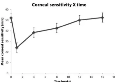
Corneal sensitivity gradually returned to preoperative levels in all treated eyes. The mean central corneal sensitivity was 52.24 (±6.44) mm before surgery, 24.08 (±7.78) mm 7 days after surgery, and 38.29 (±4.23), 42.50 (±3.02), 50.00 (±3.07), and 52.50 (±2.78) mm 1, 2, 3, and 4 months after CXL, respectively. Normal levels of corneal sensation, but not a return to baseline (preoperative) levels, were noted 2 months after surgery (p=0.000). Statistical analysis showed a signifi-cant decrease in corneal sensitivity for up to 3 months after surgery. Preoperative levels were observed at 3 (p=0.081) and 4 months after surgery (p=0.817; Figure 1).
Four months after corneal CXL, 3 eyes (7.9%) experienced 1-line improvement in postoperative BSCVA. It did not change in 33 eyes (86.84%) and decreased by 1 line in 2 eyes (5.2%). No statistically signi-ficant difference was noted between preoperative and postoperative BSCVA values (p=0.571).
Changes in corneal sensitivity following cross-linking for progressive early-stage keratoconus
8 6 Arq Bras Oftalmol. 2014;77(2):84-7
Figure 1. Changes in corneal sensitivity over time.
Table 1. Mean corneal pachymetry values, spherical equivalents,
topo-graphic K-readings (Kmax andKmin), intraocular pressure, and endothelial
cell counts before and after corneal cross-linking
Before corneal CXL
Four months after
surgery p-value
Pachymetry values 478 ± 24.8 µm (440-508 µm)
454 ± 29.06 µm (420-488 µm)
0.00
Spherical equivalents -2.08 (±0.85) D -1.92 (±0.84) D 0.00
Kmax 51.45 ± 1.77 D 50.18 ± 1.64 D 0.00
Kmin 45.49 ± 1.79 D 45.48 ± 1,81 D 0.860
Intraocular pressure 11.66 ± 1.23 mmHg 11.50 ± 1.24 mmHg 0.279
Endothelial cell counts 2280.26 ± 258 mm2 2287 ± 270 mm2 0.549
Four months after surgery, comparison of preoperative and posto-perative results showed a statistically significant difference between some of them. The mean thickness as assessed by central corneal pa chymetry decreased from 478 ± 24.8 µm to 454 ± 29.06 µm (p=0.00). The mean spherical equivalent decreased from -2.08 ± 0.85 D to -1.92 ± 0.84 D (p=0.00), and the mean maximum topographic K-readings (Kmax) dropped from 51.45 ± 1.77 D to 50.18 ± 1.64 D (p=0.00). No other data showed significant differences before and after surgery. The mean minimum topographic K-readings (Kmin) chan ged from 45.49 ± 1.79 D to 45.48 ± 1.81 D (p=0.860). The mean intraocular pressure changed from 11.66 ± 1.23 mmHg to 11.50 ± 1.24 mmHg (p=0.279), and the endothelial cell counts changed form 2280.26 ± 258 to 2287 ± 270 cells/mm2 (p=0.549). The mean patient
data before and after cross-linking are shown in table 1.
No intraoperative, early postoperative, or late postoperative com-plications were observed in this series of patients. After 4 months, all corneas remained transparent, without any scar in the stroma.
DISCUSSION
Many corneal surgical procedures have been associated with a tem porary decrease in corneal sensitivity due to amputation or laser ablation of the corneal nerves. This damage may contribute to the development of dry eye, impaired epithelial wound healing, increased epithelial permeability, and ocular surface changes after the pro -cedure. In such cases, the corneal sensitivity usually returns to nor mal within 3-12 months after surgery(9,13).
Increased visibility of nerve fibers by slit lamp biomicroscopy has been demonstrated in patients with keratoconus. Corneal nerves pass between the stroma and epithelium at sites of early degradative change. The keratocytes wrap around the nerves as they pass through an otherwise acellular Bowman’s layer. Localized nerve thickening develops in the epithelium and stresses the epithelial architecture(14).
In vivo laser scanning confocal microscopy has revealed decreased
innervation and decreased cell density in all layers of the keratoco-nic cornea(15).The sub-basal nerve plexus architecture isaltered with
fragmentation of the plexus, increased sub-basal nerve tortuosity, decreased central nerve fiber density, and a correlation between the decrease in nerve density decrease and severity of disease(16-18).
Ano-ther study(19) reported decreased sensitivity in all corneal zones of
pa-tients with keratoconus, and the decrease depended on the severity of the condition. Injured nerve fibers may quickly regenerate and may exhibit abnormal responses because of the altered expression of ion channel proteins in the regenerating nerve terminals(20).In the current
study group, according to the Amsler-Krumeich classification(10) of
ke ratoconus disease, all eyes were considered to be in early-stage keratoconus: 32 eyes (84.2%) were in stage I and 6 eyes (15.8%) were in stage II. Therefore, faster corneal sensitivity recovery was expected. Corneal CXL with riboflavin and UVA irradiation is a technique of a minimally invasive nature that is used for the stabilization of diffe-rent types of corneal ectatic disorders. This technique augments the mechanical rigidity of the cornea by inducing cross-links at the corneal stroma(3-5). Removal of the epithelium has been recommended as an
initial step in the CXL procedure because its lipophilic nature decreased the diffusion of riboflavin into the corneal stroma(4,5). Moreover, the
epithelium may block the UV rays(3). The photosensitizer riboflavin is
applied to the de-epithelialized surface of the cornea and is allowed to penetrate the corneal stroma(3-6). Subsequent exposure of the cornea
to UVA light is believed to result in photodynamic cross-linking when riboflavin, excited by UVA, creates free radicals leading to collagen cross-linking(4,5).
An in vivo confocal laser scanning microscopy study(16) has
de-mons trated that the anterior subepithelial stroma was recolonized by ner ve fibers, with the restoration of corneal sensitivity, 6 months after CXL using the epithelium-off technique. An animal study(17) of corneal
sensitivity and innervation after CXL in rabbits has also shown that corneal sensitivity was significantly decreased 3 days after UVA irra-diation with de-epithelialization treatment. Corneal sensitivity in the central region of the cornea decreased to the lowest level 7 days after treatment and returned to normal 3 months after the procedure; the corneal nerve fiber density appeared normal after 6 months. A recent prospective, interventional case series(21) evaluated the effects of CXL
on corneal innervation, sensitivity, and tear function in patients with progressive early-stage keratoconus. The study detected prominent hypoesthesia 1 month after surgery, which gradually recovered by 9 months after surgery; the time course was similar for both sensi-tivity restoration and corneal nerve regeneration. Another study(22)
evaluated the changes in corneal tactile sensitivity following corneal collagen cross-linking in patients with progressive keratoconus and concluded that corneal CXL performed in keratoconus patients in-duced a considerable decrease in corneal sensitivity. This decrease was greater in the first week after the procedure, with progressive recovery for up to 6 months of follow-up. Our results demonstrated that normal levels of corneal sensation that were not yet baseline (preoperative) levels were observed two months after surgery, and significant transient hypoesthesia was observed for as long as 3 months after CXL. The difference in the time of corneal sensation recovery between the different studies may be related to other factors such as the degree of ectasia, contact lens use, and systemic disease that can affect epithelial healing or corneal sensitivity.
Contact lens wear decreases corneal sensitivity(23). To minimize any
Lago AM, et al.
8 7
Arq Bras Oftalmol. 2014;77(2):84-7 Corneal sensation is also decreased in patients with diabetes and
mild to moderate somatic neuropathy, and it progresses with the severity of neuropathy. These findings have important clinical im pli-cations with regard to the development of corneal abnormalities in patients with diabetes(24). Therefore, patients with systemic di sease
that can affect epithelial healing and corneal sensitivity, such as dia-betes, were excluded from our study.
An important finding of this study was a significant decrease in corneal thickness after corneal CXL, which may also have contributed to the decrease in corneal sensitivity. Our group(25) recently described
transient corneal thinning after corneal CXL with UVA irradiation and a hypo-osmolar riboflavin solution in thin corneas (<400 µm) for pro-gressive keratoconus. In our analysis of changes in corneal thickness over time, pachymetric values decreased until 1 month after surgery and appeared to increase thereafter. Six months after treatment, no statistically significant difference was found between postoperative and baseline values. The physiology of this initial thinning and subse-quent rethickening remains unclear. Epithelial removal may increase the rate of water evaporation from the stroma and renders the cornea vulnerable to thinning because the stroma has no dehydration resis-tance(26). Epithelial remodeling, anatomical and structural changes in
corneal collagen fibrils(27), and keratocyte apoptosis(28) may also be
im plicated. Surgeons must be aware about postoperative thinning of the cornea after CXL, particularly in thinner corneas. Ours results suggest that further studies are necessary to evaluate the relationship between the decrease incorneal thickness and transient corneal hy -poesthesia after CXL.
A few potential limitations were apparent in this study, with the small sample of studied eyes and the absence of a control group of patients being the major ones. However, because all patients pre -sented with bilateral progressive keratoconus, it was considered une thical to leave the disease untreated in one eye. Esthesiometry is a reproducibly accurate measure of corneal sensation. The most popular device for this purpose is the Cochet-Bonnet esthesiometer, which comprises a calibrated nylon filament for mechanical stimula-tion(11).It has limitations in its sensitivity as a test, but it is the most
prac tical method available(12). The conclusions of this study should be
interpreted within the limits of this method of evaluation.
Touching the cornea triggers one of the most sensitive protective reflexes of the human body. The threshold of sensitivity, particularly in the center of the cornea, is exceedingly low; therefore, pathological changes can be diagnosed early and precisely and can be used for diagnosis and follow-up and to assess the prognosis of various cor-neal disorders. The loss of normal corcor-neal sensation may compromise the protective blink reflex, delay epithelial wound healing, decrease tear flow, and show an association with neurotrophic keratitis, sterile corneal melts, and infectious keratitis(11,28).
CONCLUSIONS
The results of this study emphasized the slow and gradual recovery of corneal sensitivity after CXL, as observed in other studies pu -blished in the literature. These observations can help to direct studies to better define the risk and management of persistent epitheliopa-thy and dry eye symptoms after CXL.
REFERENCES
1. Rabinowitz YS. Keratoconus. Surv Ophthalmol. 1998;42(4):297-319.
2. Romero-Jiménez M, Santodomingo-Rubido J, Wolffsohn JS. Keratoconus: a review. Cont Lens Anterior Eye. 2010;33(4):157-66.
3. Wollensak G, Spoerl E, Seiler T. Ribofloavin/ultraviolet-A-induced collagen cosslinking for the treatment of keratoconus. Am J Ophthalmol. 2003;135(5):620-27.
4 Spoerl E, Huhle M, Seiler T. Induction of cross-links in corneal tissue. Exp Eye Res. 1998; 66(1):97-103.
5. Wollensak G, Spoerl E, Seiler T. Stress-strain measurements of human and porcine corneas after riboflavin-ultraviolet-A-induced cross-linking. J Cataract Refract Surg. 2003;29(9):1780-5.
6. Hafezi F, Kanellopoulos J, Wiltfang R, Seiler T. Corneal collagen cross-linking with ribo-flavin and ultraviolet A to treat induced keratectasia after laser in situ keratomileusis. J Cataract Refract Surg. 2007;33(12):2035-40.
7. Salgado J, Khoramnia R, Lohmann C, Winkler von Mohrenfels C. Corneal collagen cross linking in post-LASIK keratectasia. Br J Ophthalmol. 2011;95(4):493-7. 8. Marfurt CF, Cox J, Deek S, Dvorscak, L. Anatomy of the human corneal innervation.
Exp Eye Res. 2010;90(4):478-92.
9. Erie JC, McLaren JW, Hodge DO, Bourne WM. Recovery of corneal subbasal nerve density after PRK and LASIK. Am J Ophthalmol. 2005;140(6):1059-64.
10. Alió JL, Shabayek MH. Corneal higher order aberrations: a method to grade kerato-conus. J Refract Surg. 2006;22(6):539-45.
11. Cochet P, Bonnet R, L’ésthésie cornéenne. Sa mesure clinique. Ses variations physio-logiques et pathophysio-logiques. Clin Ophtalmol. 1960;4(1):3-27.
12. Murphy PJ, Lawrenson JG, Patel S, Marshall J. Reliability of the non-contact cor-neal aesthesiometer and its comparison with the Cochet–Bonnet aesthesiometer. Ophthal Physiol Opt. 1998;18(6):532-9.
13. Nassaralla BA, McLeod SD, Boteon JE, Nassaralla JJ. The effect of hinge position and depth plate on the rate of recovery of corneal sensation following LASIK. Am J Ophthalmol. 2005;139(1):118-24.
14. Dogru M, Karakaya H, Ozçetin H, Erturk H, Yucel A, Ozmen A, et al. Tear function and ocualr surface changes in keratoconus. Ophthalmology. 2003;110(6):1110-8. 15. Niederer RL, Perumal D, Sherwin T, McGhee NJ. Laser scanning in vivo confocal
microscopy reveals reduced innervation and reduction in cell density in all layers of the keratoconic cornea. Invest Ophthalmol Vis Sci. 2013;49(7):2964-70.
16. Mazzotta C, Traversi C, Baiocchi S, Caporossi O, Bovone C, Sparamo M, et al. Corneal healing after riboflavin ultraviolet-A collagen cross-linking determined by confocal laser scanning microscopy in vivo: early and late modifications. Am J Ophthalmol. 2008;146(4):527-3.
17. Xia Y, Chai X, Zhou C, Ren Q. Corneal nerve morphology and sensitivity changes after ultraviolet A/riboflavin treatment. Exp Eye Res. 2011;93(4):541-7.
18. Kymionis GD, Diakonis VF, Kalyvianaki M, Pallikaris IG. One-year follow-up of corneal confocal microscopy after corneal cross-linking in patients with post laser in situ keratomileusis ectasia and keratoconus. Am J Ophthalmol. 2009;147(5):774-8. 19. Bleshoy H. Corneal sensitivity in keratoconus. J Br Contact Lens Assoc. 1986;9:9-12. 20. Belmonte C, Acosta MC, Gallar J. Neural basis of sensation in intact and injured
corneas. Exp Eye Res. 2004;78(3):513-25.
21. Kontadakis GA, Kymionis GD, Kankariya VP, Pallikaris AI. Effect of corneal collagen cross-linking on corneal innervation, corneal sensitivity, and tear function of patients with keratoconus. Ophthalmology. 2013;120(5):917-22.
22. Wasilewski D, Mello HR, Moreira H. Impact of collagen crosslinking on corneal sensi-tivity in keratoconus patients. Cornea 2013;(7):899-902.
23. Martin XD, Safran AB. Corneal hypoesthesia. Surv Ophtalmol. 1988;33(1):28-40; erratum p.217.
24. Mitra T, Panagiotis A, Kallinikos NE, Andrew JM, Boulton RA. Corneal sensitivity is reduced and relates to the severity of neuropathy in patients with diabetes. Diabetes Care. 2007;(30):1895-7.
25. Nassaralla BA, Vieira DM, Machado ML, Figueiredo MN, Nassaralla JJ Jr. Corneal thickness changes during corneal collagen cross-linking with UV-A irradiation and hypo-osmolar riboflavin in thin corneas. Arq Bras Oftalmol. 2013;76(3):155-8.
26. Wollensak G, Aurich H, Pham D-T, Wirbelauer C. Hydration behavior of porcine cornea cross-linked with riboflavin and ultraviolet A. J Cataract Refract Surg. 2007;33(3):516-21. 27. Hafezi F, Mrochen M, Iseli HP, Seiler T. Collagen cross linking with ultraviolet-A and
hypo-osmolar riboflavin solution in thin corneas. J Cataract Refract Surg. 2009;35(4): 621-24. 28. Wollensak G, Spoerl E, Reber F, Seiler T. Keratocyte cytotoxicity of