chance of cure.(3) However, even in clinical stage IA (T1N0M0), 30-40% of patients will die as a consequence of the progression of the cancer,(4-6) especially due to systemic recurrence. Therefore, the identification of the different histological types and of prognostic factors is important and has the purpose of selecting patients for receiving appropriate treatment, in addition to identi-fying tumors at higher risk of recurrence. These selected patients would potentially benefit from therapeutic approaches directed at the histo-logical type, as well as from adjuvant therapies aimed at avoiding cancer recurrence and fatal outcomes. The intention of such classification systems is to provide light microscopy criteria that allow pathologists to establish consistent diagnoses among themselves. With such disci-pline, comparatively, treatment protocols can then be established. When the criteria proposed by these classification systems are observed,
Classification
Carcinomas account for nearly 95% of all cases of lung cancer, whereas sarcomas and lymphomas account for most of the remainder. Clinically, lung carcinomas are classified as non-small cell lung cancer (NSCLC) or non-small cell lung cancer. According to the current World Health Organization classification,(1) NSCLC includes the following histological types: squamous cell carcinoma; adenocarcinoma; and large cell carcinoma. In the same classification, small cell carcinomas are considered neuroendocrine carcinomas, as are the following histological types: typical carcinoid tumor, atypical carcinoid tumor and large cell neuroendocrine carcinoma. Accounting for 75-80% of all cases, NSCLC is the more common of the two types.(2) Clinical staging and identification of the histological type are fundamental to establishing the thera-peutic strategy for treating such patients. In earlier stages, surgical resection offers a real
Role of immunohistochemistry in the diagnosis of lung cancer*
Papel da imuno-histoquímica no diagnóstico do câncer de pulmãoVera Luiza Capelozzi
Abstract
The role of immunohistochemistry is to recognize antigens and, consequently, to identify and classify specific cells within a cell population whose morphology is heterogenous or apparently homogenous. The visualization of the antigen-antibody complex is made possible through the addition of either a fluorochrome conjugate or an enzyme to the antibody, which is then viewed under microscopy. Immunohistochemistry can be used in the routine diagnosis of lung cancer, in order to identify biological markers (diagnostic and prognostic). The essential immunohistochemistry panels will be discussed in this review.
Keywords: Immunohistochemistry; Tumor markers, biological; Lung neoplasms.
Resumo
O propósito da imuno-histoquímica é reconhecer antígenos e assim identificar e classificar células específicas dentro de uma população celular morfologicamente heterogênea (ou aparentemente homogênea). A visualização do complexo antígeno-anticorpo é possível pela adição de um fluorocromo conjugado ao anticorpo, que pode então ser observado ao microscópio, ou alternativamente uma enzima, cujo produto de reação pode igualmente ser visualizado. A imuno-histoquímica pode ser aplicada na rotina diagnóstica complementar do câncer de pulmão para a identificação de marcadores biológicos diagnósticos e prognósticos. Os painéis imuno-histoquímicos mínimos necessários para a complementação diagnóstica serão discutidos nesta revisão.
Descritores: Imunoistoquímica; Marcadores biológicos de tumor; Neoplasias pulmonares.
* Study carried out in the Department of Pathology, Faculdade de Medicina Universidade de São Paulo – FMUSP, University of São Paulo School of Medicine – São Paulo, Brazil.
Correspondence to: Vera Luiza Capelozzi. Av. Dr. Arnaldo, 455, Sala 1143, CEP 01246-903, São Paulo, SP, Brasil. Tel 55 11 3061-7427. E-mail: vcapelozzi@lim05.fm.usp.br
Financial support: None.
tumors as one of the four major histological types: squamous cell carcinoma, adenocarci-noma, neuroendocrine carcinoma and large cell carcinoma. When technical problems related to sampling, artifactual problems or problems related to undifferentiated cells do not allow a definitive classification, every effort should be made to identify, at least, the histogenesis (carcinoma vs. sarcoma vs. lymphoma) and separate undifferentiated large cell tumors from undifferentiated small cell tumors. It is true that, in some situations, it is not possible to make even these broad categorizations, due to lack of analyzable material. In this context, immunohis-tochemistry is an indispensable complementary tool, as we will here discuss in detail.
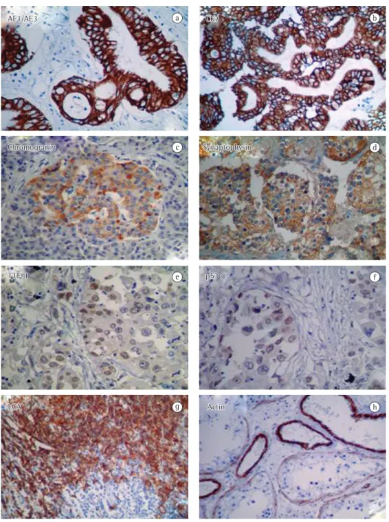
Although the reliability rate for neuroendo-crine carcinoma, especially small cell carcinoma, is approximately 85%, in 15% of the cases, it is necessary to perform an immunohistochem-istry panel including the following markers(1,7): high- and low-molecular-weight cytokeratins (AE1/AE3, Figure 1a); cytokeratins specific for primary pulmonary origin (CK7, Figure 1b); cytokeratins for primary gastrointestinal tract origin (CK20); markers of neuroendocrine differ-entiation (chromogranin, synaptophysin and CD56; Figures 1c and 1d); TTF-1 (Figure 1e); p53 (Figure 1f); and markers of lymphoid origin (leukocyte common antigen; Figure 1g). markers of mesenchymal origin (vimentin and smooth muscle actin; Figure 1h); The use of CK7 and CK 20 also offers the opportunity to establish whether the tumor is a primary pulmonary lesion (CK7+ and CK20−) or a metastatic lesion of the digestive tract (CK7− and CK20+).
Neuroendocrine differentiation
Once a diagnosis of neuroendocrine tumor has been established, it is necessary to classify the tumor as a high-grade neuroendocrine tumor (small cell carcinoma or large cell neuroendo-crine carcinoma) or a low-grade neuroendoneuroendo-crine tumor (typical carcinoid tumor or atypical carci-noid tumor).(7)
Typical carcinoid tumor or low-grade
neuroendocrine carcinoma
Typical carcinoid tumor or low-grade neuroendocrine carcinoma is characterized by 0 or 1 mitoses per 2 mm2, absence of necrosis, the diagnostic reliability among experienced
pathologists is 90%.(1) The high inter-rater vari-ability is found in the identification of large cell carcinoma in cases of squamous cell carcinoma and poorly differentiated adenocarcinoma. In this context, immunohistochemistry represents a tool that can provide a clear distinction among the various types.
Immunohistochemistry
The objective of performing immunohis-tochemistry is to recognize cell constituents (antigens) and, consequently, to identify and classify specific cells within a cell population whose morphology is heterogenous or appar-ently homogenous. The visualization of the antigen-antibody complex is made possible through the addition of either a fluorochrome conjugate or an enzyme to the antibody, which is then viewed under microscopy. In some cases, the antibodies are not entirely specific and can cross-react with more than one antigen, or there can be false-positive and false-negative results, which are caused by high levels of background staining and absorption. In addition, some cells can express antigens typically associated with completely different types, such as prostate-specific antigen in the pancreas or lymphoid antigens in lung tumors. Therefore, appropriate sections of control tissues (such as lymph nodes or tonsils for lymphoid antigens) are always processed together with lung sections, and these controls are always checked before the evalua-tion of a lung tumor.
Immunohistochemistry can be used in the routine diagnosis of lung cancer in order to identify biological markers (diagnostic and prog-nostic). Most pathological anatomy laboratories have the essential immunohistochemistry panels necessary to complement the diagnosis.
Figure 1 illustrates an immunohistochem-istry panel for epithelial markers (cytokeratin AE1/AE3 and cytokeratin 7—CK7), neuroendo-crine markers (chromogranin, synaptophysin, thyroid transcription factor-1 [TTF-1] and p53) and mesenchymal markers (leukocyte common antigen and smooth muscle actin).
Biological diagnostic markers
AE1/AE3
Chromogranin c Synaptophysin d
e
TTF-1 p53 f
g
LCA Actin h
a CK7 b
and CK7+); primary digestive tract tumor (CK20+); primary thyroid tumor (positive for thyroglobulin); primary prostate tumor (posi-tive for prostate-specific antigen—PSA); primary breast tumor (positive for estrogen receptors, progesterone receptors and Br-est); and primary ovarian tumor (CA125+). Primary or metastatic squamous cell carcinomas of the lung are usually positive for high-molecular weight cytokeratin 34βE12, cytokeratin 5/6 (CK5/CK6) and low-molecular weight cytokeratin 35βH11—few cases are TTF-1+ and CK7+.(1) To date, no immuno-histochemistry markers to differentiate primary carcinomas from metastatic carcinomas have been identified. One group of authors demon-strated the importance of p63 protein expression, as well as of CK5/CK6 and CK7 expression and surfactant protein A (SP-A) expression, in the classification of 42 types of NSCLC in autopsy and surgical resection specimens.(8) The authors also showed the usefulness of these markers in differentiating squamous cell carcinomas from adenocarcinomas based on the implications for prognosis and treatment. All adenocarcinomas were p63−, 9/16 (56.2%) were CK5/CK6+, 16/17 (94.1%) were CK7+, and 4/15 (26.6%) were SP-A+. Of the squamous cell carcinomas, 1 case was CK7+ and SP-A+, and 14/18 (77.8%) were p63+. The authors concluded that p63 protein expression is useful in differentiating between the two histological types.
Bronchioloalveolar carcinoma
Among the various histological subtypes of adenocarcinoma, the one that should be prioritized, in terms of identification, is bron-chioloalveolar carcinoma, due to its prognostic implications. The most significant histological characteristic of bronchioloalveolar carcinoma is growth along the alveolar walls, without destruction of the lung architecture. Although histological recognition of bronchioloalveolar carcinoma does not require the use of comple-mentary techniques, immunohistochemical studies typically reveal expression of TTF-1 and SP-A.
Biological prognostic markers
Immunohistochemistry can be used in the routine diagnosis of bronchogenic carcinoma in order to identify biological prognostic markers. neuroendocrine structures (rosettes, trabeculae
and solid nests) and chromogranin expression in the cytoplasm.
Atypical carcinoid tumor or
low-grade neuroendocrine carcinoma
Atypical carcinoid tumor or low-grade neuroendocrine carcinoma is defined by 2-10 mitoses per 2 mm2, necrosis, neuroendo-crine structures (rosettes, trabeculae and solid nests) and chromogranin expression in the cytoplasm.
Large cell neuroendocrine carcinoma
Large cell neuroendocrine carcinoma is defined by numerous mitoses (typically more than 30 per 2 mm2); large nuclei (25-35 µm); an organoid structure, similar to that of a carcinoid tumor; extensive areas of necrosis; and positivity (in more than 25% of the cells) for chromogranin, synaptophysin and other neuroendocrine markers.
Small cell neuroendocrine carcinoma
Small cell neuroendocrine carcinoma is defined by numerous mitoses, an organoid structure, a nuclei between 16-23 µm, dense heterochromatin, invisible nucleoli, an invis-ible or scarce cytoplasm and positivity for the neuroendocrine markers chromogranin, synap-tophysin and TTF-1.
Non-neuroendocrine differentiation
Non-small cell carcinoma
positive impact on survival among patients who underwent surgery. The authors also found that lymph node involvement was strongly associated with high p53 and AgNOR expression, as well as that p53 and AgNOR are major parameters in determining prognosis in such patients. More recently,(12) it was demonstrated that immuno-histochemical expression of p53 correlates with lower survival but not with gene mutations in resected tumors.
c-erbB-1
The c-erbB-1 proto-oncogene, when linked to the membrane, encodes a growth receptor (170-kDa tyrosine kinase) that is the receptor for the epidermal growth factor receptor (EGFR). In the normal lung, c-erbB-1, through its protein product, acts to stimulate epithelial cell prolif-eration and to promote airway maturation during embryonic development. This proto-on-cogene has been shown to be overexpressed in NSCLC, especially in squamous cell carcinomas (in 65-90% of the cases reported). The use of c-erbB-1 gene overexpression as a marker for clinical prognosis is controversial, some studies showing an association with low survival and others showing no such association.
c-erbB-2
The c-erbB-2 proto-oncogene belongs to the c-erbB family of receptors linked to the tyrosine kinase membrane, it is structurally related to the c-erbB-1 proto-oncogene. The protein that encodes c-erbB-2 is known as p185c-erbB-2, or HER2. This protein is expressed in the epithelial cells of the airways of the normal lung, playing a role in the growth and differentiation of the normal lung epithelium. It has been observed that HER2 is produced in conjunction with EGFR in many lung adenocarcinomas. One group of authors(13) recently demonstrated that survival is strongly associated with cyclin D1, c-erbB-2 and VEGF expression in resected primary adenocar-cinomas and their metastases.
MMP-9
Before there are metastases, a cell or a group of cells must detach themselves from the primary tumor, penetrate the adjacent tissue, survive and grow in the host tissue. Squamous cell carci-noma is the only known histological subtype Although there is no unanimous panel to detect
lung cancer in our diagnostic routine, we have been using p53, c-ERB, vascular endothelial growth factor (VEGF), cyclooxygenase (COX) and matrix metalloproteinase 9 (MMP-9).
p53
submitted to surgery due to bronchial carci-noid tumor and monitored for more 10 years in two university hospitals.(18) The authors used the following data to predict the individual risk for node metastases or distant metastases: age; gender; tumor-node-metastasis staging; tumor diameter; tumor location (central or peripheral); immunohistochemical index for p53, Ki67 and Bcl-2; and density of newly formed microvessels and fibers of the collagen-elastic system. The incidence of lymph node metastases and distant metastases was 11% and 5%, respectively. The univariate analysis associated biomolecular markers with node metastases. The multivariate analysis identified neoangiogenesis as a predic-tive marker for node metastasis. Other important interactions were demonstrated by one group of authors(19) who, while studying how the charac-teristics of the tumor cell relate to stroma for proliferative factors, p53, vascular microdensity and metalloproteinases, controlled for extent of the primary lesion (T1 to T4), in limited (non-metastatic) NSCLC and advanced ((non-metastatic) NSCLC, suggested that different interactions between the tumor cells and the stromal cells control the metastases and, therefore, control the biological risk for NSCLC.
VEGF
Angiogenesis represents an important factor since it allows tumor growth, tumor invasion and metastases. The genes or molecules that participate in tumor invasion and metastases are regulating factors that have a positive or nega-tive character. The VEGF promotes the formation of blood vessels at the beginning of the develop-ment (vasculogenesis) and plays a central role in the growth of new blood vessels (angiogenesis). Its role in tumor growth is fundamental, since, without vascularization, tumors cannot exceed 1-2 mm in diameter or in thickness. In addition, angiogenesis is indispensable for the formation of metastases. Two of the greatest inducers of angiogenesis produced in lung cancer are VEGF and fibroblast growth factor.(20) There have been various studies showing the relationship between high VEGF levels and poor prognosis in cases of NSCLC.(13,20) One group of authors(21) recently demonstrated the importance of VEGF as a prognostic marker in adenocarcinomas and their hematogenous metastases.
stromal proportion and PCNA, Ki-67, and p53 proteins in patients with resected adenocarcinoma of the lung. Mod Pathol. 2000;13(5):511-20.
11. Carvalho PE, Antonãngelo L, Bernardi FD, Leão LE, Rodrigues OR, Capelozzi VL. Useful prognostic panel markers to express the biological tumor status in resected lung adenocarcinomas. Jpn J Clin Oncol. 2000;30(11):478-86.
12. Massoni Neto LM, Bianchi CP, Ab’Saber AM, Parra ER, Takagaki T, Pereira JC, et al. p53 immunostaining is correlated with reduced survival and is not correlated with gene mutations in resected pulmonary large cell carcinomas. Braz J Med Biol Res. 2007;40(8):1045-53. 13. Saito DM, Franco MR, Parra ER, Ab’saber AM, Farhat
C, Eher E, et al. Cell cycle regulator in primary lung adenocarcinoma and its haematogenous metastases. Histopathology. 2007;50(4):525-7.
14. Pinto CA, Carvalho PE, Antonângelo L, Garippo A, Da Silva AG, Soares F, et al. Morphometric evaluation of tumor matrix metalloproteinase 9 predicts survival after surgical resection of adenocarcinoma of the lung. Clin Cancer Res. 2003;9(8):3098-104.
15. Yamaguchi NH, Lichtenfels AJ, Demarchi LM, da Silva AP, Garippo AL, Alves VF, et al. COX-2, MMP-9, and Noguchi classification provide additional prognostic information about adenocarcinoma of the lung. A study of 117 patients from Brazil. Am J Clin Pathol. 2004;121(1):78-86.
16. Martins SJ, Takagaki TY, Silva AG, Gallo CP, Silva FB, Capelozzi VL. Prognostic relevance of TTF-1 and MMP-9 expression in advanced lung adenocarcinoma. Lung Cancer. 2009;64(1):105-9.
17. das Neves Pereira JC, da Silva AG, Soares F, Ab’Saber AM, Schmidt A, Rodrigues OR, et al. Nuclear and environment morphometric profile in tumor size and nodal metastasis of resected typical pulmonary carcinoid. Pathol Res Pract. 2004;200(6):459-67.
18. Das-Neves-Pereira JC, Bagan P, Milanez-de-Campos JR, Capelozzi VL, Danel C, Jatene FB, et al. Individual risk prediction of nodal and distant metastasis for patients with typical bronchial carcinoid tumors. Eur J Cardiothorac Surg. 2008;34(3):473-7; discussion 477-8.
19. Minamoto H, Antonângelo L, da Silva AG, Gallo CP, de Andrade e Silva FB, Fenezelian S, et al. Tumour cell and stromal features in metastatic and non-metastatic non-small cell lung carcinomas. Histopathol. 2003;43(5):427-43
20. Fontanini G, Vignati S, Boldrini L, Chinè S, Silvestri V, Lucchi M, et al. Vascular endothelial growth factor is associated with neovascularization and influences progression of non-small cell lung carcinoma. Clin Cancer Res. 1997;3(6):861-5.
21. Parra ER, Park JY, Saito DM, Takagaki TY, Rodrigues OR, Capelozzi VL. Prognostic index expression of cyclin-D1, cerbB-2 and VEGF: metastases vs corresponding primary cancers and metastatic vs non-metastatic adenocarcinomas. Histol Histopathol. 2008;23(8):987-93.
Final considerations
Immunohistochemistry represents an impor-tant complementary tool for the routine diagnosis of lung cancer and for the identification of the different histological types and prognostic factors. Its purpose is to categorize patients in order to ensure appropriate and specific treat-ment, as well as to identify tumors at higher risk of recurrence and fatal outcomes. The essential immunohistochemistry panel recommended for the diagnosis and prognosis includes expression of the following markers: CK7; CK20; COX-2; TTF-1, chromogranin; synaptophysin, CD56; PSA; CA125; p53; c-erbB-2; MMP-9; and VEGF.
References
1. Travis WD; World Health Organization; International Agency for Research on Cancer; International Association for the Study of Lung Cancer; International Academy of Pathology; et al. Pathology and genetics of tumours of the lung, pleura, thymus, and heart. Lyon: IARC Press, 2004.
2. Hinson KF, Miller AB, Tall R. An assessment of the World Health Organization classification of the histologic typing of lung tumors applied to biopsy and resected material. Cancer. 1975;35(2):399-405.
3. Anelli A. Quimioterapia em câncer de pulmão de pequenas células. In: Younes RN, editor. Tumores Torácicos. Rio de Janeiro: Medsi. 1997. p. 269-80. 4. Nunes RN, Gross JL, Deheinzelin D. Follow-up in
lung cancer: how often and for what purpose? Chest. 1999;115(6):1494-9.
5. Mountain CF. Revisions in the International System for Staging Lung Cancer. Chest. 1997;111(6):1710-7. 6. Naruke T, Tsuchiya R, Kondo H, Asamura H, Nakayama
H. Implications of staging in lung cancer. Chest. 1997;112(4 Suppl):242S-248S.
7. Farhat CA, Parra ER, Rogers AV, Elian SN, Sheppard MN, Capelozzi VL. Using electron microscopy and multivariate cluster analysis to determine diagnosis and prognosis in cases of neuroendocrine lung carcinoma. J Bras Pneumol. 2008;34(10):804-11.
8. Camilo R, Capelozzi VL, Siqueira SA, Del Carlo Bernardi F. Expression of p63, keratin 5/6, keratin 7, and surfactant-A in non-small cell lung carcinomas. Hum Pathol. 2006;37(5):542-6.
9. Mao L, Hruban RH, Boyle JO, Tockman M, Sidransky D. Detection of oncogene mutations in sputum precedes diagnosis of lung cancer. Cancer Res. 1994;54(7):1634-7.
About the authors
Vera Luiza Capelozzi