ABSTRACT
effects of etching and adhesive applications on
the bond strength between composite resin and
glass-ionomer cements
Tijen PAMIR1, Bilge Hakan ŞEN2, Özgür EVCIN3
1- DDS, PhD, Professor, Department of Restorative Dentistry, School of Dentistry, Ege University, Bornova, Izmir, Turkey. 2- DDS, PhD, Professor, Department of Endodontology, School of Dentistry, Ege University, Bornova, Izmir, Turkey.
3- DDS, PhD, Private Dentist, (Former Research Assistant in Department of Restorative Dentistry), School of Dentistry, Ege University, Bornova, Izmir, Turkey.
Corresponding address: Tijen Pamir - Ege University School of Dentistry, Department of Restorative Dentistry, 35100 - Bornova - Izmir - Turkey - Phone: +90-232-388-03-28 - Fax: +90-232-388-03-25 - E-mail: [email protected]
Received: March 13, 2012 - Modiication:August 16, 2012 - Accepted: September 14, 2012
O
bjective: This study determined the effects of various surface treatment modalities on the bond strength of composite resins to glass-ionomer cements. Material and Methods: Conventional (KetacTM Molar Quick ApplicapTM) or resin-modiied (PhotacTM Fil QuickAplicapTM) glass-ionomer cements were prepared. Two-step etch-rinse & bond adhesive
(AdperTM Single Bond 2) or single-step self-etching adhesive (AdperTM PromptTM L-PopTM)
was applied to the set cements. In the etch-rinse & bond group, the sample surfaces were pre-treated as follows: (1) no etching, (2) 15 s of etching with 35% phosphoric acid, (3) 30 s of etching, and (4) 60 s of etching. Following the placement of the composite resin (FiltekTM Z250), the bond strength was measured in a universal testing machine and the
data obtained were analyzed with the two-way analysis of variance (ANOVA) followed by the Tukey’s HSD post hoc analysis (p=0.05). Then, the fractured surfaces were examined by scanning electron microscopy. Results: The bond strength of the composite resin to the
conventional glass-ionomer cement was signiicantly lower than that to the resin-modiied glass-ionomer cement (p<0.001). No signiicant differences were determined between the
self-etching and etch-rinse & bond adhesives at any etching time (p>0.05). However, a greater bond strength was obtained with 30 s of phosphoric acid application. Conclusions:
The resin-modiied glass-ionomer cement improved the bond strength of the composite
resin to the glass-ionomer cement. Both etch-rinse & bond and self-etching adhesives may be used effectively in the lamination of glass-ionomer cements. However, an etching time of at least 30 s appears to be optimal.
Key words: Glass-ionomer cements. Composite resins. Acid etching. Dental adhesives.
INTRODUCTION
The extensive range of tooth-colored materials for esthetic restorations on the market are mainly branches of two separate groups – glass-ionomer cements and composite resins - each with its own sub-groups16. Following their introduction
in the late 1960s, composite resins have gained general respect, due to their esthetically pleasing appearance and stability within the oral environment. However, they have several disadvantages, such as polymerization shrinkage, potential failure of adhesion leading to secondary caries, and a relatively
high co-eficient of thermal expansion in vitro12. In
contrast, some researchers have suggested that glass-ionomer cements offer stronger inhibition of secondary caries compared with composite resin7,28, since they act as a reservoir for luoride release17.
The reaction of glass-ionomer cements display a similar behaviour to dentin under thermal stimuli2.
This feature plays an important role during the mechanical and thermal loading of the material. In contrast, resin materials show a deformation different from that of the dentin under mechanical and thermal stress20. Therefore, due to its
glass-ionomer cement has been recommended as a restorative material for the lamination (sandwich) technique16,29.
The lamination technique can be applied for all deep and undermined cavities where composite resin is the primary choice. In particular cases where one or more margins of restoration exist on the dentin, lamination over a glass-ionomer cement is strongly recommended to enhance adhesion to the dentin and limit microleakage. In this technique, the glass-ionomer cement represents the lost dentin, although composite resin is used instead of enamel. Two types of glass-ionomer cements,
i.e., conventional or resin-modiied, can be used
for this purpose, even though they have differences in the adhesion mechanism, setting reaction, and sensitivity to the moisture of the materials. The expectations from the lamination technique are to combine the advantages of both glass-ionomer cements and composite resins to enhance the clinical serviceability of the restoration16,20. When the current
diversity of adhesive systems and differences in adhesion and setting mechanisms of the restorative materials are taken into consideration, the bonding of the laminated materials to each other becomes important, as well as the bonding of any material to the tooth structures. Until now however, researchers have not reached a consensus on the type and duration of surface treatment modalities over glass-ionomer cements. Therefore, in this study, our null hypotheses were that adhesive type and duration of the acid-etching procedure would not affect the bond strength of the composite resin to glass-ionomer cements. Furthermore, the type of glass-ionomer
cement would not critically inluence the bonding
quality.
MATERIAL AND METhODS
Study design and surface treatment modalities over the glass-ionomer cement (GIC) samples were performed as presented in Figure 1. One hundred
and ifty specimens - 75 each of conventional (C-GIC)
(Ketac™ Molar Quick Applicap™, 3M eSPe AG Dental Product, Seefeld, Germany) and resin-modified glass-ionomer cement (RM-GIC) (Photac™ Fil Quick Applicap™, 3M eSPe AG Dental Product, Seefeld, Germany) - were prepared in Delrin (acetal resin) moulds containing cylinders 4 mm in diameter and 6 mm long. During the setting, the bottoms and tops of the moulds were covered with cellulose acetate strips and glass microscope slides under hand pressure, to produce a smooth surface. For the C-GIC, the assembly was held in place for 10 min, whereas the RM-GIC was light-cured for 20 s on each side of the material in a LeD (light-emitting diode) dental curing apparatus (Bluephase, Ivoclar/Vivadent, Schaan, Liechtenstein) having 1100 mW/cm2 light intensity.
After polymerization, the cylinders were removed from the moulds, and the bonding surfaces were
ground lat with 600-grit Al2O3 papers (Buehler,
Lake Bluff, IL, USA). Both the C-GIC and RM-GIC samples were divided into 5 groups, each consisting of 15 samples.
Following the surface treatment procedure, the samples were placed into second split Delrin moulds with holes 4 mm in diameter and 12 mm in depth, and composite resin material Filtek Z250 (3M eSPe AG Dental Products, Seefeld, Germany) was incrementally added on top of the GIC samples. A LeD curing unit was used to polymerize the composite resin layers for 20 s at a LOF (low-power curing mode with 650 mW/cm2 light intensity) plus 15
s at a HIF mode (high-power with 1100 mW/cm2 light
intensity). All experimental procedures were carried out at room temperature, and cylindrical specimens of each material were fabricated according to the manufacturer’s instructions. After curing, the second split mould was removed, and samples were kept in a 100% humidity incubator at 37°C for 48 h.
The shear bond strength of each specimen was measured in a universal testing machine (Shimadzu Autograph, AGS-J 5 kN, Shimadzu Corporation, Tokyo, Japan) at a cross-head speed of 0.5 mm/ min. The knife edge blade was applied vertically against the specimens to load 0.5 mm away from the bonding area of the materials. The specimens were loaded until failure and the failure load was converted to failure stress the TRAPeZIUM2 software program (Shimadzu Corporation, Tokyo, Japan) on the computer connected to a universal testing machine. Mean and standard deviations were calculated, and the two-way analysis of variance (ANOVA) was used
to determine the signiicance of differences among
the GICs and the surface treatment modalities. Pair-wise comparisons were performed with the Tukey’s
HSD post hoc analysis (p=0.05).
Visual examination of the failure modes of the bonding specimens was accomplished by viewing all of the de-bonded specimens under a light microscope
at x10 or x20 magniication. Accordingly, the samples
were sorted into 3 groups: Fractures were called “adhesive failure” when the composite resin was removed from the glass-ionomer surfaces without residual debris, “cohesive failure” when fracture occurred inside the restoratives, and “mixed failure” when both occurred. Scanning electron microscopy (SeM) (JeOL-5200, Tokyo, Japan) was used to observe the fractured surfaces of three samples from each group.
RESULTS
As a result of the statistical analysis, it was determined that the interaction between the GICs
Glass- ionomer cements
Surface treatment modalities Self-etch
(No rinse)
Etch, rinse & bond All in one Adper™ Prompt™ L-Pop™
(3M ESPE Dental Products,
Seefeld-Germany) Scotchbond™ Etch + Adper™ Single Bond 2
(3M ESPE Dental Products,Seefeld-Germany)
%35 phosphoric acid
No etch 15 s 30 s 60 s
Ketac Molar Quick Aplicap The capsules were activated and mixed using VariMix at medium frequency for 7 s. Then GICs were placed into
the mould and waited 10 min before any surface
treatment application.
Adper Single Bond 2 was directly brushed
onto the moist samples at 2-3 consecutive coats
for 15 s, and dried gently for 5 s. Then light cured with LED at LOF
mode for 10 s.
35% phosphoric acid was applied onto the GIC samples for 15 s,
washed and rinsed, blew excess water off
leaving the GIC samples moist. Adper Single Bond 2 application was same as described previously.
Acid etching with 35% phosphoric acid for 30 sec,
and then Adper Single
Bond 2 application in same manner.
Acid etching with 35% phosphoric acid for 60 sec,
and then Adper Single
Bond 2 application
in same manner.
Adper Prompt L-Pop was activated. Adhesive was brushed onto the
surface of the GIC samples, massaged it in for
15 s applying pressure. Gentle stream of air was used thoroughly dry the adhesive
to a thin ilm.
Then second application was
performed without massage
and dried gently. The adhesive was
light-cured with LED at LOF mode for 10 s.
Photac Fil Quick Aplicap
The capsules activated and mixed using VariMix at high frequency for 9 s.
Then GICs were placed into the
mould incrementally. Each layer was cured for 20 s at
SOF mode of LED.
Acid etching procedures and
Adper Single Bond application
were same as that of the Ketac Molar Quick Aplicap group.
Procedure was same as that of the Ketac Molar Quick Aplicap
group.
Procedure was same as that of the Ketac Molar Quick Aplicap
group.
Procedure was same as that of the Ketac Molar Quick Aplicap
group.
Adper Prompt L-Pop application
procedure was same as that of the Ketac Molar Quick Aplicap
group.
GIC=glass-ionomer cement; LED=light-emitting diode; LOF=low-power curing mode with 650 mW/cm2 light intensity
is, for both types of glass-ionomer cements used in this study, the effects of any surface treatment were similar. The mean shear bond strengths, standard deviations, and statistical differences for each group
are presented in Table 1. There were no signiicant
differences in the bond strengths between the “self-etch” and “etch & rinse” adhesives at any time period
(p>0.05). When 35% phosphoric acid pretreatments
of the “etch & rinse” adhesive system were compared for various application times, the highest bond strength values were obtained at 60 s for the Ketac Molar and 30 s for the Photac Fil. However, there
were no statistically signiicant differences between
30 and 60 s of the etching application time with 35% phosphoric acid for both types of GICs (p>0.05). Furthermore, the bond strength of the composite
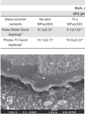
resin (RC) to the C-GIC was signiicantly lower than
that to the RM-GIC (p<0.001). SeM evaluation of the fractured surfaces indicated the presence of cracks in some interfaces between the C-GIC & adhesive & the RC (Figure 2). In contrast, good interlocking adhesion was exhibited between the RM-GIC and
the adhesive (Figure 3).
Figures 4 and 5 show the results of the failure mode for the shear strengths of the specimens bonded to the C-GIC and RM-GIC, respectively. Adhesive failure was observed only when the composite resin was bonded to the C-GIC Ketac Molar. Most of the C-GIC samples exhibited mixed-type failure modes (Figure 4), whereas cohesive failure was the dominant mode in the RM-GIC groups (Figure 5). Figure 6 shows the adhesive coating and small deposits of RC on a fractured area of a sample where the self-etch adhesive system Prompt L-Pop was used. Mixed-type sample failure is exhibited in Figure 7, where 35% phosphoric acid was applied for 60 s.
DISCUSSION
Lamination of the composite resin over the glass-ionomer material has been recommended as a viable restoration alternative, especially in areas of higher microleakage risk, such as with the coronal restoration of an endodontically treated tooth. Coronal leakage is one of the most important factors in determining the long-term success of endodontic treatment10,19. It has been noted that the placement of an intraoriice barrier following the root canal
filling is beneficial in delaying and preventing coronal microleakage22. A laboratory study1
demonstrated that a conventional glass-ionomer cement, with its superior sealing ability, would be
the best intraoriice barrier material. Furthermore,
during root canal therapy, a large amount of dentin is generally lost due to the nature of this treatment. Since glass-ionomer cement is often known as a biomimetic material, with mechanical properties similar to those of the dentin29,it can replace the lost
dentin. Due to these favorable properties mentioned above, glass-ionomer cements were often applied in combination with composite resins in the coronal restoration of endodontically treated teeth.
Another high-risk area for microleakage in
Surface treatment modalities Self-etch
(No rinse)
Etch, rinse & bond
35% phosphoric acid
Glass-ionomer cements
No etch MPa±(SD)
15 s MPa±(SD)
30 s MPa±(SD)
60 s MPa±(SD)
All in one MPa±(SD)
Ketac Molar Quick Applicap*
6.1±(2.3)a 5.1±(1.6)b,c 7.1±(1.7)a,b 8.0±(2.1)c 6.0±(2.2)
Photac Fil Quick Applicap*
10.1±(2.7)d 10.0±(2.3)e,f 12.7±(2.9)d,e 12.0±(2.8)f 12.8±(2.5)
Table 1- Mean bond strength (MPa) of resin composite to two different types of glass-ionomer cements treated with various surface treatment procedures of different adhesive systems. Same symbol and letters within a group indicate statistically
signiicant differences
Figure 2- Scanning electron microscopy (SEM)
operative dentistry is the gingival region of a tooth. The heterogeneous nature of the dentin and the
dificulties of moisture control in this area cause
complications for perfect sealing by the dentinal bond27. In all large and deep cavities with one
or more restoration margins located within the dentin, the lamination technique is recommended to enhance adhesion and to limit microleakage, if
composite resin is preferred as the primary illing
material16,24.In this situation, the practitioner can
leave the glass-ionomer cement exposed to the oral environment in the gingival area. even if microleakage persists in this area, the possibility
of luoride recharge and release from the
glass-ionomer cement offers the potential of preventing secondary caries. Furthermore, with this technique, practitioners can take advantage of the major
beneits of glass-ionomer cement - i.e., its stress absorbing nature, cation releasing property, different setting reaction compensating shrinkage, good ionic adhesion and low modulus of elasticity5.
The bond strength between glass-ionomer cement and composite resin is certainly important for both the retention of the resin restoration and prevention of microleakage. Although the need for enamel and dentin pretreatment has been well-established in the literature, the need for surface treatment over the GIC before composite resin lamination in sandwich restorations remains controversial. In past research, there was no consensus on the necessity of acid-etching over the glass-ionomer surface to improve the bond Figure 3- Good interlocking adhesion between
resin-modiied glass-ionomer cement (RM-GIC) and adhesive
Figure 4- Fracture modes of the specimens bonded to Ketac Molar Quick Applicap according to various surface treatment modalities
Figure 5- Fracture modes of the specimens bonded to Photac Fil Quick Applicap according to various surface treatment modalities
Figure 6- Prompt L-Pop adhesive coating and small deposits of composite resin (RC) on the conventional glass-ionomer cement (C-GIC) surface
strength of composite resin11,23,25.McLean, et al.15
(1985) advocated the etching procedure for 60 s to obtain closer contact and mechanical interlocking between the bonding agent and the porosity created by acid-etching of the cement surface. However, some investigators have rejected the acid-etching procedure, since it leads to a decrease in the cohesive strength of the cement21,25.Sheth,
et al.21 (1989) suggested that acid-etching of the
glass-ionomer cement would only undermine the cement surface, and hence cohesive failure of this weakened zone would be determined instead of a “true” interfacial resin bond strength. However, microleakage results of their study did not reveal any differences between the etched and non-etched samples. Further, in the SeM evaluation of the current study, we did not observe subsurface deterioration in the glass-ionomer cement surfaces where acid-etching was applied.
Researchers, who even suggested the etching procedure, have not reached a consensus on standardizing the etching time yet. Some authors have restricted the etching process to 15 s, because the surface deterioration of the cements occurs with a prolonged time4.There are studies
recommending 30 and/or 60 s of etching time for a desirable bonding effect8,13,25.All of these previous
studies were carried out with a 37% phosphoric acid application. From past to present, however, restorative materials, bonding systems, and their application procedures have been greatly improved. Therefore, the restorative materials and the surface treatment modalities of the glass-ionomer
cement are still critically important. Our indings
demonstrated that the etching process improved the bond strength of the composite resin. However, when the “etch & rinse” system was used, 30 s seemed to be the optimal etching time of 35% phosphoric acid for both the C-GIC and RM-GIC.
The indings of this study are not in agreement
with those of other studies3,9,26,30 which have
suggested that the etching process did not improve the bond strength of the RC to the GIC. In the aforementioned study, however, etching had been applied for 15 s. Our opinion is that this application
period may not be adequate to determine signiicant
differences between the etched and non-etched GIC samples. Indeed, we did not observe any statistical differences between the “no etch” and 15 s groups (Table 1). On the contrary, the effect
of etching time became signiicant at 30 s, when
the procedure was carried out with 35% phosphoric acid. The effect of the “self-etch adhesive” to the GICs was not different from that of the “etch & rinse” system since the bond strengths obtained
with both adhesive systems were not signiicantly
different. In this study, failure modes within each GIC group seemed to be similar, with the exception
of the specimens of the PhotacFil group, which were etched with 35% phosphoric acid for 30 s (Figures 4 and 5). Only cohesive failure was seen in this application group (Figure 5). This result
might conirm the strong bonding between the RC
and RM-GIC, especially when 35% phosphoric acid etching was applied for 30 seconds.
Indeed, lamination over the RM-GIC seems to be more effective, since the bond strength of the RC
to the RM-GIC was signiicantly higher compared
with that of the C-GIC. This is in agreement with the results of previous reports26,30.It has been suggested
that a similarity in compositions of both materials and curing mechanisms by the free-radical initiator system might be responsible for the increased bond strengths4,6,14.SeM photographs of the current study
indicated good interlocking adhesion between the RM-GIC and RC. Furthermore, fracture types of the specimens bonded to the PhotacFil Quick Applicap were dominantly cohesive within the GIC.
Although in vitro studies highlighted the promising results of the lamination technique, Opdam, et al.18 (2007) reported an increased failure
rate for closed-sandwich restorations after 9 years. However, their study was based on the database of Classes I and II composite resin restorations placed with and without the RM-GIC lining. According to their treatment protocol, even though the cavity walls were etched with 38% phosphoric acid before the GIC liners were placed, a bonding agent was applied to the surface of the GIC without the etching procedure. Besides, it is known that a strong acid application to the tooth structures was not proposed prior to the GIC placement. Those factors might be the reason for the failure in their sandwich
restorations. With the indings of our laboratory
study, it seems that acid-etching of a glass-ionomer base will improve the bonding capability of a composite resin to the RM-GIC.
Some researchers claimed that the technique of a glass-ionomer sample preparation affected the bond strength results9,21. Their suggestion was that
the smooth, almost glazed surfaces of non-etched GIC, which were produced when the cement was allowed to set against a glass and/or mylar strip, would lead to an inferior bond compared with that of etched surfaces. In this study, the surfaces of the GIC samples were roughened with 600-grit Al2O3 discs before the surface treatment modalities were applied, since glazed cement surfaces cannot be reproduced in clinical conditions.
CONCLUSIONS
Within the limitations of this study, the following conclusions may be drawn:
either two-step “etch, rinse & bond” or single-step “self-etch” adhesive systems may be preferred in the bonding of the RC to the GICs.
For an etching procedure via 35% phosphoric acid, the optimal etching time seems to be 30 s.
ACKNOwLEDgMENTS
This study was supported mainly by ege University Research Funds (project number
2005-Diş-09). The content is solely the responsibility of
the authors and does not necessarily represent the
oficial views of Ege University.
REFERENCES
1- Çelik EU, Yapar AGD, Ateş M, Şen BH. Bacterial microleakage of
barrier materials in obturated root canals. J endod. 2006;32:1074-6.
2- Davidson CL. Advances in glass-ionomer cements. J Appl Oral Sci. 2006;14:3-9.
3- Della Bona A, Pinzetta C, Rosa V. effect of acid etching of glass ionomer cement surface on the microleakage of sandwich restorations. J Appl Oral Sci. 2007;15:230-4.
4- Farah CS, Orton VG, Collard SM. Shear bond strength of chemical and light-cured glass ionomer cements bonded to composite resins. Aust Dent J. 1998;43:81-6.
5- Ferrari M. Use of glass ionomers as bondings, linings, or bases. In: Davidson CL, Mjör IA, eds. Advances in glass-ionomer cements. Berlin: Quintessence Publishing; 1999. p.137-48.
6- Fortin D, Vargas MA, Swift eJ Jr. Bonding of composite resins to
resin-modiied glass ionomers. Am J Dent. 1995;8:201-4.
7- Gama-Teixeira A, Simionato MR, elian SN, Sobral MA, Cerqueira Luz MA. Streptococcus mutans-induced secondary caries adjacent to glass ionomer cement, composite resin and amalgam restorations in vitro. Braz Oral Res. 2007;21:368-74.
8- Garcia-Godoy F, Draheim RN, Titus HW. Shear bond strength of a posterior composite resin to glass ionomer bases. Quintessence Int. 1988;19:357-9.
9- Gopikrishna V, Abarajithan M, Krithikadatta J, Kandaswamy D. Shear bond strength evaluation of composite resin bonded to GIC using three different adhesives. Oper Dent. 2009;34:467-71.
10- Heling I, Goril C, Slutzky H, Kopolovic K, Zalkind M,
Slutzky-Goldberg I. endodontic failure caused by inadequate restorative procedures: review and treatment recommendations. J Prosthet Dent. 2002;87:674-8.
11- Hinoura K, Moore BK, Philips RW. Tensile bond strength between glass ionomer cement and composite resins. J Am Dent Assoc. 1987;114:167-72.
12- Jandt KD, Sigusch BU. Future perspectives of resin based materials. Dent Mater. 2009;25:1001-6.
13- Joynt RB, Williams D, Davis eL, Wieczkowski G Jr. effects of etching time on surface morphology and adhesion of a posterior resin to glass-ionomer cement. J Prosthet Dent. 1989;61:310-4. 14- Li J, Liu Y, Liu Y, Söremark R, Sundström F. Flexural strength
of resin-modiied glass ionomer cements and their bond strength
to dental composites. Acta Odontol Scand. 1996;54:55-8. 15- McLean JW, Powis DR, Prosser HJ, Wilson AD. The use of glass-ionomer cements in bonding composite resin to dentine. Br Dent J. 1985;158:410-4.
16- Mount GJ, Tyas MJ, Ferracane JL, Nicholson JW, Berg JH,
Simonsen RJ. A revised classiication for direct tooth-colored
restorative materials. Quintessence Int. 2009;40:691-7. 17- Okuyama K, Murata Y, Pereira PN, Miguez PA, Komatsu H, Sano H. Fluoride release and uptake by various dental materials
after luoride application. Am J Dent. 2006;19:123-7.
18- Opdam NJ, Bronkhorst eM, Roeters JM, Loomans BA. Longevity and reasons for failure of sandwich and total-etch posterior composite resin restorations. J Adhes Dent. 2007;9:469-75. 19- Ray HA, Trope M. Periapical status of endodontically treated
teeth in relation to the technical quality of the root illing and the
coronal restoration. Int endod J. 1995;28:12-8.
20- Saito S, Tosaki S, Hirota K. Characteristics of glass ionomer cement. In: Davidson CL, Mjör IA, eds. Advances in glass-ionomer cements. Berlin: Quintessence Publishing; 1999. p.15-50. 21- Sheth JJ, Jensen Me, Sheth PJ, Versteeg J. effect of etching glass-ionomer cements on bond strength to composite resin. J Dent Res. 1989;68:1082-7.
22- Slutzky-Goldberg I, Slutzky H, Goril C, Smidt A. Restoration
of endodontically treated teeth review and treatment recommendations. Int J Dent. 2009;2009:150251.
23- Sneed WD, Looper SW. Shear strength of a composite resin to an etched glass ionomer. Dent Mater. 1985;1:127-8.
24- Stockton LW, Tsang ST. Microleakage of Class II posterior composite restorations with gingival margins placed entirely within dentin. J Can Dent Assoc. 2007;73:255.
25- Taggart Se, Pearson GJ. The effect of etching on glass polyalkenoate cement. J Oral Rehabil. 1991;18:31-42.
26- Taher NM, Ateyah NZ. Shear bond strength of resin modiied
glass ionomer cement bonded to different tooth-colored restorative materials. J Contemp Dent Pract. 2007;8:25-34.
27- Tay FR, Pang KM, Gwinnett AJ, Wei SH. A method for microleakage evaluation along the dentin/restorative interface. Am J Dent. 1995;8:105-8.
28- Torii Y, Itota T, Okamoto M, Nakabo S, Nagamhe M, Inoue K.
Inhibition of artiicial secondary caries in root by luoride-releasing
materials. Oper Dent. 2001;26:36-43.
29- Tyas MJ. Clinical evaluation of glass-ionomer cement restorations. J Appl Oral Sci. 2006;14:10-3.