419
Giant oesophageal liposarcoma: case report
Radiol Bras. 2008 Nov/Dez;41(6):419–421 Relato de Caso • Case Report
Giant oesophageal liposarcoma: case report*
Lipossarcoma gigante de esôfago: relato de casoRichard Page1, Ranjeet Narlawar2, John Holemans3, John Gosney4, Richard Warwick5, Hany Elsayed6
We describe imaging findings of a oesophageal liposarcoma in a 66 year old man. The computed tomography scan was performed after a chest radiograph showed a large posterior mediastinal mass. Oesophageal liposarcomas are rare tumours. They can achieve large size before they become symptomatic. Our patient was successfully managed with complete surgical removal.
Keywords: Liposarcoma; Oesophagus; Computed tomography scan.
São descritos os achados de imagem de um lipossarcoma de esôfago em um paciente do sexo masculino, de 66 anos de idade. Foi feita tomografia computadorizada, após radiografias de tórax terem mostrado massa mediastinal posterior. Lipossarcomas de esôfago são tumores raros. Eles podem atingir grandes dimensões antes de se tornarem sintomáticos. O paciente foi tratado com sucesso, com remoção cirúrgica completa do tumor.
Unitermos: Lipossarcoma; Esôfago; Tomografia computadorizada. Abstract
Resumo
* Study developed at Liverpool Heart and Chest Hospital NHS Trust, Liverpool, United Kingdom.
1. ChM, FRCS(CTh), Medical Doctor of the Department of Cardiothoracic Surgery of the Liverpool Heart and Chest Hospi-tal NHS Trust, Liverpool, United Kingdom.
2. MBBS, DNB, FRCR, Medical Doctor of the Department of Radiology of the Liverpool Heart and Chest Hospital NHS Trust, Liverpool, United Kingdom.
3. BSc, MBBS, FRCP, FRCR, Medical Doctor of the Depart-ment of Radiology of the Liverpool Heart and Chest Hospital NHS Trust, Liverpool, United Kingdom.
4. BSc(Hons), MD, FRCPath, Medical Doctor of the Depart-ment of Histopathology of the Liverpool University Hospitals NHS Trust, Liverpool, United Kingdom.
5. MUDr, MRCS, Medical Doctor of the Department of Cardiothoracic Surgery of the Liverpool Heart and Chest Hospi-tal NHS Trust, Liverpool, United Kingdom.)
6. MSc, MRCS, Medical Doctor of the Department of Cardiothoracic Surgery of the Liverpool Heart and Chest Hospi-tal NHS Trust, Liverpool, United Kingdom.
Mailing address: Dr J Holemans. Department of Radiology, Liverpool Heart and Chest Hospital NHS Trust, Thomas Drive, Liverpool L14 3PE, UK. E-mail: [email protected]
Received September 9, 2008. Accepted after revision Sep-tember 22, 2008.
A chest x-ray was performed, which re-vealed a large posterior mediastinal tumour (Figure 1). The lung fields were normal. Computed tomography (CT) scan of the chest and abdomen was performed for further characterisation. CT scan showed a large, well defined, mixed density, but
Page R, Narlawar R, Holemans J, Gosney J, Warwick R, Elsayed H. Giant oesophageal liposarcoma: case report. Radiol Bras. 2008;41(6):419–421.
present imaging findings with histological correlation of such a rare entity.
CASE REPORT
A 66 year old man presented with chronic cough and mucoid expectoration.
0100-3984 © Colégio Brasileiro de Radiologia e Diagnóstico por Imagem INTRODUCTION
Gastrointestinal liposarcomas are rare tumours, with oesophagus being the least common site. Imaging findings of very few cases have been described in literature. These tumours have varied imaging ap-pearances. Although imaging can not accu-rately differentiate between benign and malignant tumours, it has a pivotal role in staging and management of liposarcomas, especially oesophageal due to their close proximity to mediastinum and spine. We
420
Page R et al.
Radiol Bras. 2008 Nov/Dez;41(6):419–421
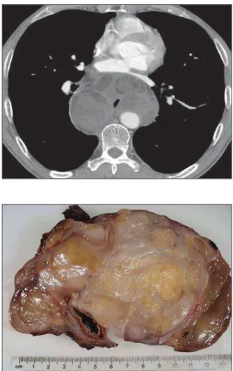
predominantly fat density mass in the pos-terior mediastinum. The mass was engulf-ing the oesophagus all around (Figure 2). There was mass effect and displacement of the posterior wall of the left atrium. The tu-mour was encasing the thoracic aorta for at least 180 degrees. The fat plane between the mass and the adjacent mediastinal structures was effaced. More distally, the mass caused complete collapse of the oesophageal lumen. There was no medias-tinal or hilar lymphadenopathy. The lung fields were normal. No abnormality de-tected within the abdomen. Due to mixed nature of the lesion, possibility of the ma-lignant lesion in the posterior mediastinum was raised.
On further questioning, he gave the his-tory of few months of difficulty in swallow-ing for which upper gastrointestinal endos-copy was performed in other hospital. It showed changes of Barrett’soesophagus; no other significant abnormality was noted. Patient underwent thoracotomy. The mass was well encapsulated, it was encir-cling the oesophagus, was seen free from the surrounding mediastinal structures. Complete removal of the tumour with distal oesophagectomy and oesophagogas-trectomy was performed. Post operative re-covery was uneventful.
Examination of the resected specimen revealed a fatty tumour that clearly arose from the submucosal region of the oeso-phageal wall, growing and expanding out-side the oesophagus to encircle it and form a thinly-encapsulate mass 150 × 140 × 75 mm in diameters (Figure 3). The mucosa overlying the tumour was normal.
Microscopic examination (Figure 4) showed lobules of fatty tissue separated by a collagenous and myxoid stroma. There was marked variation in the size of the adipocytes with scattered uni- and multi-vacuolated lipoblasts, some of them appar-ently entrapped within the fibrous septa. These lipoblasts had large, atypical, hyper-chromatic nuclei. Mitotic figures were present, numbering between 5 and 10 per ten high-power microscopic fields, but there was no necrosis. These features indi-cated the tumour to be a well differentiated (grade 1) liposarcoma of sclerosing subtype. The thin capsule of the lesion was intact and excision was considered complete.
DISCUSSION
Gastro-intestinal (GI) liposarcomas constitute about 0.1% to 5.8% of all the li-posarcomas at autopsy and the oesophagus is the least common site. Oesophageal
li-posarcomas constitutes 1.2% to 1.5% of all GI liposarcomas(1). First case was reported
in 1983(2) and since then only 15 other
cases have been reported in the world lit-erature so far. They are slowly growing tumours arising from the mucosa or
submu-Figure 2. Contrast enhanced axial CT scan showing a large lobulated mass with multiple areas showing fat density be-hind the left atrium. Note the effacement of the posterior wall of the left atrium and en-casement of the aorta.
Figure 3. Gross photograph showing the lobulated fatty mass arising from the oesopha-geal wall, compressing the oesophageal lumen and ex-panding outside the oesophagus into the mediastinum. The mu-cosa overlying it is intact.
421
Giant oesophageal liposarcoma: case report
Radiol Bras. 2008 Nov/Dez;41(6):419–421
cosal soft tissue layers of the oesophagus. Pathologically, the tumour is either well-differentiated liposarcoma or myxoid histotype(1).
There are varied clinical presentations of oesophageal liposarcoma. Mean age var-ies between 43 to 73 years and male sex being affected slightly more than female sex. Most patients complain dysphagia. They may remain asymptomatic, until the tumour grow to a very large size and cause invasion or mass effect on the adjacent structures. Weight loss, fever, odyno-phagia, respiratory distress, epigastric pain(3) which may or may not be associated
with gastro-intestinal bleeding. There has been even a case report where there was oral protrusion of the tumour.
CT scan features are similar to those re-ported in liposarcoma elsewhere in the body. Due to fatty nature of this lesion, CT scan mostly reveals a large, predominantly fat density lesion mixed with varying amount of soft tissue. Most of the reported cases showed intra or extra luminal polypoid mass(1,4,5). Proximal, mid as well
as distal oesophageal involvement have been reported. Presence of fat may indicate lipomatous tumour such as lipoma, atypi-cal lipoma and liposarcoma. CT scanning not only is helpful in characterising the le-sion but also helps in studying the medias-tinal structures in detail, as these tumours are known to cause compression or inva-sion of the adjacent structures.
Although, CT scanning remains the most important diagnostic modality, other modalities like barium swallow, trans-oesophageal ultrasound scanning,
endos-copy, magnetic resonance imaging (MRI) may also be performed. MRI may show high signal intensity within the fatty tissue on T1 weighted images, which suppresses on in phase and out of phase imaging or on fat suppressed images(6). In addition, MRI
helps in better assessment of involvement of the adjacent structures as compared with CT scanning.
Poorly differentiated tumours, which pathologically tend to be more cellular with less fat per cell component, are likely to have high CT numbers. CT number is not sufficient to distinguish well-differentiated liposarcoma from benign lipoma(7) and
his-topathologic examination is always neces-sary as much for diagnosis as prognosis in these cases(8).
Imaging modalities are limited in their capability to tell the malignant potential of the tumour. Hence complete resection is most commonly advised treatment op-tion(6,9,10). There are various surgical
op-tions. They vary from endoscopic resec-tion, simple enucleation to more invasive transthoracic, trascervical or transgastric approach. Survival depends on histologic subtypes, grade of malignancy, location and surgical radicality(4). Survival between
7 months to 104 months has been reported. A previous study reported a case of recur-rence 25 years after the first episode in a 68 year old woman(11).
In conclusion, oesophageal liposarco-mas are rare tumours with varied presen-tation. Even if CT and MRI features are typical, they are not unique. There are vari-ous differential diagnoses of fat containing masses in the mediastinum. Surgical
resec-tion is the treatment of choice and only histological examination can make confi-dent diagnosis.
Acknowledgement
The authors would like to acknowledge the contribution of Mr Mike Poullis and Dr Klaus Irion.
REFERENCES
1. Yang B, Shi PZ, Li X, et al. Well-differentiated liposarcoma of esophagus. Chin Med J (Engl). 2006;119:438–40.
2. Mansour KA, Fritz RC, Jacobs DM, et al. Pedun-culated liposarcoma of the esophagus: a first case report. J Thorac Cardiovasc Surg. 1983;86:447– 50.
3. Liakakos TD, Troupis TG, Tzathas C, et al. Pri-mary liposarcoma of esophagus: a case report. World J Gastroenterol. 2006;12:1149–52.
4. Mica L, Gianom D, Bode B, et al. Rare cause of dysphagy: giant polypoid esophageal well-differ-entiated liposarcoma. Case Rep Gastroenterol. 2007;1:7–14.
5. Di Mascio L, Gamble L, Wajed S, et al. Intussus-cepting giant liposarcoma of the oesophagus. J Postgrad Med. 2006;52:231–2.
6. Chung JJ, Kim MJ, Kim JH, et al. Imaging find-ings of giant liposarcoma of the esophagus. Yonsei Med J. 2003;44:715–8.
7. Taira N, Kinoshita S, Miyake T, et al. Primary liposarcoma of the anterior mediastinum – case report and review of literature. Jpn J Thorac Cardiovasc Surg. 1998;46:450–4.
8. Bonnette P, Jouan J, Colchen A, et al. Myxoid li-posarcoma of the mediastinum. Rev Mal Respir. 2000;17:109–11.
9. Boggi U, Viacava P, Naccarato AG, et al. Giant pedunculated liposarcomas of the esophagus: lit-erature review and case report. Hepatogastroen-terology. 1997;44:398–407.
10. Bréhant O, Pessaux P, Hennekinne-Mucci S, et al. Giant pedunculated liposarcoma of the esopha-gus. J Am Coll Surg. 2004;198:320–1. 11. Beaudoin A, Journet C, Watier A, et al. Giant