59
Pseudotumoral mesenteric panniculitis
Radiol Bras. 2010 Jan/Fev;43(1):59–61 Case Report • Relato de Caso
Pseudotumoral mesenteric panniculitis: tomographic
findings in a case*
Paniculite mesentérica pseudotumoral: aspectos tomográficos de um caso
Eduardo Miranda Brandão1, Thales Paulo Batista2, Jonas José da Silva Júnior3, Francisco Igor Bulcão de Macêdo4, Paulo Henrique Domingues Miranda Brandão5
Mesenteric panniculitis is a rare inflammatory process of the mesentery whose etiology is unknown that in only very few cases may present as an abdominal pseudotumor. The authors report a rare case, emphasizing the tomographic findings of mesenteric panniculitis that initially presented as a pseudotumor involving the peripancreatic region.
Keywords: Sclerosing mesenteritis; Mesenteric panniculitis; Pseudotumor; Pancreas.
A paniculite mesentérica representa um processo inflamatório do mesentério de ocorrência rara e etiologia desconhecida, que apenas em alguns poucos casos pode se manifestar sob a forma de pseudotumores ab-dominais. Descreve-se, enfatizando os aspectos tomográficos, um raro caso de paniculite mesentérica que se apresentou inicialmente como um pseudotumor que envolvia a região peripancreática.
Unitermos: Mesenterite esclerosante; Paniculite mesentérica; Pseudotumor; Pâncreas.
Abstract
Resumo
* Study developed at the Center of Oncology of Hospital Uni-versitário Oswaldo Cruz – Universidade de Pernambuco (CEON/ HUOC-UPE), Recife, PE, Brazil.
1. Master in Surgery, Oncologist Surgeon, Professor in the Discipline of Oncology at Faculdade de Ciências Médicas – Uni-versidade de Pernambuco (FCM-UPE), Recife, PE, Brazil.
2. MD, Oncologist Surgeon, Resident in Surgical Oncology at Hospital Universitário Oswaldo Cruz – Universidade de Pernam-buco (HUOC-UPE), Recife, PE, Brazil.
3. MD, Radiologist at Hospital Universitário Oswaldo Cruz – Universidade de Pernambuco (HUOC-UPE), Recife, PE, Brazil.
4. Medical Student, Faculdade de Ciências Médicas – Univer-sidade de Pernambuco (FCM-UPE), Recife, PE, Brazil.
5. Medical Student, Faculdade Boa Viagem – Instituto Ma-terno Infantil Professor Fernando Figueira (FBV-IMIP), Recife, PE, Brazil.
Mailing address: Dr. Thales Paulo Batista. Rua Desembarga-dor Góis Cavalcante, 100, Ed. Costa do Sol, ap. 1206, Parna-mirim. Recife, PE, Brazil, 52060-140. E-mail: t.paulo@bol. com.br
Received February 20, 2008. Accepted after revision June 26, 2008.
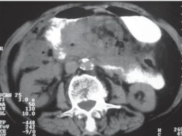
creatic fat planes with loss in the definition of the pancreatic head (5.2 × 4.9 × 3 cm) (Figure 1). Other findings, i.e densification of the mesentery and displacement of me-senteric vessels, besides thickening of in-testinal loops and subtle ascites are showed on Figure 2.
All laboratory tests including amylase and lipase measurements and tumor mark-ers were within normal limits, i.e. CEA = 0.655 U/ml and CA19.9 = 21.3 U/ml. Up-per digestive endoscopy demonstrated ex-trinsic compression of the second portion of the duodenum without mucosal involve-ment, while colonoscopy demonstrated a concentric, abrupt narrowing of the trans-verse colon near the splenic angle. The muscosa of this region presented hyemia and was friable. Biopsy was per-formed during the examination.
The attempt to collect a specimen for bi-opsy by means of percutaneous puncture was unsuccessful and while waiting for the biopsy to be performed the patient pro-gressed with constipation, recurrent low fever, anorexia and vomiting in association with significant decrease in the volume of the abdominal mass at the daily clinical examinations.
During the work-up serial amylase mea-surements remaided normal and a new CT Brandão EM, Batista TP, Silva Júnior JJ, Macêdo FIB, Brandão PHDM. Pseudotumoral mesenteric panniculitis: tomographic findings in a case. Radiol Bras. 2010;43(1):59–61.
The present report is aimed at describ-ing a rare case of mesenteric panniculitis that initially presented as a pseudotumor in-volving the peripancreatic region with emphasis to tomographic findings.
CASE REPORT
A 73-year-old female patient initially di-agnosed with abdominal tumor was re-ferred for oncologic evaluation due to an intense and persistent epigastric pain in association with occasional nausea and vomiting for the past 30 days. Additionally, the patient reported recurrent and low fe-ver, anorexia and a non-quantified weight loss. She also reported a similar, but less intense and self-limited event four months ago, however no significant comorbidity was observed.
On physical examination, a 15 cm in di-ameter painful partially mobile, ill-defined epigastric tumor was palpated. Neither sig-nificant lymph nodes enlargement nor rel-evant findings were observed at gyneco-logical and rectal examination.
Abdominal ultrasound (US) showed an agglomeration of thickened intestinal loops in the left hypochondrium, besides an ab-dominal computed tomography (CT) study revealed increased density of
peripan-0100-3984 © Colégio Brasileiro de Radiologia e Diagnóstico por Imagem
INTRODUCTION
60
Brandão EM et al.
Radiol Bras. 2010 Jan/Fev;43(1):59–61 was ordered and showed an ill-defined,
infiltrating lesion located in the retroperi-toneum and anterior pararenal space, in-volving the pancreas, duodenum and me-sentery. Additionally, focal areas of lique-faction and involvement without invasion of mesenteric vessels, besides the effect of extrinsic compression on the duodenum were observed (Figure 3).
An exploratory laparotomy was per-formed and demonstrated a small amount of citrine fluid in the pelvis and a tumor-like fat accumulation with an intense edema in the mesenteric root. Also, there was a fibrotic involvement of the transverse mesocolon causing lumen stenosis in the region near the splenic angle, and periduo-denal fibrotic reaction with bridles to the parietal peritoneum of the right hypochon-drium. After the opening of the epiploon retrocavity and subsequent Kocher-Vautrin and Cattell-Braasch maneuvers, the normal macroscopic aspect of the whole pancreas could be observed. Lysis of the bridles and adhesions were performed to clear the transverse colon lumen, with abdominal fluid drainage and freezing biopsy of the mesenteric tumor-like lesion which dem-onstrated an inflammatory and fibrotic component with no neoplastic involve-ment.
The patient was given antibiotics for 48 hours and symptomatic medication for five additional days, with progressive improve-ment. She was discharged at the day 32 (5th
postoperative day) and remains asymptom-atic after 12-month follow-up.
Neither aerobic bacterial growth nor the presence of neoplastic cells was found at the abdominal fluid analysis. Histology of specimens taken during colonoscopy dem-onstrated only an inflammatory infiltrate mimicking a mild chronic colitis. Finally, the biopsy of the mesenteric tumor-like lesion described a miscellaneous fat necrosis, chronic inflammatory infiltrate and different degrees of fibrosis in the several samples.
DISCUSSION
Mesenteric panniculitis represents a rare, non-specific inflammatory reaction of the mesentery whose etiology still remains
unknown. Only in few cases, the disease presents in the form of abdominal pseudo-tumors(1,2).
Histologically, mesenteric lipodystro-phy, mesenteric panniculitis and retractile mesenteritis have been considered as pro-gressive stages of a single mesenteric in-flammatory and non-specific process, char-acterized by a mix of variable degrees of fat necrosis, chronic inflammatory infiltrate and fibrosis(1–3). These three main terms characterize the predominance of each of such components, respectively, in the dif-ferent phases of the disease. However, con-sidering that a certain degree of these three components is found in all of the cases, considering their progressive character for mesenteric fibrosis, the term “sclerosing
Figure 1. Pseudotumor involving the peripancreatic region. One can observe the increased density of the peripancreatic fat planes and loss in definition of the pancreatic head.
Figure 2. Increased mesenteric density and displacement of mesenteric ves-sels. Linear bands with soft tissue density can be observed at the center of the lesion, representing fibrotic reaction within the mesenteric fat tissue in-flammatory process.
61
Pseudotumoral mesenteric panniculitis
Radiol Bras. 2010 Jan/Fev;43(1):59–61 mesenteritis” is generically considered as most appropriate(1,3).
The most frequent clinical findings in-clude pain and abdominal mass(1,2), gener-ally in association with other non-specific, constitutional signs and symptoms(2). Labo-ratory and sonographic findings are equally non-specific(1,2). Barium studies demon-strate only indirect signs of parietal in-volvement with preserved mucosal pattern. On the other hand, computed tomography presents a good anatomopathological cor-relation and is frequently employed in the diagnosis and follow-up of these cases. Also, magnetic resonance imaging presents additional benefits because of the better characterization of tissues and non-inva-siveness in the evaluation of small-caliber vessels(2), which can also be achieved with multi-detector computed tomography with intravenous contrast injected by an infusion pump.
The main findings in the tomographic evaluation of mesenteric panniculitis are the following: presence of heterogeneous mass with thickened fat density, displace-ment and ectasia of mesenteric vessels, intestinal loops distortion and presence of a tumor pseudocapsule. Other findings in-clude increased mesenteric density, cysts development, fat ring sign, calcifications and lymph nodes enlargement(1,2,4,5).
Mesenteric panniculitis can be satisfac-torily studied with abdominal CT as method of choice(4), the findings depend-ing on the disease stage(2). At the acute and subacute phases of lipodystrophy and pan-niculitis, there is a predominance of hetero-geneous mass effect with a slightly
in-creased fat density as a result of the inflam-matory process, and without direct vessels involvement, besides development of lin-ear bands resulting from the fibrotic retrac-tion. On the other hand, at the chronic phase of the disease, such masses acquire a more homogeneous appearance and in-creased density typical of fibrotic reac-tion(2).
The fat ring sign and the tumor pseudo-capsule represent the preservation of the fat adjacent to the mesenteric vessels and the development of peripheral bands with soft tissue density separating the healthy mesen-tery from the inflammatory process, respec-tively. Both strongly suggest the diagnosis of mesenteric panniculitis, however these findings tend to disappear with the progres-sion of the fibrotic process(2).
Furthermore, for the correct diagnosis of this disease, it is necessary to rule out the presence of pancreatitis, intestinal inflam-matory disease and Weber-Christian dis-ease(2). The differential diagnoses also must include Whipple’s disease, carcinoid tu-mors, inflammatory paseudotutu-mors, lei-omyosarcoma, abscesses, hematomas, fatty neoplasms, mesenteric carcinomatosis, Non-Hodgkin lymphoma, retroperitoneal fibromatosis, secondary fibrosing peritoni-tis, mesenteric edema and other causes of intestinal obstruction(1–3).
Many treatment methods have been em-ployed, but none of them have shown to be indistinctly appropriate(1,2,4,5). The role of surgery remains restricted to biopsies and management of complications(2). In most of cases, the patients progress with complete or partial resolution of the condition with
the process stabilization, with very rare cases of fatal outcome(1–3).
Considering the non-specific character-istics of this condition, the diagnosis of pseudotumoral mesenteric panniculitis re-quires laparotomy and biopsies for clinical, radiological, surgical and histological cor-relation(1,3). However, the knowledge of clinical and radiological characteristics can avoid unnecessary surgeries, considering the favorable clinical course of the disease in most of cases.
In the present report, the appropriate correlation of clinical, radiological, surgi-cal and histologisurgi-cal findings has led to the diagnosis of mesenteric panniculitis with a tendency to progress to fibrosis where, except for the tumor pseudocapsule, all the mentioned tomographic findings were ob-served.
REFERENCES
1. Sheikh RA, Prindiville TP, Arenson D, et al. Scle-rosing mesenteritis seen clinically as pancreatic pseudotumor: two cases and a review. Pancreas. 1999;18:316–21.
2. Moreira LBM, Pinheiro RA, Melo ASA, et al. Pa-niculite mesentérica: aspectos na tomografia com-putadorizada. Radiol Bras. 2001;34:135–40. 3. Emory TS, Monihan JM, Carr NJ, et al.
Scleros-ing mesenteritis, mesenteric panniculitis and me-senteric lipodystrophy: a single entity? Am J Surg Pathol. 1997;21:392–8.
4. Varela C, Fuentes M, Rivadeneira R. Procesos in-flamatorios del tejido adiposo intraabdominal, causa no quirúrgica de dolor abdominal agudo: hallazgos en tomografía computada. Rev Chil Radiol. 2004;10:28–34.