Mesenteric lymph reperfusion
exacerbates spleen injury caused
by superior mesenteric artery
occlusion shock
L.L. Li*, C.H. Zhang*, J.C. Liu, L.N. Yang, C.Y. Niu and Z.G. Zhao
Institute of Microcirculation, Hebei North University, Zhangjiakou, Hebei, China
Abstract
The intestinal lymph pathway plays an important role in the pathogenesis of organ injury following superior mesenteric artery occlusion (SMAO) shock. We hypothesized that mesenteric lymph reperfusion (MLR) is a major cause of spleen injury after SMAO shock. To test this hypothesis, SMAO shock was induced in Wistar rats by clamping the superior mesenteric artery (SMA) for 1 h, followed by reperfusion for 2 h. Similarly, MLR was performed by clamping the mesenteric lymph duct (MLD) for 1 h, followed by reperfusion for 2 h. In the MLR++SMAO group rats, both the SMA and MLD were clamped and then released for reperfusion for 2 h. SMAO shock alone elicited: 1) splenic structure injury, 2) increased levels of malondialdehyde, nitric oxide (NO), intercellular adhesion molecule-1, endotoxin, lipopolysaccharide receptor (CD14), lipopolysaccharide-binding protein, and tumor necrosis factor-a, 3) enhanced activities of NO synthase and myeloperoxidase, and 4) decreased activities of superoxide dismutase and ATPase. MLR following SMAO shock further aggravated these deleterious effects. We conclude that MLR exacerbates spleen injury caused by SMAO shock, which itself is associated with oxidative stress, excessive release of NO, recruitment of polymorphonuclear neutrophils, endotoxin translocation, and enhanced inflammatory responses.
Key words: Mesenteric lymph reperfusion; Superior mesenteric artery; Shock; Spleen injury; Endotoxin; Inflammation
Introduction
Superior mesenteric artery occlusion (SMAO) shock is a severe pathological event that results from intestinal ischemic insult due to superior mesenteric artery occlusion and subsequent reperfusion injury. Because the intestinal tract is the largest ‘‘bacteria storeroom’’ in the body, the intestinal barrier dysfunction induced by ischemia/reperfu-sion (I/R) injury leads to bacterial/endotoxin translocation (BET). The resulting inflammatory cascade causes dysfunc-tion and structural damage of remote organs (1-3). Recent investigations suggest that mesenteric lymph produced following hemorrhagic shock and acute pancreatitis is a source of factors leading to multiple organ injury and dysfunction (4,5). Additional studies reported that the lymphatic system is a pathway underlying the spread of lung and gut injury after intestinal ischemia/reperfusion (6), that mesenteric lymph duct (MLD) ligation improved survival rate (7), and that mesenteric lymph drainage could block the
‘‘gut-lymph’’ pathway, improve intestinal barrier function, and attenuate distant organ injury incurred by intestinal I/R (8). Our previous studies showed that mesenteric lymph reperfusion (MLR) after SMAO shock could exacerbate SMAO shock-induced organ damage of the lung, kidney, liver, and myocardium (9) and that endotoxin translocation was involved in the process (10). However, the mechanism needs further research.
Several studies have suggested that disturbances of immune function play an important role in imbalance of inflammation response and organ injury from shock, trauma, and sepsis (11-13). The spleen is the major immune organ and the center of cellular and humoral immunity. Previous studies have reported that SMAO shock caused spleen injury (3). In addition, splenic macrophages are a source of tumor necrosis factor (TNF) after intestinal I/R, and post-injury alteration in immune function is manifested by
Correspondence: C.Y. Niu and/or Z.G. Zhao, Institute of Microcirculation, Hebei North University, 11 Diamond South Road, Hebei, Zhangjiakou 075000, China. E-mail: [email protected] and/or [email protected]
*These authors contributed equally to this study.
depressed ability of splenocytes to increase production of IL-2 after stimulation with Con A (14). It has also been found that inhibiting tyrosine kinases in the spleen could prevent tissue damage after intestinal I/R (15). These studies demonstrated that the spleen injury following intestinal I/R or SMAO shock was a contributor to the remote organ injury and immune dysfunction. However, how spleen activity evolves and what role spleen plays in the process by which MLR exacerbates organ inflammation response induced by SMAO shock are worthy of investigation. We hypothesized that MLR exacerbates spleen injury caused by SMAO shock. To test this hypothesis, SMAO shock and MLR were produced in male Wistar rats. Splenic morphology and splenic levels of endotoxin, free radical indices, ATPase activity, myeloperoxidase (MPO) activity, and proinflamma-tory factors were measured.
Material and Methods
Animals and experimental groups
Twenty-four adult, male, specific pathogen-free Wistar rats weighing 280-350 g (Animal Breeding Center of the Chinese Academy of Medical Sciences, Beijing, China) were randomly assigned to four groups receiving MLR, SMAO, MLR++SMAO or sham treatment (all n=6). All animals were maintained in accordance with the Guide for the Care and Use of Laboratory Animals, and the Hebei North University Animal Care and Use Committee approved all experiments. Prior to experimentation, all rats were fasted for 12 h, but allowed free access to water.
SMAO shock model
Rats were subjected to a nonlethal model of SMAO shock as previously described (9,10,16). Briefly, after rats were anesthetized with 50 mg/kg pentobarbital sodium, femoral surgery was performed as follows. The right femoral vein and artery were isolated using aseptic techniques and 500 U/kg heparin sodium was injected through the femoral vein to systematically inhibit coagula-tion. Polyethylene tubing connected to a biological signal acquisition system (RM6240BD, Chengdu Instrument, China) was inserted in the femoral artery and used to monitor mean arterial pressure. Subsequently, a 5-cm midline laparotomy was performed, and the superior mesenteric artery (SMA) was separated from the accom-panying mesenteric lymphatic trunk. After 10 min of stabilization, the SMA was occluded by placing an atraumatic hemostatic clamp close to its origin for 1 h. Rats in the SMAO group were then reperfused for 2 h. In the MLR group, the MLD was clamped for 1 h followed by reperfusion for 2 h. In the MLR++SMAO group, occlusion of both the SMA and the MLD was maintained for 1 h followed by reperfusion for 2 h. Rats in the sham-treatment group were anesthetized and subjected to femoral and abdominal surgery as described above, excluding the occlusion of both the SMA and MLD.
Collection of splenic tissue
At 2 h after reperfusion, under strict aseptic condi-tions, the spleens were immediately obtained from the rats. Subsequently, the splenic tissue was divided into three parts. Part I was fixed in 4% formaldehyde for the observation of splenic morphology; part II was placed in 9 times its volume of pyrogen-free water for measurement of endotoxin; part III was placed in 5 times its volume of 46C normal saline for the determination of malondialde-hyde (MDA), superoxide dismutase (SOD), nitric oxide (NO), nitric oxide synthase (NOS), ATPase, MPO, intercellular adhesion molecule-1 (ICAM-1), lipopolysac-charide receptor (CD14), lipopolysaclipopolysac-charide-binding pro-tein (LBP), and TNF-a.
Observation of splenic morphology
The formaldehyde-fixed splenic tissue was embedded in paraffin, sectioned at a thickness of 4-6mm, and
stained with hematoxylin and eosin. Histological change was examined by light microscopy (90i, Nikon, Japan), and photographed using an image collection and analysis system (Eclipse, Nikon). All morphological observations were performed by a forensic pathologist who had no prior knowledge of the experimental conditions.
Measurement of endotoxin
A standard curve was constructed using standard endotoxin products (National Institute for the Control of Pharmaceutical and Biological Products, China) as pre-viously described (10). The splenic tissue was homo-genized using a glass homogenizer and deproteinized by incubation at 706C in a water bath for 10 min followed by centrifugation at 3700gfor 10 min using a Labofuge 400R-type low-temperature centrifuge (Heraeus, Germany). The endotoxin level in the splenic supernatant was determined through interference tests and recovery tests as described in the Chinese Pharmacopoeia (2005 edition). Endotoxin data were collected and analyzed automatically using a 32-well tube detector (ATi 320-06 kinetic tube reader, Lab Kinetics Ltd., UK). During the experiments, all appliances that came into contact with samples or reagents also were sterile and pyrogen-free, and all procedures were performed in a safety cabinet (Thermo Electron, USA).
Measurement of free radical indices
Part III of the splenic tissue was homogenized for 30 s using a tissue homogenizer (FJ-200, Shanghai Specimen and Model Factory, China) and then centrifuged at 3100g
xanthine oxidase (16,19) methods, respectively, following the manufacturer’s instructions (Jiancheng Biotechnology Research Institute). MDA level is reported as nmol/mg protein and SOD activity as U/mg protein.
Measurement of NO level and NOS activity
The NO level and NOS activity were assayed by the nitrate reductase (20,21) and chromogenic (21,22) meth-ods, respectively, following the manufacturer’s instruc-tions (Jiancheng Biotechnology Research Institute). The results are reported as NOmmol/g protein and NOS U/g
protein.
Determination of ATPase activity
The activities of Na+-K+-ATPase, Ca2++-ATPase, Mg2++-ATPase, and Ca2++-Mg2++-ATPase in splenic homogenates were determined as previously described by measuring phosphorus content (23) using an ATPase activity detection kit (Jiancheng Biotechnology Research Institute) (16,21). The results are reported as ATPase U/mg protein.
Determination of MPO activity
MPO activity in the splenic homogenate was mea-sured by the hydrogen peroxide method (24) using an MPO kit from Jiancheng Biotechnology Research Institute, as previously described (16,21,25). Results are reported as MPO U/g protein.
Enzyme-linked immunosorbent assay (ELISA)
The levels of ICAM-1, CD14, LBP, and TNF-a in
splenic homogenates were determined by a rat ELISA kit (Jiangsu Hope Biotechnology Ltd., China) according to the manufacturer’s instructions. Antibodies were obtained from R&D Systems (USA). The SPSS 11.0 statistical software (Polar Engineering and Consulting Inc., USA) was used to draw the standard curve. The results were standardized against the total protein content of homo-genates.
Statistical analysis
Data are reported as means±SD and were analyzed using the SPSS version 11.0 software. One-way analysis of variance (ANOVA) was used to identify differences within groups, and paired t-tests were used to identify differences between groups. The Kruskal-Wallis test was used to analyze data that was not suitable for one-way ANOVA. P,0.05 was considered to be statistically significant.
Results
Morphological changes in splenic tissues
As shown in Figure 1, spleens in the sham and MLR rats had normal structure with no obvious change in width of splenic cords and sinuses. In contrast, spleens in the
SMAO shock rats had congestion in the splenic sinuses and fundamentally normal splenocytes, while, in the SMAO++MLR group, rats had swollen splenocytes and extensive congestion of the splenic sinuses.
Free radical indices in splenic homogenates
There were no significant differences in MDA level or SOD activity in splenic homogenates of the MLR and sham groups. However, compared with the sham or MLR
Figure 1. Effect of mesenteric lymph reperfusion (MLR) on splenic histomorphology in rats with superior mesenteric artery occlusion (SMAO) shock (H&E staining).AandB, Sham group.
C and D, MLR group. E and F, SMAO group. G and H, MLR++SMAO group. Magnification bars:A,C,E,G: 100mm;B,
groups, MDA levels were higher and SOD activities were lower in the SMAO and SMAO++MLR groups, respec-tively. Moreover, the deleterious changes in MDA level and SOD activity were greater in the SMAO++MLR group than in the SMAO group (see Figure 2).
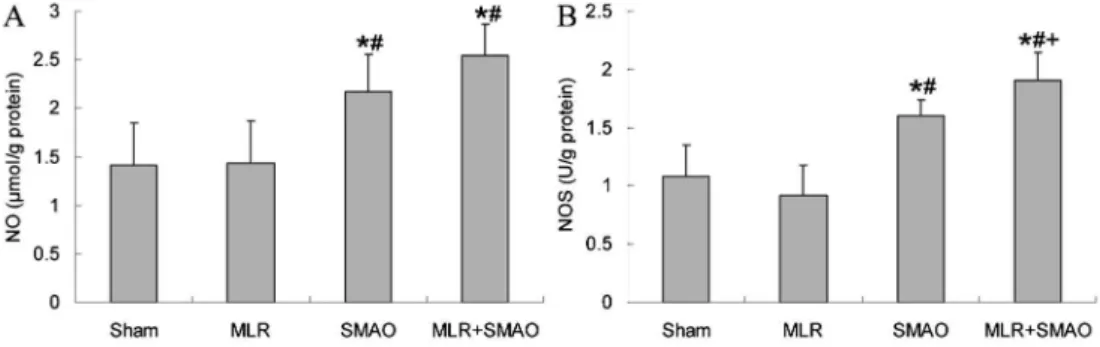
NO levels and NOS activity in splenic homogenates
As shown in Figure 3, NO levels and NOS activity in splenic homogenates of the sham and MLR groups were similar. In contrast, NO levels and NOS activity in the SMAO and SMAO++MLR groups were significantly higher than in the sham and MLR groups. Finally, these indices were lower in the SMAO++MLR group than in the SMAO group.
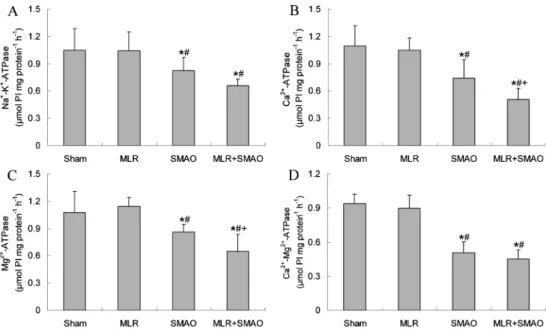
ATPase activity in splenic homogenates
As shown in Figure 4, there were no differences in the activities of Na+-K+-ATPase, Ca2++-ATPase, Mg2++
-ATPase, or Ca2++-Mg2++-ATPase observed in the MLR and sham groups. The corresponding ATPase activities in the SMAO and SMAO++MLR groups were significantly lower than in the sham and MLR groups; and the activities of Ca2++-ATPase and Mg2++-ATPase in the SMAO++MLR group were further decreased compared to those in the
SMAO group. No statistically significant differences were observed in Na+-K+-ATPase and Ca2++-Mg2++-ATPase in
the SMAO and SMAO++MLR groups.
ICAM-1 levels and MPO activity in splenic homogenates
The changes in ICAM-1 levels and MPO activity were similar to those seen in the NO levels and NOS activity. There were no statistically significant differences in ICAM-1 levels or MPO activity in the MLR and sham groups. ICAM-1 levels and MPO activity in the SMAO group were significantly higher than in the sham and MLR groups and were significantly higher in the SMAO++MLR group than in each of the other three groups (Figure 5).
Indices related to endotoxin translocation and inflammatory response in splenic homogenates
There were no statistically significant differences in endotoxin, CD14, LBP, or TNF-acontent of homogenates
from the sham and MLR groups (Figure 6). These indices were higher in the SMAO and MLR++SMAO groups than in the sham and MLR groups, and, except for TNF-a, they
were further increased in the MLR++SMAO compared with the SMAO group.
Figure 2.Effect of mesenteric lymph reperfusion (MLR) on the indices related to oxidative stress in splenic homogenate of rats with superior mesenteric artery occlusion (SMAO) shock. Data are reported as means±SD, n=6.A, Malondialdehyde (MDA) level.B, Superoxide dismutase (SOD) activity. *P,0.05vssham group;#P
,0.05vsMLR group;+P
,0.05vsSMAO group (one-way ANOVA).
Figure 3.Effect of mesenteric lymph reperfusion (MLR) on the nitric oxide (NO) level and nitric oxide synthase (NOS) activity in splenic homogenate of rats with superior mesenteric artery occlusion (SMAO) shock. Data are reported as means±SD, n=6.A, NO level.
B, NOS activity. *P,0.05vssham group;#P
,0.05vsMLR group;+P
Discussion
To our knowledge, this is the first study to report the effects of MLR on the spleens of rats subjected to SMAO shock. Our results showed that simultaneous occlusion and reperfusion of the SMA and MLD exacerbated spleen damage based on the morphological observations, suggesting that mesenteric lymph plays an important role in the pathogenesis of spleen organ injury after SMAO shock. Due to the fact that the spleen is a major organ of cellular and humoral immunity, and that immune dysfunc-tion is a major mechanism of inflammadysfunc-tion response imbalance and organ injury, the additional spleen injury induced by MLR following SMAO might reflect another
mechanism of multiple organ injury accompanying that of shock alone.
The next question to answer was how MLR exacer-bates spleen injury. The present study showed that the MDA level in splenic tissue was significantly higher, and SOD activity significantly lower, in rats subjected to both MLR and SMAO than in rats subjected to SMAO shock alone, suggesting that exacerbated spleen injury after MLR may be caused, at least in part, by oxygen-free radicals and the subsequent inflammatory cascade. Our findings are consistent with previous evidence linking the production and release of reactive oxygen species to the pathogenesis of intestinal I/R-induced remote organ damage (26,27).
Figure 4.Effect of mesenteric lymph reperfusion (MLR) on the adenosine triphosphatase (ATPase) activity in splenic homogenate of rats with superior mesenteric artery occlusion (SMAO) shock. Data are reported as means±SD, n=6.A, Na+-K+-ATPase activity.
B, Ca2++-ATPase activity.C, Mg2++-ATPase activity.D, Ca2++-Mg2++-ATPase activity. *P,0.05vssham group;#P
,0.05vsMLR group; +P,0.05vsSMAO group (one-way ANOVA).
Figure 5.Effect of mesenteric lymph reperfusion (MLR) on the indices related to adhesion and recruitment of polymorphonuclear neutrophils in splenic homogenate of rats with superior mesenteric artery occlusion (SMAO) shock. Data are reported as means±SD, n=6.A, Intercellular adhesion molecule-1 (ICAM-1) level.B, Myeloperoxidase (MPO) activity. *P,0.05vssham group;#P
,0.05vs
MLR group;+P
It has been demonstrated that the NO-induced inflam-matory cascade effects have been proposed to be a major contributor to systemic inflammatory response syndrome (SIRS) and multiple organ failure after shock (28), because NO can induce and increase the release of free radicals (29), thereby causing cell membrane damage and leading to organ dysfunction. In this study, we found that the NO level and NOS activity in splenic tissue of rats subjected to MLR +SMAO shock were higher than that of the sham and MLR groups. NOS activity in the MLR++SMAO group increased more than in the SMAO group. These results suggest that MLR may promote the synthesis and release of NO, resulting in an increased inflammatory response and leading to spleen injury.
It has been reported that membrane pump dysfunction plays an important role in spleen injury in rabbits following acute renal failure subjected to hypodermic injection of HgCl2or intramuscular injection of glycerin (21). However,
it is not clear whether membrane pump dysfunction is involved in the spleen injury following SMAO shock. The present data showed that SMAO shock depressed the activities of Na+- K+-ATPase, Ca2++-ATPase, Mg2++ -ATPase, and Ca2++-Mg2++-ATPase in splenic tissue, suggesting that membrane pump dysfunction is related to the spleen damage that occurs in rats subjected to SMAO shock. Along with other mechanisms exacerbating spleen injury after SMAO shock, MLR added to decreases in Ca2++-ATPase and Mg2++-ATPase activities.
It has been shown that increased ICAM-1 promotes poly-morphonuclear neutrophil (PMN) adhesion, aggregation,
and sequestration in tissue to result in SIRS and organ injury (30,31). Meanwhile, MPO activity is another marker that correlates with the number of PMN in tissue injury and is frequently utilized to evaluate tissue PMN activation (32,33). The study data showed that reperfusion after ligation of the MLD increased the ICAM-1 level and MPO activity in splenic tissue following SMAO shock. These results suggest that increased ICAM-1 level might induce recruitment of PMN by splenic tissue, as well as increased MPO activity, which is direct evidence of PMN sequestration. As a result, increased TNF-a appeared, suggesting that MLR enhanced the
inflammatory cascade in spleen tissue through PMN adhesion in rats subjected to SMAO shock.
The intestinal tract is the largest ‘‘bacteria storeroom’’ in the body, and dysfunction of the gut barrier due to ischemia can lead to BET from the intestine to the entire body in severe pathological conditions, such as hemorrhagic and traumatic shock and acute pancreatitis (4,5). Our previous results showed that the plasma endotoxin level in rats subjected to SMAO shock plus MLR was higher than in the rats subjected to only SMAO shock, suggesting that the lymphatic system plays an important role in enterogenous BET (10). In the present study, we found that the endotoxin level in splenic tissue was the highest in the MLR++SMAO group, indicating that the exacerbation of SMAO-induced organ injury by MLR is related to intestinal endotoxin translocation via the lymphatic pathway.
It has been shown that the increased levels of LBP and membrane CD14 enhanced the responses of both blood monocytes and tissue macrophages to endotoxin
Figure 6.Effect of mesenteric lymph reperfusion (MLR) on the indices related to endotoxin (ET) translocation and inflammatory response in splenic homogenate of rats with superior mesenteric artery occlusion (SMAO) shock. Data are reported as means±SD, n=6.A, ET level.B, Lipopolysaccharide receptor (CD14) level.C, Lipopolysaccharide-binding protein (LBP) level.D, Tumor necrosis factor-a(TNF-a) level. *P,0.05vssham group;#P
(34). Therefore, the inflammatory response of tissue injury is mediated by endotoxin in an LBP- and CD14-dependent manner. We found that SMAO shock caused increased levels of LBP and CD14 in the spleen, and that MLR after SMAO exacerbated these changes. These data suggest that LBP and CD14 are an important link between endotoxin and inflammatory response during MLR-aggravated spleen injury following SMAO shock.
In summary, MLR after SMAO exacerbated spleen injury by increasing oxidative stress and NO release, causing cell membrane pump dysfunction, increased adhesion and recruitment of PMNs, as well as endotoxin
translocation. These results suggest that the mesenteric lymph plays an important role in the pathogenesis of inflammatory response and multiple organ injury after SMAO shock through spleen injury.
Acknowledgments
Research supported by grants from the Key Scientific and Technological Project of Hebei Province (#09276101D-31, #11276103D-84), and the Innovative Talents Support Program of Hebei Province (#CPRC047,
#CPRCII26).
References
1. Moore FA. The role of the gastrointestinal tract in postinjury multiple organ failure.Am J Surg1999; 178: 449-453, doi: 10.1016/S0002-9610(99)00231-7.
2. Deitch EA. Gut-origin sepsis: evolution of a concept.
Surgeon2012; 10: 350-356, doi: 10.1016/j.surge.2012.03. 003.
3. Kawasaki T, Chaudry IH. The effects of estrogen on various organs: therapeutic approach for sepsis, trauma, and reperfusion injury. Part 2: liver, intestine, spleen, and kidney.J Anesth2012; 26: 892-899, doi: 10.1007/s00540-012-1426-2.
4. Deitch EA. Gut lymph and lymphatics: a source of factors leading to organ injury and dysfunction.Ann N Y Acad Sci
2010; 1207 (Suppl 1): E103-E111, doi: 10.1111/j.1749-6632.2010.05713.x.
5. Fanous MY, Phillips AJ, Windsor JA. Mesenteric lymph: the bridge to future management of critical illness.JOP2007; 8: 374-399.
6. Cavriani G, Domingos HV, Soares AL, Trezena AG, Ligeiro-Oliveira AP, Ligeiro-Oliveira-Filho RM, et al. Lymphatic system as a path underlying the spread of lung and gut injury after intestinal ischemia/reperfusion in rats.Shock2005; 23: 330-336, doi: 10.1097/01.shk.0000157303.76749.9b.
7. Badami CD, Senthil M, Caputo FJ, Rupani BJ, Doucet D, Pisarenko V, et al. Mesenteric lymph duct ligation improves survival in a lethal shock model.Shock2008; 30: 680-685, doi: 10.1097/SHK.0b013e318173edd1.
8. He GZ, Zhou KG, Zhang R, Wang YK, Chen XF. Impact of intestinal ischemia/reperfusion and lymph drainage on distant organs in rats. World J Gastroenterol 2012; 18: 7271-7278, doi: 10.3748/wjg.v18.i48.7271.
9. Zhang CH, Niu CY, Zhao ZG, Zhang YP, Han R, Zhang J. [Effect of mesenteric lymph reperfusion on multiple organs injury in superior mesenteric artery occlusion shock rats].
Zhongguo Bingli Shengli Zazhi2008; 24: 2103-2107. 10. Yang LN, Zhao ZG, Zhao YQ, Liu ZJ, Niu CY, Zhang J.
[Role of endotoxin translocation on mesenteric lymph reperfusion aggravating multiple organ injury in SMAO shock rats]. Zhongguo Ying Yong Sheng Li Xue Za Zhi
2012; 28: 74-78.
11. Boontham P, Chandran P, Rowlands B, Eremin O. Surgical sepsis: dysregulation of immune function and therapeutic implications. Surgeon 2003; 1: 187-206, doi: 10.1016/ S1479-666X(03)80018-5.
12. Jiang LN, Yao YM, Sheng ZY. The role of regulatory T cells in the pathogenesis of sepsis and its clinical implication.
J Interferon Cytokine Res2012; 32: 341-349, doi: 10.1089/ jir.2011.0080.
13. Ni Choileain N, Redmond HP. Cell response to surgery.
Arch Surg2006; 141: 1132-1140, doi: 10.1001/archsurg. 141.11.1132.
14. Cohen S, Lahat N, Kinarty A, Bitterman H.Ex vivosecretion of tumor necrosis factor and interleukin-2 by rat splenocytes after intestinal ischemia and shock.Lymphokine Cytokine Res1992; 11: 215-220.
15. Pamuk ON, Lapchak PH, Rani P, Pine P, Dalle Lucca JJ, Tsokos GC. Spleen tyrosine kinase inhibition prevents tissue damage after ischemia-reperfusion. Am J Physiol Gastrointest Liver Physiol 2010; 299: G391-G399, doi: 10.1152/ajpgi.00198.2010.
16. Zhao ZG, Niu CY, Shang AM, Tian JM, Han R, Zhang CH, et al. Mesenteric lymph reperfusion may exacerbate brain injury in a rat model of superior mesenteric artery occlusion shock.Neural Regen Res2010; 5: 683-689.
17. Bautista J, Corpas R, Ramos R, Cremades O, Gutierrez JF, Alegre S. Brain mitochondrial complex I inactivation by oxidative modification. Biochem Biophys Res Commun
2000; 275: 890-894, doi: 10.1006/bbrc.2000.3388. 18. Rochette L, Tatou E, Vergely C, Maupoil V, Bouchot O,
Mossiat C, et al. Regional heterogeneity of decreased myocardial norepinephrine and increased lipid peroxidation levels in patients with end-stage failing heart secondary to dilated or ischemic cardiomyopathy. J Heart Lung Transplant2008; 27: 767-774, doi: 10.1016/j.healun.2008. 03.025.
19. Morild E, Olmheim JE. Pressure variation of enzymatic reaction rates: IV. Xanthine oxidase and superoxide dismutase.Physiol Chem Phys1981; 13: 483-491. 20. Ding AH, Nathan CF, Stuehr DJ. Release of reactive
nitrogen intermediates and reactive oxygen intermediates from mouse peritoneal macrophages. Comparison of activating cytokines and evidence for independent produc-tion.J Immunol1988; 141: 2407-2412.
21. Zhao ZG, Niu CY, Zhang YP, Han R, Hou YL, Wang XR, et al. The mechanism of spleen injury in rabbits with acute renal failure. Ren Fail 2011; 33: 418-425, doi: 10.3109/ 0886022X.2011.568145.
Quartarone C, et al. Recombinant human erythropoietin inhibits iNOS activity and reverts vascular dysfunction in splanchnic artery occlusion shock.Br J Pharmacol 1999; 127: 482-488, doi: 10.1038/sj.bjp.0702521.
23. Fedorova OV, Lakatta EG, Bagrov AY. Endogenous Na, K pump ligands are differentially regulated during acute NaCl loading of Dahl rats.Circulation2000; 102: 3009-3014, doi: 10.1161/01.CIR.102.24.3009.
24. Anderson BO, Brown JM, Shanley PF, Bensard DD, Harken AH. Marginating neutrophils are reversibly adherent to normal lung endothelium.Surgery1991; 109: 51-61. 25. Zhao ZG, Zhang LL, Niu CY, Zhang J. Exogenous normal
lymph reduces liver injury induced by lipopolysaccharides in rats.Braz J Med Biol Res2014; 47: 128-134, doi: 10.1590/ 1414-431X20133280.
26. Yang X, Bai H, Cai W, Li J, Zhou Q, Wang Y, et al. Lycium barbarum polysaccharides reduce intestinal ischemia/reper-fusion injuries in rats.Chem Biol Interact2013; 204: 166-172, doi: 10.1016/j.cbi.2013.05.010.
27. Sapalidis K, Papavramidis TS, Gialamas E, Deligiannidis N, Tzioufa V, Papavramidis S. The role of allopurinol’s timing in the ischemia reperfusion injury of small intestine.J Emerg Trauma Shock 2013; 6: 203-208, doi: 10.4103/0974-2700.115346.
28. Takizawa Y, Kitazato T, Ishizaka H, Kamiya N, Tomita M, Hayashi M. Effect of aminoguanidine on ischemia/reperfu-sion injury in rat small intestine.Biol Pharm Bull2011; 34: 1737-1743, doi: 10.1248/bpb.34.1737.
29. Krauss H, Sosnowski P, Biczysko M, Biczysko W, Majewski P, Jablecka A, et al. Effects of L-arginine and NG-nitro L-arginine methyl ester (L-NAME) on ischemia/reperfusion injury of skeletal muscle, small and large intestines.Chin J Physiol2011; 54: 7-18, doi: 10.4077/CJP.2011.AMK011. 30. Power C, Wang JH, Sookhai S, Wu QD, Redmond HP.
Proinflammatory effects of bacterial lipoprotein on human neutrophil activation status, function and cytotoxic potential
in vitro.Shock2001; 15: 461-466, doi: 10.1097/00024382-200115060-00009.
31. Xu DZ, Lu Q, Adams CA, Issekutz AC, Deitch EA. Trauma-hemorrhagic shock-induced up-regulation of endothelial cell adhesion molecules is blunted by mesenteric lymph duct ligation. Crit Care Med2004; 32: 760-765, doi: 10.1097/ 01.CCM.0000114815.88622.9D.
32. Zaets SB, Xu DZ, Lu Q, Feketova E, Berezina TL, Malinina IV, et al. Recombinant factor XIII mitigates hemorrhagic shock-induced organ dysfunction.J Surg Res 2011; 166: e135-e142, doi: 10.1016/j.jss.2010.12.001.
33. Reino DC, Pisarenko V, Palange D, Doucet D, Bonitz RP, Lu Q, et al. Trauma hemorrhagic shock-induced lung injury involves a gut-lymph-induced TLR4 pathway in mice.PLoS One2011; 6: e14829, doi: 10.1371/journal.pone.0014829. 34. Heumann D, Adachi Y, Le Roy D, Ohno N, Yadomae T,