375 Study of optimization of images in interventional fluoroscopy
Radiol Bras. 2009 Nov/Dez;42(6):375–378 Original Article • Artigo Original
Study of optimization of images in interventional
fluoroscopy*
Estudo de otimização de imagens em fluoroscopia intervencionista
Alexandre Parizoti1, Thomaz Ghilardi Netto2
OBJECTIVE: The objective of the present study is to analyze the optimization of fluoroscopic image quality and patient entrance surface air kerma rate in interventional radiology procedures, utilizing a phantom adapted for fluoroscopy. MATERIALS AND METHODS: The authors utilized a phantom developed for evaluating conventional radiological images adapted for fluoroscopy through the addition of two catheters with different diameters, both of them utilized in interventional radiology. The patient entrance surface air kerma rate was determined with the aid of this phantom. RESULTS: The evaluation of technical parameters for different exposure modes of a digital fluoroscopic imaging system has allowed the determination of the air kerma rate, enabling the optimization of the image quality in interventional procedures. The decrease in the patient entrance surface air kerma rate may achieve 67%. CONCLUSION: The optimization of fluoroscopic image quality achieved with a phantom allows reducing the patient entrance surface air kerma with no significant loss of diagnostic performance.
Keywords: Images optimization; Fluoroscopy; Phantom; Dose reduction.
OBJETIVO: O objetivo deste trabalho é o de estudar a otimização da qualidade da imagem fluoroscópica e a taxa de kerma no ar de entrada na superfície do paciente em procedimentos de radiologia intervencionista, utilizando-se de um objeto simulador adaptado para fluoroscopia. MATERIAIS E MÉTODOS: Foi utilizado um objeto simulador desenvolvido para avaliação de imagens em radiologia convencional. O objeto simulador foi adaptado para fluoroscopia mediante incorporação de dois cateteres com diferentes espessuras, ambos uti-lizados em radiologia intervencionista. Os níveis de taxa de kerma no ar de entrada na superfície do paciente foram determinados utilizando-se este objeto simulador. RESULTADOS: A avaliação dos parâmetros técni-cos para diversos modos de exposição de um equipamento fluoroscópico com digitalização de imagens permitiu estabelecer os indicadores de taxa de kerma no ar, que permitem a otimização da qualidade das imagens em procedimentos intervencionistas. A redução na taxa de kerma no ar de entrada na superfície do paciente pode chegar a 67%. CONCLUSÃO: A otimização da qualidade da imagem utilizando-se um objeto simulador possibilita reduzir a taxa de kerma no ar de entrada na superfície do paciente, sem perda considerável de informação diagnóstica.
Unitermos: Otimização de imagens; Fluoroscopia; Objeto simulador; Redução de dose.
Abstract
Resumo
* Study developed at Department of Physics and Mathemat-ics, Faculdade de Filosofia Ciências e Letras de Ribeirão Preto da Universidade de São Paulo (FFCLRP-USP), Ribeirão Preto, SP, Brazil.
1. Medical Physicist, Master in Physics Applied to Medicine and Biology, Department of Physics and Mathematics - Facul-dade de Filosofia Ciências e Letras de Ribeirão Preto da Univer-sidade de São Paulo (DFM/FFCLRP-USP), Ribeirão Preto, SP, Brazil.
2. Titular Professor, Coordinator for the Unit of Medical Physics at Hospital das Clínicas da Faculdade de Medicina de Ribeirão Preto da Universidade de São Paulo (HCFMRP-USP), Ribeirão Preto, SP, Brazil.
Mailing address: Dr. Alexandre Parizoti. Rua Eliza dos Reis Lourenço, 175, Parque Primavera. São Joaquim da Barra, SP, Brazil, 14025-670. E-mail: [email protected]
Received April 20, 2009. Accepted after revision August 27, 2009.
ent exposure modes, by utilizing a phantom adapted for interventional procedures(6).
MATERIALS AND METHODS
The experimental procedure was per-formed with a BV Pulsera C-arm type fluo-roscopic system (Philips Medical Systems; Eindhoven, The Netherlands), with images digitization. The phantom utilized in the present study was adapted for fluoroscopy, consisting of a patient simulator and a re-alistic-analytic phantom(7), as shown on
Figure 1, simulating human anatomy struc-tures(8), relevant for radiological
proce-dures. The adaptation of the realistic-ana-lytic phantom consisted of the implantation
Parizoti A, Ghilardi Netto T. Study of optimization of images in interventional fluoroscopy. Radiol Bras. 2009;42(6):375– 378.
among radiologic procedures involving the highest radiation exposure for both patients and medical professionals involved. Ac-cording to the ALARA principle(1), it is
extremely important to implement pro-grams of quality assurance and radiopro-tection(2,3) with the objective of optimizing
the imaging quality and that the radiation exposure rates are brought down as low as feasible. Therefore, it is essential to evalu-ate the parameters involved in the opti-mized production of radiographic im-ages(4,5) with the aid of phantoms.
Thus, the main objective of the present study was to evaluate fluoroscopic images, as well as the effect of the variation in the entrance surface air kerma rate for
differ-0100-3984 © Colégio Brasileiro de Radiologia e Diagnóstico por Imagem
INTRODUCTION
376
Parizoti A, Ghilardi Netto T
Radiol Bras. 2009 Nov/Dez;42(6):375–378
Figure 1. Geometrical distribution of the realistic-analytic phantom compo-nents.
Figure 2. Realistic-analytic phantom adapted for fluoroscopy.
of two catheters utilized in pelvic and ce-rebral vascular interventions, respectively with 1.65 mm and 0.8 mm thicknesses, as shown on Figure 2. The air kerma rates were determined by a 10x5-60 ionization chamber (Radcal Corp.; Monrovia, USA) coupled with an electrometer 9015 model (Radcal Corp.; Monrovia, USA).
The measurements of entrance surface air kerma rates were performed with 38 mm-thick aluminum plates, likewise in procedures of constancy tests(9), besides
the phantom simulating the patient. In both cases, the same geometric configuration adopted for the performance typical stud-ies was utilized with a 100 cm-distance between the x-ray tube and the image in-tensifier, and the ionization chamber po-sitioned at a 30 cm-distance from the im-age intensifier input, and the test body at a 20 cm-distance from the ionization chamber, that is to say in the middle point between the focal point and the image in-tensifier(9).
The evaluation of image quality was performed with the realistic-analytic phan-tom duly adapted for fluoroscopy with the two catheters implantation. Such images evaluation was performed with the assis-tance of medical radiologists and was fo-cused on the conditions of visualization of
each structure. The phantom adapted for fluoroscopy was utilized in the acquisition of about a hundred fluoroscopic images covering the full range of exposure modes available in the system as follows: three low-definition fluoroscopy (LDF) and six high-definition fluoroscopy (HDF).
RESULTS
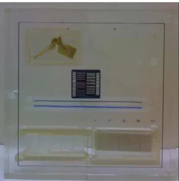
Figure 3 presents a picture of the phan-tom adapted for fluoroscopy, as well as the structures of interest for the interventional procedure that allowed the visualization of images besides the catheters C1 and C2: (a) the nylon caps simulating tumors; (b) the air-acrylic wedge-shaped phantom simulat-ing the body cavities; (c) the acrylic-PVC wedge shaped simulating bone structures; (d) aluminum spheres representing the bone tissue borders; (e) the device for evaluating resolution in line pairs/mm; (g) iron spheres for analyzing magnification; (h) tin wire for field evaluation; (j) half thoracic vertebra. Thus, one can evaluate thickness differences of a specific material or materials of very close effective atomic numbers, as well as visualizing and estab-lishing bone structures limits, visualizing and guiding extremely thin catheters uti-lized in cerebral interventional procedures,
and estimating limits for the system spatial resolution.
The images of the phantom adapted for fluoroscopy obtained on different exposure modes demonstrate that there is no signifi-cant loss of diagnostic information for the different exposure modes as shown on Fig-ure 4. In other words, in most of cases, the variation in the exposure conditions which reduces the dose to the patient does not affect the fluoroscopic image, neither con-demn nor impede the interventional proce-dure. This fact is evidenced by the obser-vation of Figure 4 in correlation with Table 1, where the image A was obtained with a mean air kerma rate of 4.6 ± 0.1 mGy/ minute, while the image C was obtained with a mean air kerma rate of 1.58 ± 0.01 mGy/minute. In this case, the reduction in the mean air kerma rate achieves 67%.
377 Study of optimization of images in interventional fluoroscopy
Radiol Bras. 2009 Nov/Dez;42(6):375–378
Table 1 Phantom entrance surface air kerma rate.
Exposure mode
LDF
HDF
Continuous 1/2 dose 1/4 dose
Continuous 1/2 dose 1/4 dose Pulsed
Voltage (kVp)
68 69 69
68 69 70 70 70 70
Amperage (mA)
2.4 1.3 0.7
5.8 3.1 1.6 1.8 3.0 4.4
Mean air kerma rate (mGy/min)
4.6 ± 0.1 3.07 ± 0.01 1.58 ± 0.01
10.9 ± 0.1 6.73 ± 0.01 3.68 ± 0.02 4.16 ± 0.07 6.93 ± 0.01 10.18 ± 0.01 3 frames/s
5 frames/s 8 frames/s
Table 2 Patient entrance surface air kerma rate.
Exposure mode
LDF
HDF
Continuous 1/2 dose 1/4 dose
Continuous 1/2 dose 1/4 dose Pulsed
Voltage (kVp)
68 69 69
68 69 69 69 69 69
Amperage (mA)
2.4 1.3 0.7
5.7 3.0 1.5 1.7 2.9 4.3
Mean air kerma (mGy/min)
3.37 ± 0.03 2.88 ± 0.01 1.50 ± 0.01
10.63 ± 0.09 6.37 ± 0.06 3.38 ± 0.01 3.98 ± 0.01 6.46 ± 0.01 9.53 ± 0.01 3 frames/s
5 frames/s 8 frames/s
Figure 4. Effect of the variation in the air kerma rate on image A – continuous mode; B – half dose; C – quarter dose; D – pulsed (8 frames/s).
Figure 3. Radiographic image of the realistic-analytic phantom adapted for fluoroscopy.
Table 1 presents the entrance surface air kerma rate obtained with the phantom for the different exposure modes, demonstrat-ing the alternatives for reduction of the dose to the patient during the examination. The abbreviations LDF and HDF corre-spond to the low- and high-definition fluo-roscopic exposure modes, respectively.
Table 2 presents the measurements of the typical entrance surface air kerma rates obtained with aluminum plates as sug-gested by the constancy test protocols, so one can compare the values on Tables 1 and 2, demonstrating the equivalence between the phantom adapted for fluoroscopy and the 38 mm-thick aluminum plates in terms of entrance surface air kerma rates.
DISCUSSION
Figure 3 shows the image of the phan-tom adapted for evaluating fluoroscopy equipment. The images obtained at differ-ent exposure modes did not demonstrate any significant loss of diagnostic informa-tion, highlighting that the exposure modes with low frame rate pulsed fluoroscopy and the continuous mode of quarter dose fluo-roscopy constitute the options with lowest exposure to the patients.
The values obtained for entrance sur-face air kerma rate with the phantom
in-cluded on Table 1 are approximately equiva-lent to the ones observed with the alumi-num plates shown on Table 2. In some cases, the reduction in the entrance surface air kerma rate for the patient may achieve
67% according to the procedure to be per-formed.
378
Parizoti A, Ghilardi Netto T
Radiol Bras. 2009 Nov/Dez;42(6):375–378
a phantom adapted for fluoroscopy and demonstrates the equivalence between the phantom and the patient.
CONCLUSIONS
The study of optimization of images in interventional fluoroscopy has demon-strated the relevance of a method for opti-mizing images quality and dose to the pa-tients in this type of procedure.
The utilization of a phantom adapted for fluoroscopy allows the evaluation of the images quality besides determining the patient entrance surface air kerma rate.
The phantom allows the evaluation of the patient entrance surface air kerma rate as well as determining which exposure modes reduce the dose to the patient with no significant loss of diagnostic informa-tion. The evaluation of images obtained with the phantom, in cerebrovascular interventional procedures included, allows
the achievement of fluoroscopic images optimization.
Acknowledgements
The authors thank Coordenação de Aperfeiçoamento de Pessoal de Nível Su-perior (Capes) for the financial support and the technical staff Hospital das Clínicas da Faculdade de Medicina de Ribeirão Preto da Universidade de São Paulo (HCFMRP-USP).
REFERENCES
1. International Commission on Radiological Pro-tection. Recommendations of the International Commission on Radiological Protection. ICRP Publication 26. Annals of the ICRP. 1977;1(3).
2. Brasil. Ministério da Saúde. Secretaria de Vigi-lância Sanitária. Diretrizes de proteção radioló-gica em radiodiagnóstico médico e odontológico. Portaria no 453, de 1o de junho de 1998. Diário
Oficial da União, Brasília, 2 de junho de 1998. 3. Secretaria de Estado da Saúde. Uso, posse e
ar-mazenamento de fonte de radiação ionizante no âmbito do Estado de São Paulo. Resolução
SS-625. Diário Oficial do Estado, São Paulo, 14 de dezembro de 1994.
4. Pina DR, Ghilardi Netto T, Rocha SL, et al. Cons-trução de um fantoma homogêneo para padroni-zação de imagens radiográficas. Radiol Bras. 2000;33:41–4.
5. Pina DR, Duarte SB, Ghilardi Netto T, et al. Op-timization of standard patient radiographic im-ages for chest, skull and pelvis exams in conven-tional x-ray equipment. Phys Med Biol. 2004;49: N215–26.
6. Parizoti A. Otimização de imagens e proteção radiológica em fluoroscopia [dissertação de mes-trado]. Ribeirão Preto: Universidade de São Pau-lo; 2008.
7. Pina DR, Duarte SB, Ghilardi Netto T, et al. Phan-tom development for radiographic image optimi-zation of chest, skull and pelvis examination for nonstandard patient. Appl Radiat Isot. 2009;67: 61–9.
8. Pina DR, Duarte SB, Morceli J, et al. Develop-ment of phantom for radiographic image optimi-zation of standard patient in the lateral view of chest and skull examination. Appl Radiat Isot. 2006;64:1623–30.