Risks of radiographic procedures for neonates admitted
to a public hospital in Belo Horizonte, MG, Brazil*
Riscos dos exames radiográficos em recém-nascidos internados em um hospital público de Belo Horizonte, MG
Marco Aurélio de Sousa Lacerda1
, Teógenes Augusto da Silva2
, Helen Jamil Khoury3 , José Nelson Mendes Vieira4
, João Paulo Kawaoka Matushita5
OBJECTIVE: The present study was aimed at: a) evaluating radiographic procedures and estimating entrance surface air kerma in preterm neonates submitted to chest and abdominal radiography at the unit of neonatology in a public hospital of Belo Horizonte, MG, Brazil; b) estimating the dose to organs and respective risks for cancer as a result from radiation exposure. MATERIALS AND METHODS: Records of patients admitted to the unit of neonatology of this hospital in the period between May and September 2004 were reviewed. Anthropometric data, admission/discharge dates and radiographic studies performed were recorded for each of the patients. The calculation of the entrance surface air kerma was based on the x-ray tube output and irradiation parameters adopted for examinations. Dose to organs was calculated with the aid of the software PCXMC, and the lifetime risk for cancer, with the software IREP. RESULTS: Mean entrance surface air kerma per examination was below the diagnostic reference levels recommended by the European Community. In the most severely irradiated patients, liver, breast and stomach were the organs at highest risk for cancer, with maximum excess relative risk of respectively 3.4%, 2.3%, and 1.7%. CONCLUSION: The present study demonstrated the need for optimization of radiographic procedures in order to reduce the risks for neonates that, in spite of being considered to be low as compared with the benefits, should be reduced to values as low as reasonably achievable.
Keywords: Patient dosimetry; Radiological protection; Pediatric radiology; Cancer risk.
OBJETIVO: Os objetivos deste trabalho são: a) avaliar os procedimentos radiográficos e estimar o valor do kerma no ar na superfície de entrada nos recém-nascidos prematuros submetidos a exames de tórax e abdome, realizados no setor de neonatologia de um hospital público de Belo Horizonte; b) estimar as doses nos ór-gãos e os respectivos riscos de ocorrência de câncer nesses órór-gãos em decorrência das exposições à radiação. MATERIAIS E MÉTODOS: Foram analisados os prontuários dos pacientes internados no setor de neonatolo-gia desse hospital durante o período de maio a setembro de 2004, anotando-se os dados antropométricos, data de internação/alta, exames de raios X realizados. O kerma no ar na superfície de entrada foi determinado a partir do rendimento do tubo de raios X e dos parâmetros de irradiação utilizados nos exames. As doses nos órgãos foram estimadas com o software PCXMC e o risco, durante o restante da expectativa de vida,
com o software IREP. RESULTADOS: O valor médio do kerma no ar na superfície de entrada por exame foi
abaixo do nível de referência da publicação da Comunidade Européia. Para o paciente mais severamente irradiado, os órgãos mais suscetíveis à ocorrência de câncer foram fígado, mama e estômago, com valores máximos de excess relative risk, respectivamente, de 3,4%, 2,3% e 1,7%. CONCLUSÃO: Foi constatada a
necessidade de otimização dos procedimentos radiográficos com vista à diminuição do risco para os recém-nascidos, que apesar de ser considerado baixo (comparativamente ao benefício), deve ser sempre diminuído para valores tão baixos quanto razoavelmente exeqüíveis.
Unitermos: Dosimetria do paciente; Proteção radiológica; Radiologia pediátrica; Risco de câncer.
Abstract
Resumo
* Study developed at Centro de Desenvolvimento da Tecnolo-gia Nuclear – Comissão Nacional de EnerTecnolo-gia Nuclear (CDTN-CNEN), Belo Horizonte, MG, Brazil.
1. PhD, Assistant Researcher at Centro de Desenvolvimento da Tecnologia Nuclear – Comissão Nacional de Energia Nuclear (CDTN-CNEN), Belo Horizonte, MG, Brazil.
2. PhD, Titular Researcher at Centro de Desenvolvimento da Tecnologia Nuclear – Comissão Nacional de Energia Nuclear (CDTN-CNEN), Belo Horizonte, MG, Brazil.
3. PhD, Titular Professor, Department of Nuclear Energy at Universidade Federal de Pernambuco (DEN-UFPE), Recife, PE, Brazil.
INTRODUCTION
Diagnostic radiology is considered as the main artificial radiation source to which human beings are exposed, being respon-sible for about 14% of the total annual ab-Lacerda MAS, da Silva TA, Khoury HJ, Vieira JNM, Matushita JPK. Risks of radiographic procedures for neonates admitted to a public hospital in Belo Horizonte, MG, Brazil. Radiol Bras. 2008;41(5):325–329.
4. MD, Radiologist, Assistant Professor at Faculdade de Me-dicina da Universidade Federal de Minas Gerais (UFMG), Belo Horizonte, MG, Brazil.
5. PhD, Associate Professor at Faculdade de Medicina da Universidade Federal de Minas Gerais (UFMG), Belo Horizonte, MG, Brazil.
Mailing address: Dr. Marco Aurélio de Sousa Lacerda. Comis-são Nacional de Energia Nuclear, Centro de Desenvolvimento da Tecnologia Nuclear, Unidade de Pesquisa e Produção de Radio-fármacos (UPPR). Avenida Presidente Antônio Carlos, 6627, Prédio 28, sala 02, Pampulha. Belo Horizonte, MG, Brasil, 31270-901. Caixa Postal 941. E-mail: [email protected]
sorbed radiation dose as a result of overall radiation exposure to the general popula-tion(1). Considering that any radiation
ex-posure can induce a risk for harmful effects, it is indispensable that a request for radio-logical examinations is preceded by a care-ful evaluation of risks versus benefits(2).
Special attention should be paid to ra-diographic examinations in children, con-sidering their higher susceptibility to the harmful effects of radiation as compared with the rest of the population(3). In units
of neonatology, particularly in cases where the patients are typically found in adverse clinical circumstances, the request for a number of radiographic studies may repre-sent a significant increase in the risk for these patients(4).
Studies developed in units of neonatol-ogy have demonstrated a great variation in radiographic technique conditions (volt-age, filtration, load, screen-film combina-tion, etc.) and, consequently, in the ab-sorbed-dose to newborn patients(5–8). In this
sense, an optimization of radiographic pro-cedures, particularly with the application of quality criteria recommended by the Euro-pean Community(9,10), can significantly
re-duce dose to patients without impairing the quality of radiographic images.
The estimation of the entrance surface air kerma rate (Ke) for inpatients of units of neonatology can be performed with the utilization of thermoluminescent dosim-eters, dose-area product mdosim-eters, or could be indirectly evaluated on the basis of radio-graphic technique parameters. This latter method, employing radiographic technique parameters in association with measure-ments of the x-ray equipment output is usually appropriate for this purpose(6).
Based on Ke, magnitudes related to risks, such as dose to organs, can be ob-tained with appropriate conversion coeffi-cients shown in tables available in the lit-erature(11,12) or by means of some
softwares(13–16). So, based on the dose to
or-gan, the risk of an exposed individual for developing a determined type of cancer (in the irradiated organ) as compared with a non-exposed individual can be determined with the aid of appropriate models avail-able in the literature(17).
The present study encompasses two ob-jectives: a) to evaluate radiographic
proce-dures and Ke in preterm neonates submit-ted to chest and abdominal radiography in the unit of neonatology (not ICU) of a pub-lic hospital in Belo Horizonte, MG, Brazil; b) to estimate dose to organs and respec-tive risks for developing cancer in these organs as a result of the radiation exposure.
MATERIALS AND METHODS
Records of inpatients of the unit of neo-natology in a public hospital of Belo Horizonte in the period between May and September 2004 were reviewed and the following individual data were recorded for the purposes of the present study: a) iden-tification number; b) sex; c) weight; d) height; e) admission date; f) discharge date; g) chest and abdominal radiographic stud-ies performed. Chest and abdomen account for 75% of radiographic procedures in neo-nates performed in this hospital.
The equipment utilized for radiographic images acquisition was a portable, mono-phase Movix 120 system with 1.5 mm alu-minum filtration and full-wave rectifica-tion exclusively utilized in the unit of neo-natology. All the images were acquired with the neonates in their respective incu-bators. Estimation of Ke values was based on the x-ray tube output. For this purpose, measurements of air kerma were performed with a MDH 10X5-6 ionization chamber (Radcal Corp.; Monrovia, USA) with an electrometer MDH 9015 (Radcal Corp.; Monrovia, USA), both previously cali-brated. The ionization chamber was posi-tioned on the center of the radiation field at a distance of 100 cm from the focus, and at 20 cm from the floor. Based on the x-ray tube output and irradiation parameters uti-lized in the examinations, the authors could estimate the incident air kerma (Ki) and Ke, by means of the following equations:
Ki = Ri . Q . (Dref / DFP)² (1)
Ke = Ki . BSF (2)
where: Ri = x-ray tube output for the radio-graphic technique employed, in mGy/mAs; Q = tube current (I) by exposure time (t) employed in the examination, in milliam-pere/second (mA.s); Dref = distance where the output was measured (1 m); DFP = fo-cus-skin distance, in meter, estimated by the difference between the focus-film
dis-tance (FFD) and the patient equivalent di-ameter (De) (equation 3)(18); BSF =
non-dimensional retroscattering factor. This is a function of the field size, equipment fil-tration and radiographic technique em-ployed. A fixed value of 1.16 for BSF was adopted in the present study(19).
De = 2 . [W / (H . pi)]1/2 (3)
where: H = patient’s height in meter; W = patient’s weight in grams.
Based on Ki, patient´s characteristics and radiographic techniques employed, the dose to the most exposed organs was evalu-ated by means of the software PCXMC(14),
developed by Finnish Centre for Radiation and Nuclear Safety. So, the patient´s life-time risk for developing cancer was esti-mated for some of the most exposed or-gans, by means of the software IREP (In-teractive RadioEpidemiological Pro-gram)(17), developed by National Institute
of Cancer in the United States of America. The IREP operation is based on risk mod-els (excess relative risk – ERR; a measure-ment of change in the relative risk for can-cer or death for a group of individuals ex-posed to a known radiation dose as com-pared with a non-exposed group), on a magnitude denominated assigned share (AS), defined by the equation 4, for a spe-cific age where the cancer was diagnosed. In the present study, the AS was calculated by the IREP for the most severely irradiated individual at each year subsequent to the radiation exposure (totaling 50 years) and converted into ERR, a procedure similar to the one adopted by Thierry-Chef et al.(20).
AS = ERR / (1 + ERR) (4)
RESULTS
Considering that, in the hospital evalu-ated, simultaneous irradiation of both re-gions (chest and abdomen) in a same ex-amination is frequent, the results were jointly reported as a single chest/abdomi-nal acquisition. So, dose to organs were es-timated assuming an irradiation field cov-ering both regions.
Table 1 Statistical analysis of weight, height, hospitalization period, number of studies per patient and estimated Ke for neonates in the Unit of Neonatology of a hospital in Belo Horizonte.
Mean Minimum 1st quartile Median 3rd quartile Maximum Weight (kg) 2.26 0.53 1.69 2.25 2.84 4.80 Height (cm) 44.5 30.0 42.0 45.0 48.0 62.0 H.P. (days) 16.0 1.0 4.0 10 21.5 137 n 3.9 0.0* 0.0* 2.0 5.0 50.0
Ke (mGy)
0.075 0.070* 0.074 0.075 0.077 0.080
n, number of studies per patient; H.P., hospitalization period.
* Minimum Ke corresponds to the lowest kerma rate among the patients admitted to the hospital and submitted to examinations. The patients who had not been submitted to examinations and therefore were not exposed to radiation were not included in the statistical analysis of Ke.
Table 3 Estimated minimum, mean and maximum values for doses (DT) to some of the most exposed
organs of neonates in a public hospital of Belo Horizonte.
Organ Stomach Lung Ovary Testicle Breast Liver Thyroid Minimum 0.040 0.028 0.020 0.032 0.050 0.035 0.010 Mean 0.045 0.031 0.022 0.049 0.056 0.037 0.027 Maximum 0.049 0.037 0.025 0.065 0.078 0.042 0.055 DT (mSv)
parametric values (voltage, load, time, fo-cus-film distance) usually adopted by the technicians for the chest/abdominal radio-graphic examinations of the neonates were, respectively: 53 kV, 1,5 mA.s, 50 ms, 95 cm.
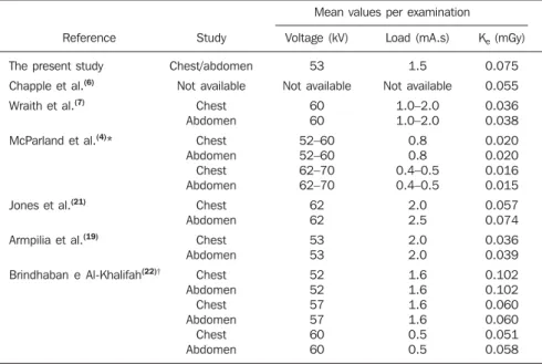
Table 2 shows a comparison between ra-diographic technique parameters and mean values for Ke, per study found for the neo-nates in the present study and those re-ported in the literature.
Table 3 presents minimum, mean and maximum doses to some of the most ex-posed organs, estimated per study.
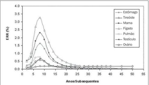
Figure 1 shows ERR variation with the time subsequent to the exposure estimated for some of the organs of the most severely exposed neonate (i.e. the patients submit-ted to 50 examinations).
DISCUSSION
The statistical analysis of weight and height of the neonates, as well as hospital-ization period presented on Table 1, cor-roborates the preterm condition of the in-patients in the unit of neonatology and the necessity of special care to be provided to these neonates. Such special care is trans-lated into a significant number of radio-graphic studies per patient. On average, the neonates were submitted to 3.9 chest/ab-dominal radiographic examinations, over a mean 16-day hospitalization period. It is important to note that one of the patients was submitted to an exceptionally high number of examinations (50) over a 137-day hospitalization period.
The analysis of radiographic technique parameters demonstrates that the x-ray tube voltage (kV) utilized for chest examination is below that recommended by the Euro-pean Community (60–65 kV). On the other hand, the exposure time utilized is higher than recommended (4 ms), and the focus-film distance is shorter than recommended by the European Community (100–150 cm). Also, additional copper filters have not been utilized during the procedures as recommended by the European Commu-nity(10).
The analysis of Table 2 demonstrates that the mean Ke per examination found in the present study is below the reference level suggested by the European Commu-nity(10) (0.080 mGy). However, this value
is higher than the mean values reported by the majority of studies in the litera-ture(4,6,7,18,20).
The fact that the mean Ke per examina-tion has been higher than the reference level recommended by the European Com-munity, despite the non-optimization of the irradiation parameters. Can be partially explained by the low x-ray equipment out-put. Previous results of quality control tests Table 2 Mean values for irradiation technique parameters and Ke, per examination in the present study
and in other studies in the literature.
Reference
The present study Chapple et al.(6)
Wraith et al.(7)
McParland et al.(4)*
Jones et al.(21)
Armpilia et al.(19)
Brindhaban e Al-Khalifah(22)†
Study Chest/abdomen Not available Chest Abdomen Chest Abdomen Chest Abdomen Chest Abdomen Chest Abdomen Chest Abdomen Chest Abdomen Chest Abdomen
Mean values per examination
Voltage (kV) 53 Not available 60 60 52–60 52–60 62–70 62–70 62 62 53 53 52 52 57 57 60 60 Load (mA.s) 1.5 Not available 1.0–2.0 1.0–2.0 0.8 0.8 0.4–0.5 0.4–0.5 2.0 2.5 2.0 2.0 1.6 1.6 1.6 1.6 0.5 0.5
Ke (mGy)
0.075 0.055 0.036 0.038 0.020 0.020 0.016 0.015 0.057 0.074 0.036 0.039 0.102 0.102 0.060 0.060 0.051 0.058
have shown that the equipment time and voltage accuracy and reproducibility are in compliance with the technical performance standards established by the Brazilian au-thorities(23). However, the half-value layer
at 80 kV (1.75 mm of Al) was significantly below of the minimum value established by the mentioned technical standard (2.3 mm of Al); that is to say, the x-ray tube output is low, even with the utilization of inad-equate filtration. Considering that the hos-pital technicians already operate with mini-mum values for time and current, it may be concluded that besides the x-ray equipment inappropriateness for examining neonates, the optimization of the radiographic tech-niques in compliance with European Com-munity recommendations is not feasible. This fact demonstrates that, probably, the quality of radiographic images is currently impaired. But this latter assertion only could be confirmed by further studies in-volving a joint evaluation of dose and im-age quality(24).
Table 3 demonstrates that mean doses to the gonads (testicles and ovaries) and thy-roid were relatively high. This fact demon-strates that an accurate collimation of the x-ray field, limiting the irradiated area to the chest and abdomen (provided it is clini-cally acceptable), and the utilization of lead shielding in the incubators or collimator lead shields would certainly reduce the dose to at least one of these organs.
The analysis of Figure 1 demonstrates that liver (maximum ERR = 3.4%), breast
(maximum ERR = 2.3%) and stomach (maximum ERR = 1.7%) are most suscep-tible to the development of cancer. In the case of the liver, this ERR value represents an increase in the risk for cancer from 89/ 10.000 (baseline)(25) to 92/10.000. The
maximum ERR is typically observed eight years after the exposure. In the case of the thyroid, this maximum ERR persists over the patient´s lifetime. In the other organs, the ERR tends to decrease to values near zero over the patient´s lifetime. Consider-ing that these results refer to the most se-verely exposed patient, who was submitted to a number of examinations 12 times higher than the average for the unit (~ four examinations/patient), it may be concluded that the neonates’ risk for developing sev-eral types of cancer in the future as a result of these examinations is relatively low as compared with the benefits from the appro-priately justified radiographic examina-tions. That is to say that the extremely ad-verse conditions of some neonates and the relevance of the radiodiagnosis as an essen-tial clinical tool in the improvement of the management of the patient could justify this higher risk.
CONCLUSIONS
Doses and risks for preterm neonates admitted to a public hospital in Belo Horizonte were investigated. The authors observed that the mean Ke/examination found in the present study is below the
ref-erence level suggested by the European Community(10) and above the mean values
found in a considerable number of studies in the literature. However, the adoption of non-optimized radiographic technique pa-rameters imposed by the utilization of a x-ray equipment with low output and inap-propriate for examination of neonates, is probably impairing the radiographic im-ages quality. But this hypothesis could only be confirmed by further studies involving a joint evaluation of dose and image qual-ity.
It was suggested that an appropriate col-limation of the x-ray field and/or the utili-zation of lead shielding (as clinically ac-ceptable) for reducing the doses to the thy-roid and/or gonads. Liver, breast and stom-ach were most susceptible to the develop-ment of cancer. The optimization of radio-graphic procedures is essential for reduc-ing the risk for neonates that, in spite of being considered to be low as compared with the benefits, should be reduced to values as low as reasonably achievable.
REFERENCES
1. United Nations Scientific Committee on the Ef-fects of Atomic Radiation. Sources and effect of ionizing radiation. UNSCEAR Reports to the General Assembly of the United Nations, with an-nexes. New York: United Nations; 2000. 2. International Commission on Radiological
Protec-tion. 1990 Recommendations of the ICRP. ICRP Publication no. 60. Oxford: Pergamon Press; 1991.
3. Ron E. Cancer risks from medical radiation. Health Phys. 2003;85:47–59.
4. McParland BJ, Gorka W, Lee R, et al. Radiology in the neonatal intensive care unit: dose reduction and image quality. Br J Radiol. 1996;69:929–37. 5. Fletcher EWL, Baum JD, Draper G. The risk of diagnostic radiation of the newborn. Br J Radiol. 1986;59:165–70.
6. Chapple CL, Faulkner K, Hunter EW. Energy im-parted to neonates during x-ray examinations in a special care infant unit. Br J Radiol. 1994;67: 366–70.
7. Wraith CM, Martin CJ, Stockdale EJN, et al. An investigation into techniques for reducing doses from neonatal radiographic examinations. Br J Radiol. 1995;68:1074–82.
8. Wilson-Costello D, Rao PS, Morrison S, et al. Ra-diation exposure from diagnostic radiographs in extremely low birth weight infants. Pediatrics. 1996;97:369–74.
9. European Commission. Quality criteria for diag-nostic radiographic images in paediatrics. CEC XII/307/91. Brussels: CEC; 1992.
10. European Commission. European guidelines on quality criteria for diagnostic radiographic images in paediatrics. EUR 16261 EN. Luxembourg: European Commission; 1996.
Figure 1. ERR variation with the time subsequent to the radiation exposure estimated for some of the organs of the most severely exposed neonate.
Estômago Tireóide Mama
Fígado Pulmão Testículo
11. Hart D, Jones DG, Wall BF. Normalised organ doses for paediatric x-ray examinations calcu-lated using Monte-Carlo techniques. NRPB-SR279. Chilton: National Radiological Protection Board; 1996.
12. Rosenstein M, Beck TJ, Warner GG. Handbook of selected organ doses for projections common in pediatric radiology. Rockville: U.S. Depart-ment of Health and Human Services; 1979. 13. Le Heron JC. CHILDOSE: a user’s guide.
Soft-ware and manual. Christchurch: National Radia-tion Laboratory; 1996.
14. Tapiovaara M, Lakkisto M, Servomaa A. PCXMC: a PC-based Monte Carlo program for calculating patient doses in medical x-ray exami-nations, report STUK-A139. Helsinki: Finnish Centre for Radiation and Nuclear Safety; 1997. 15. Kyriou JC, Newey V, Fitzgerald MC. Patient doses in diagnostic radiology at the touch of a button. London: The Radiological Protection Center, St. Georges Hospital; 2000.
16. Lacerda MAS, Khoury HJ, Silva TA, et al. Ra-dioproteção, dose e risco em exames radiográfi-cos nos seios da face de crianças, em hospitais de Belo Horizonte, MG. Radiol Bras. 2007;40:409– 13.
17. Land CE, Gilbert E, Smith JM, et al. Report of the NCI-CDC Working Group to revise the 1985 NIH radioepidemiological tables. DHHS Publ No. 03-5387. Bethesda: National Institute of Health; 2003. Available in: http://dceg.cancer. gov/docs/report03.pdf. Interactive RadioEpide-miological Program (IREP) v.5.3. Online software available in: http://www.irep.nci.nih.gov/
18. Lindskoug BA. Reference man in diagnostic ra-diology dosimetry. Radiat Prot Dosimetry. 1992; 43:111–4.
19. Armpilia CI, Fife IAJ, Croasdale PL. Radiation dose quantities and risk in neonates in a special care infant unit. Br J Radiol. 2002;75:590–5.
20. Thierry-Chef I, Simon SL, Miller, DL. Radiation dose and cancer risk among pediatric patients
undergoing interventional neuroradiology proce-dures. Pediatr Radiol. 2006;36 Suppl 14:159–62.
21. Jones NF, Palarm TW, Negus IS. Neonatal chest and abdominal radiation dosimetry: a comparison of two radiographic techniques. Br J Radiol. 2001;74:920–5.
22. Brindhaban A, Al-Khalifah K. Radiation dose to premature infants in neonatal intensive care units in Kuwait. Radiat Prot Dosimetry. 2004;111:275– 81.
23. Ministério da Saúde, Agência Nacional de Vigi-lância Sanitária. Radiodiagnóstico médico: de-sempenho de equipamentos e segurança. Brasí-lia: Ministério da Saúde; 2005.
24. Osibote AO, Azevedo ACP, Carvalho ACP, et al. Exposição de pacientes e qualidade da imagem em radiografias de tórax: uma avaliação crítica. Radiol Bras. 2007;40:119–22.