Alpha chain hemoglobins with electrophoretic mobility similar to that of hemoglobin S in
a newborn screening program
Marcilene Rezende Silva Shimene Mascarenhas Sendin Isabela Couto de Oliveira Araujo Fernanda Silva Pimentel Marcos Borato Viana
Universidade Federal de Minas Gerais - UFMG, Belo Horizonte, MG, Brazil
Conlict-of-interest disclosure:
The authors declare no competing inancial interest
Submitted: 9/17/2012 Accepted: 11/9/2012
Corresponding author: Marcos Borato Viana
Departamento de Pediatria da UFMG Av. Alfredo Balena, 190 – Sala 267 30130-100 Belo Horizonte, Brazil Fax: 55-31-34099770
www.rbhh.org or www.scielo.br/rbhh
DOI: 10.5581/1516-8484.20130031
Objective: To characterize alpha-chain variant hemoglobins with electric mobility similar to that of hemoglobin S in a newborn screening program.
Methods: βS allele and alpha-thalassemia deletions were investigated in 14 children who had undeined hemoglobin at birth and an electrophoretic proile similar to that of hemoglobin S when they were six months old. Gene sequencing and restriction enzymes (DdeI, BsaJI, NlaIV, Bsu36I and TaqI) were used to identify hemoglobins. Clinical and hematological data were obtained from children who attended scheduled medical visits.
Results: The following alpha chain variants were found: seven children with hemoglobin Hasharon [alpha2 47(CE5) Asp>His, HbA2:c.142G>C], all associated with alpha-thalassemia, ive with hemoglobin Ottawa [alpha1 15(A13) Gly>Arg, HBA1:c.46G>C], one with hemoglobin St Luke’s [alpha1 95(G2) Pro>Arg, HBA1:c.287C>G] and another one with hemoglobin Etobicoke [alpha212 84(F5) Ser>Arg, HBA212:c.255C>G]. Two associations with hemoglobin S were found: one with hemoglobin Ottawa and one with hemoglobin St Luke’s. The mutation underlying hemoglobin Etobicoke was located in a hybrid α212 allele in one child. There was no evidence of clinically relevant hemoglobins detected in this study.
Conclusion: Apparently these are the irst cases of hemoglobin Ottawa, St Luke’s, Etobicoke and the α212 gene described in Brazil. The hemoglobins detected in this study may lead to false diagnosis of sickle cell trait or sickle cell disease when only isoelectric focusing is used in neonatal screening. Additional tests are necessary for the correct identiication of hemoglobin variants.
Keywords: Hemoglobins, abnormal; Anemia, Sickle Cell; Neonatal screening; alpha-thalassemia; Polymerase chain reaction
Introduction
Hemoglobinopathies are a heterogeneous group of diseases caused by a disruption in the normal pattern of expression of genes encoding the globin chains. They are classiied fundamentally into two groups: a) structural variants, in which one or more amino acids are replaced in one of the polypeptide chains and b) thalassemias, in which an imbalance occurs in the production of one or more globin chains(1).
The hemoglobin variants result from substitutions of amino acids in the α, β, γ or δ chain tetramers of hemoglobins (Hbs) A, F and A2. The variants are caused by changes of DNA nucleotides, such as deletions, insertions or point mutations in one of the globin genes(2).
Hb S is the most frequent variant in humans. A point mutation in the b globin gene leads to the exchange of a single amino acid in the sixth position of the polypeptide chain (b6; glutamic acid → valine). Cells with Hb S undergo the sickling phenomenon, caused by low oxygen tension, acidosis and dehydration(3).
In the Globin Gene Server (http://globin.bx.psu.edu/cgi-bin/hbvar/counter) there were 1153 Hb variants registered as of 20 October 2012. The consequences of the structural changes upon the physicochemical properties of the molecule are dependent on the nature of the mutation and the place where it occurs. The consequences can be hemolytic anemia, when the change determines instability of the hemoglobin tetramer, altered oxygen transport if there is an increase or decrease in the afinity of Hb for oxygen or reduced synthesis of a globin chain, resulting in a form of thalassemia(4).
Carriers of “rare” Hb variants are most often asymptomatic. Associated with other hemoglobinopathies and thalassemias, these Hbs may result in a serious illness(5-7). Furthermore, there are dozens of Hb variants that can be mistaken for Hb S because they have similar isoelectric points, leading to a false diagnosis of sickle cell disease or trait if not properly conirmed(2,6-10).
Methods
The original sample from which the present study is derived consisted of 118 children who had indeterminate Hb results at birth and who, at six months of life, had an Hb proile identical or similar to that of Hb S. They were identiied by the Neonatal Screening Program in Minas Gerais, Brazil from June 1998 to June 2008. Hb Stanleyville-II, in 96 children, has already been thoroughly described(11). The present report comprises 14 patients, including one with Hb Etobicoke for which further information is provided, in addition to what was previously reported(12). Beta chain variants are not part of the current study.
All families signed written consent forms when they returned for this cross-sectional study. The study was approved by the Research Ethics Committees of the institutions involved and conducted according to the Declaration of Helsinki (revision 2008).
Isoelectric focusing (IEF) and high performance liquid chromatography (HPLC) were performed in neonatal screening. At six months of life, only IEF was carried out as the conirmatory method.
Genomic DNA was used to detect the βS allele through allele-specific polymerase chain reaction (PCR)(13) or through polymerase chain reaction-restriction fragment length polymorphism (PCR-RFLP) with the restriction endonuclease DdeI. Deletional alpha thalassemia types 3.7 and 4.2 were detected using multiplex gap-PCR(14).
The Hb Hasharon mutation was detected using PCR-RFLP with the restriction enzyme TaqI. The product of the multiplex gap PCR, which had detected the presence of alpha-thalassemia, was used in the restriction enzyme reaction to identify the Hb Hasharon mutation. The primers of the multiplex gap-PCR were used in two different reactions, one that ampliied LIS1 and HBA2 genes and another that ampliied the hybrid gene –α3.7 and LIS1 gene (Table 1).
Gene sequencing was necessary to identify the Hb variants in the other children. HBA1 and HbA2 genes were ampliied with
primers as described previously(15). Exon 1 and part of exon 2 of the alpha genes were sequenced by nested PCR with the S1 and S18 primers(16). The rest of exon 2 and exon 3 were sequenced with the forward primer S3 (for α1 and α2)(16) and reverse primers 3.7R1 for α2 and 3.7R2 for α1 genes(15) (Table 1). Sequencing was performed on the ABI Prism 3130 (Applied Biosystems).
After sequencing, speciic PCR-RFLP tests were designed for the easy diagnosis of the respective mutations. Methodological details on the diagnosis of Hb Stanleyville-II have been described previously(17).
To assess the clinical relevance of the detected Hbs and to provide information for the families about the results of the study, medical consultations were scheduled for the 14 children in this study; only nine attended. Blood counts for the child and family members were processed using Coulter T-890 automated blood analyzer. Hb electrophoresis in agarose gel was carried out in acid and alkaline media (SPIFE kits, Helena Laboratories, Beaumont, TX). The relative concentration of Hb variants was estimated by scanning the gels from electrophoresis in the alkaline medium.
Results
Of the 14 children in this study, seven carried Hb Hasharon [alpha2 47 (CE5) Asp> His, HbA2: c.142G> C] and had co-inherited alpha-thalassemia genes (six with genotype αα/-a3.7;Hasharon and one with -a3.7/-a3.7;Hasharon). In the PCR-RFLP test, LIS1 fragment (2350 base pairs - bp) is cleaved into two fragments (1307 bp and 1043 bp) by the TaqI enzyme. Among the fragments generated by the restriction enzyme, one had 894 bp; this was present only in those patients who had the mutation encoding Hb Hasharon (Figure 1A-B).
Five children had Hb Ottawa [alpha1 15 (A13) Gly> Arg, HBA1: c.46G> C], one Hb St. Luke’s [alpha1 95 (G2) Pro> Arg, HBA1: c.287C> G] and another Hb Etobicoke [alpha212 84 (F5) Ser> Arg, HBA212: c.255C> G] as shown in the sequencings
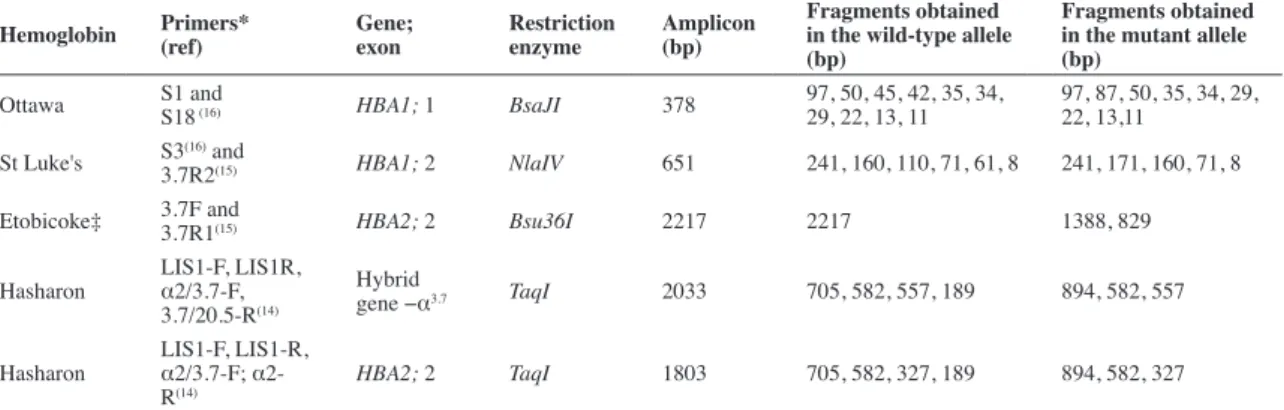
Table 1 - Primers and enzymes used for the detection of hemoglobin variants by polymerase chain reaction - restriction fragment length polymorphism
Hemoglobin Primers*
(ref)
Gene; exon
Restriction enzyme
Amplicon (bp)
Fragments obtained in the wild-type allele (bp)
Fragments obtained in the mutant allele (bp)
Ottawa S1 and
S18 (16) HBA1; 1 BsaJI 378
97, 50, 45, 42, 35, 34, 29, 22, 13, 11
97, 87, 50, 35, 34, 29, 22, 13,11
St Luke's S3(16) and
3.7R2(15) HBA1; 2 NlaIV 651 241, 160, 110, 71, 61, 8 241, 171, 160, 71, 8
Etobicoke‡ 3.7F and
3.7R1(15) HBA2; 2 Bsu36I 2217 2217 1388, 829
Hasharon
LIS1-F, LIS1R,
α2/3.7-F,
3.7/20.5-R(14)
Hybrid
gene −α3.7 TaqI 2033 705, 582, 557, 189 894, 582, 557
Hasharon
LIS1-F, LIS1-R,
α2/3.7-F;
α2-R(14)
HBA2; 2 TaqI 1803 705, 582, 327, 189 894, 582, 327
bp: base pairs
* Primers (5’...3’) S1: CAC AGA CTC AGA GAG AAC C; S3: CAC GGC AAG AAG GTG GCC GAC; S18: CGT TGG GCA TGT CGT CCA C; 3.7F: AAG TCC ACC CCT TCC TTC CTC ACC; 3.7R1:ATG AGA GAA ATG TTC TGG CAC CTG CAC TTG; 3.7R2: TCC ATC CCC TCC TCC CGC CCC TGC CTT TTC; α2/3.7-F: CCC CTC GCC AAG TCC ACC C; 3.7/20.5-R: AAA GCA CTC TAG GGT CCA GCG; α2-R: AGA CCA GGA AGG GCC GGT G; LIS1-F: ATA CCA TGG TTA CCC CAT TGA GC; LIS1-R: AGG GCT CAT TAC ATG TGG ACC C
(Figure 2A-C). Figures 1C, 3A and 3B-C illustrate, respectively, the restriction reactions for Hb Etobicoke (endonuclease
Bsu36I), Hb St. Luke’s (NlaIV endonuclease) and Hb Ottawa
(endonuclease BsaJI).
Of the Hbs identiied in this study, two were associated with Hb S: Hb Ottawa and Hb St. Luke’s (one case each). Hb Etobicoke was found in a child who had a hybrid α212 allele. Table 2 summarizes the alpha chain variants described in this study.
All these Hbs have isoelectric points similar to that of Hb S. In IEF at birth, all these children had an Hb fraction in the position of Hb S and another fraction between Hb S and Hb C, hence their initial classiication was of indeinite hemoglobin proiles. The
Figure 1 - Products of PCR and PCR-RFLP for the detection of Hb Hasharon (A & B) and Etobicoke (C)
A) Products of biplex gap-PCR for four individuals. Lanes 1 to 4 show
amplicons derived from the wild HBA2 (1803 bp) and LIS genes (2351 bp).
Lanes 5 to 8 amplicons from LIS and the -α3.7 hybrid gene (2033 bp). Lanes
1 and 5, 2 and 6, 3 and 7, 4 and 8 derive from the same children whose
HBA2 and -α3.7 genes were ampliied separately
B) Products of biplex gap-PCR, shown in Figure A, digested with TaqI for
the detection of the mutation that encodes Hb Hasharon. The fragment of 894 pb in lanes 5 and 6 indicates that these children have the mutation that
characterizes heterozygous Hb Hasharon in the -α3.7 hybrid gene
C) Products of PCR for the HBA1 gene (2213 bp: lanes 1 and 2) and for the
HBA2 gene (2217 bp: lanes 5 and 6) derived from two different individuals.
Lanes 3, 4, 7 and 8 correspond to the breakdown products of samples 1, 2, 5
and 6, respectively, digested with the endonuclease Bsu36I. Lane 7 shows a
child with the mutation that encodes Hb Etobicoke: the fragment of 2217 bp
is cleaved into two fragments of 1388 and 829 bp by Bsu36I.
1% agarose gel with ethidium bromide; MM: molecular marker; bp: base pairs; NC: blank control
Figure 2 - Gene sequencing of three children. The arrows indicate the mutations that encode (A) Hb St Luke’s (CCG>CGG; Pro>Arg), (B) Hb Ottawa (GGT>CGT; Gly>Arg), and (C) Hb Etobicoke (AGC>AGG; Ser>Arg )
Figure 3 - Products of PCR and PCR-RFLP for the detection of Hb St Luke’s (A) and Hb Ottawa (B and C)
A) Lanes 1 to 4 show amplicons derived from the exon 2 of the HBA1 gene
(651 bp). Lanes 5 to 8 show the restriction products from samples 1 to 4
with the endonuclease NlaIV. Lanes 6 and 8 illustrate two samples with the
mutation that encodes Hb St Luke’s. The fragment of 171 bp is not present in the wild gene
B) Products of simple PCR of exon 1 of HBA1 gene (378 bp)
C) Fragments of the samples shown in Figure B cleaved with the
endonuclease BsaJI. The mutation that encodes Hb Ottawa generates a
fragment of 87 bp shown in lanes 1 to 5, not present in lane 6 from a wild gene. To simplify the visualization of the result only the segment of the gel that distinguishes the two alleles is shown.
12% polyacrylamide gel with ethidium bromide; MM: molecular marker; bp: base pairs; NC: blank control
Table 2 - Alpha chain hemoglobin variants found and respective alpha-thalassemia genotypes
Hemoglobin variants and genotypes of α-thalassemia Number of children ‡
αα/-α3.7;Hasharon 6
-α3.7/-α3.7;Hasharon 1
αα/ααOttawa 5*
αα/α212Etobicoke 1¥
αα/ααSt Luke's 1*
IEF was repeated at six months of life. The results of four children with Hb Hasharon and two with Hb Ottawa were communicated by the screening program as sickle cell trait (false positive Hb AS). In the other children, the results were communicated as heterozygous “rare” Hbs in the position of Hb S. On retesting for the present study, children with Hb Hasharon were interpreted as having an Hb fraction slightly slower (cathodically) than Hb S, less than 0.5 mm from the control position. For children with Hb Ottawa and Hb St Luke’s, the Hb fraction moved slightly faster (anodically) than Hb S, also less than 0.5 mm from the control position. In the case of Hb Etobicoke, the position was exactly the same as Hb S.
In Hb electrophoresis on agarose gels (Figure 4), made as part of the medical consultation of these children, Hb Etobicoke and Hb Ottawa had Hb AS proiles in alkaline medium and Hb AA in acid pH. The child with Hb St. Luke’s co-inherited with Hb S had Hb AS proile in both pHs, because in alkaline pH Hb St. Luke’s overlaps with Hb S and in acid medium with Hb A. Hb Hasharon migrates like Hb S both in alkaline and acid media.
Discussion
Among the children studied, six had been considered by the newborn screening program as having undetermined Hb at birth and being heterozygous for Hb S (sickle cell trait) at sixth months of life, both results using IEF. All were born in the irst six years of the program. Resulting from acquired experience, children were considered in the subsequent years as carriers of “rare” Hbs to be further investigated, which materialized in the present study and in others(11,12,17). The comparison of the IEF gels at birth and at six months of life helps to correct the interpretation of data. The observation of an Hb fraction between the positions of Hb S and Hb C in the neonatal IEF, corresponding to the dimer aVariantg, draws attention to the possibility of alpha chain variants with electrophoretic mobility similar to that of Hb S. As the production of g chains decreases, children appear to be heterozygous for Hb S (aAbS) when they are six months old, when in fact, they have an alpha chain variant Hb (aVariantβA). Sickling and solubility tests are clearly negative in these cases, but require whole blood for testing. As this study demonstrates, the unequivocal identiication of the Hb variant requires molecular methods.
There was no evidence of clinical relevance of the Hbs detected in this study, even when there was an association of Hb St. Luke’s with Hb S in one child and of Hb Etobicoke with the α212 hybrid gene in another. According to the literature, Hb Hasharon would not cause clinical or hematological abnormalities unless co-inherited with alpha thalassemia(19,20) as reported by other Brazilian studies(18,19,21,22). Patients with Hb Ottawa and Hb St. Luke’s were considered normal upon clinical and hematological evaluation(8,23,24). The mutation for Hb Etobicoke occurs at codon 84 (F5) of the HBA gene (Ser>Arg). Although the nature and molecular location of the amino acid substitution would indicate the possibility of abnormal properties of the Hb, no hematologic or clinical changes have been observed(25).
Due to the small number of cases of each Hb variant, the ethnic origin of these variants could not be determined in
Figure 4 - Electrophoresis of hemoglobin in alkaline pH (above) or acid pH (below), in agarose gel. Sample 8 comes from a control AS. Sample 1 comes from the child with Hb Etobicoke; Samples 2 and 3 come from a child and his mother, both with Hb St. Luke’s associated with Hb S; Sample 4 comes from a child with Hb Ottawa; Samples 5 and 7 come from children with Hb Hasharon; and Sample 6 comes from the father of the child of Sample 5
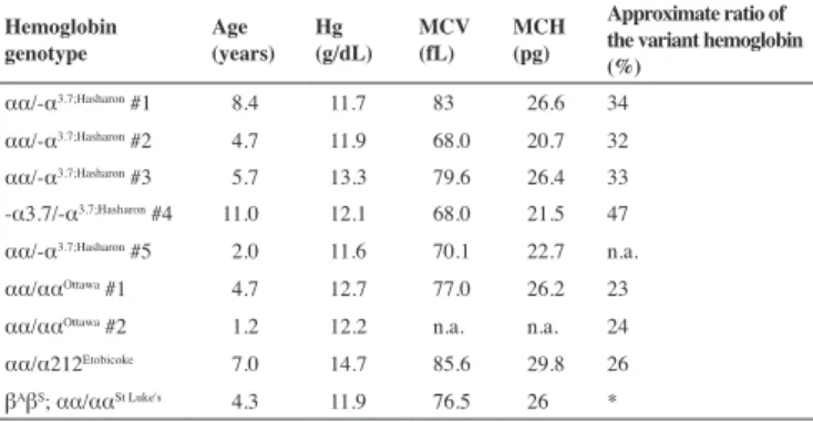
Table 3 - Hematological data of children who attended a medical examination
Hemoglobin genotype
Age (years)
Hg (g/dL)
MCV (fL)
MCH (pg)
Approximate ratio of the variant hemoglobin (%)
αα/-α3.7;Hasharon #1 8.4 11.7 83 26.6 34 αα/-α3.7;Hasharon #2 4.7 11.9 68.0 20.7 32 αα/-α3.7;Hasharon #3 5.7 13.3 79.6 26.4 33 -α3.7/-α3.7;Hasharon #4 11.0 12.1 68.0 21.5 47 αα/-α3.7;Hasharon #5 2.0 11.6 70.1 22.7 n.a. αα/ααOttawa #1 4.7 12.7 77.0 26.2 23 αα/ααOttawa #2 1.2 12.2 n.a. n.a. 24 αα/α212Etobicoke 7.0 14.7 85.6 29.8 26 βAβS; αα/ααSt Luke's 4.3 11.9 76.5 26 *
Hg: total concentration of hemoglobin in the blood; MCV: mean corpuscular volume; MCH: mean corpuscular hemoglobin
n.a.: not available
* As the fractions of Hb S and Hb St. Luke’s overlap in the alkaline pH, and Hb A and St. Luke’s at acidic pH, there is no way of estimating the relative proportion of Hb St. Luke’s
All the seven children of the present study with Hb Hasharon had alpha thalassemia associated. The relative proportions of Hb Hasharon (Table 3), estimated in gel electrophoresis in alkaline medium was, as expected, a third for cases with heterozygous alpha thalassemia (αα/-α3.7;Hasharon) and almost 50% for the single homozygous child (-α3.7/-α3.7;Hasharon). No association with alpha thalassemia was found in the children with Hb Etobicoke, Hb Ottawa and Hb St Luke’s. The relative proportion of the Hb variant stood at around 25%. The values of mean corpuscular volume (MCV) and mean corpuscular Hb (MCH) were generally lower than normal when there was co-inheritance of alpha thalassemia. They were normal for the child’s age in the other cases. Total Hb concentrations were all above the minimum reference value for the children’s ages.
this study, in contrast to Hb Stanleyville-II, which is clearly linked to African ancestry(11). Although there were reports of African ancestors in all 14 families in this study, we cannot overlook other ethnic origins as Brazil is markedly genetically heterogeneous, resulting from the admixture of Amerindian, European and African populations(26). As examples, according to the Globin Gene Server(27), Hb Hasharon has been described in people with Italian and Jewish ascent, Hb Ottawa (or Siam) in residents of Canada, Thailand and China, Hb Etobicoke in descendants of the Irish and Japanese(28) and Hb St Luke’s in the Maltese population.
PCR-RFLP tests were useful in the diagnosis or conirmation of the alpha chain variants found in the present study. Directly using the amplicons of gap-PCR for alpha thalassemia in a speciic RFLP test for Hb Hasharon facilitates diagnosis, making it simple, fast and economical, without the need for sequencing the HBA genes. In this method, however, it was essential to irst identify patients with Hb Ottawa, Hb Etobicoke and Hb St Luke’s. With standardization, it is simple to use restriction reactions carried out in this study when alpha chain variants with electrophoretic mobility similar to that of Hb S are suspected in screening tests.
Conclusion
This study describes the irst cases of Hb St. Luke’s, Hb Ottawa and Hb Etobicoke, and the α212 hybrid gene in Brazil. These Hb variants can lead to false diagnosis of sickle cell trait or sickle cell disease if only isoelectric focusing is used in neonatal screening programs. Additional methods are needed for the correct differential diagnosis and molecular analysis is essential for the identiication of the Hb variant. There was no evidence of clinical relevance for the Hbs detected in the present study, even in a child with an association of Hb St. Luke’s and Hb S. Standardized PCR-RFLP tests for the diagnosis of Hb Ottawa, Hb St Luke’s and Hb Etobicoke were devised which are simple, fast and economical making gene sequencing unnecessary.
Acknowledgments
The authors would like to thank all family members for agreeing to participate in the study and Cibele Velloso-Rodrigues, Beatriz Carvalho, and Terezinha D’Avila, of Fundação Hemominas, for their technical assistance. This work was supported by FAPEMIG (Fundação de Amparo à Pesquisa do Estado de Minas Gerais, Brazil) and Nupad (Núcleo de Ações e Pesquisa em Apoio Diagnóstico, UFMG). Marcos Borato Viana has a research grant from CNPq, Brazil.
References
1. Sonati M de F, Costa FF. The genetics of blood disorders: hereditary hemoglobinopathies. J Pediatr (Rio J). 2008;84(4 Suppl):S40-51.
2. Hocking DR. The separation and identiication of hemoglobin variants by
isoelectric focusing electrophoresis: an interpretive guide. Akron: Isolab Inc; 1997. 116 p.
3. Stuart MJ, Nagel RL. Sickle-cell disease. Lancet. 2004;364(9442):1343-60. Comment in: Lancet. 2005;365(9457):382-3.
4. Naoum PC. Hemoglobinopatias e talassemias. Sao Paulo: Sarvier; 1997. 168 p.
5. Harteveld CL, Groeneveld JH, van Dam B, Van Delft P, Akkerman N, Arkesteijn S, et al. Hb Zoeterwoude [beta23(B5)Val-->Ala)]: a new beta-globin variant found in association with erythrocytosis. Hemobeta-globin. 2005;29(1):11-7.
6. Fucharoen S, Singsanan S, Hama A, Fucharoen G, Sanchaisuriya K. Rapid molecular characterization of Hb Queens and Hb Siam: two variants easily
misidentiied as sickle Hb. Clin Biochem. 2007;40(1-2):137-40.
7. Jorge SE, Petruk AA, Kimura EM, Oliveira DM, Caire L, Suemasu CN, et al. Hb S-Sao Paulo: a new sickling hemoglobin with stable polymers and
decreased oxygen afinity. Arch Biochem Biophys. 2012;519(1):23-31.
8. Bannister WH, Grech JL, Plese CF, Smith LL, Barton BP, Wilson JB, et al. Hemoglobin St Luke’s, or alpha 2, 95 Arg (G2) beta 2. Eur J Biochem. 1972;29(2):301-7.
9. Cañizares ME, Martínez G, Hernández S, Raymond P, Colombo B. [Hemoglobinopathy S with an interaction of Hb S and Hb G-Ferrara]. Sangre (Barc). 1983;28(6):770-4. Spanish.
10. Marinucci M, Boissel JP, Massa A, Wajcman H, Tentori L, Labie D. Hemoglobin Maputo: a new beta-chain variant (alpha 2 beta 2 47 (CD 6)
Asp replaced by Tyr) in combination with hemoglobin S, identiied by high
performance liquid chromatography (HPLC). Hemoglobin. 1983;7(5):423-33.
11. Silva MR, Sendin SM, Pimentel FS, Velloso-Rodrigues C, Romanha
AJ, Viana MB. Hb Stanleyville-II [α78(EF7)Asn→Lys (α2); HbA2:
c.237C>A]: Incidence of 1:11,500 in a Newborn Screening Program in Brazil. Hemoglobin. 2012;36(4):388-94.
12. Silva MR, Sendin SM, Viana MB. Hb Etobicoke mutation in a hybrid HBA212 allele [HBA212 84 (F5) Ser>Arg; HBA212:c.255C>G]. Ann Hematol. 2012;91(12):1971-4.
13. Sanchaisuriya K, Chunpanich S, Fucharoen G, Fucharoen S. Multiplex
allele-speciic PCR assay for differential diagnosis of Hb S, Hb D-Punjab
and Hb Tak. Clin Chim Acta. 2004;343(1-2):129-34.
14. Tan AS, Quah TC, Low PS, Chong SS. A rapid and reliable 7-deletion multiplex polymerase chain reaction assay for alpha-thalassemia. Blood. 2001;98(1):250-1.
15. Liu YT, Old JM, Miles K, Fisher CA, Weatherall DJ, Clegg JB. Rapid detection of alpha-thalassaemia deletions and alpha-globin gene triplication by multiplex polymerase chain reactions. Br J Haematol. 2000;108(2):295-9.
16. Zorai A, Harteveld CL, Bakir A, Van Delft P, Falfoul A, Dellagi K, et al. Molecular spectrum of alpha-thalassemia in Tunisia: epidemiology and detection at birth. Hemoglobin. 2002;26(4):353-62.
17. Pimentel FS, Silva MR, Ferraz MH, Carvalho NO, Perone C, del Castillo DM, et al. Homozygous Hb Stanleyville-II [alpha2 78(EF7) Asn>Lys;
HBA2:c.237C>A, not C>G] associated with genotype -α 3.7/-α 3.7 in
two Brazilian families. Int J Lab Hematol. 2011;33(6):566-9.
18. Wagner SC, de Castro SM, Gonzalez TP, Santin AP, Zaleski CF, Azevedo LA, et al. Neonatal screening for hemoglobinopathies: results of a public health system in South Brazil. Genet Test Mol Biomarkers. 2010;14(4):565-9.
19. Wenning MR, Kimura EM, Costa FF, Saad ST, Gervásio S, de Jorge SB et al. Alpha-globin genes: thalassemic and structural alterations in a Brazilian population. Braz J Med Biol Res. 2000;33(9):1041-5.
20. Moradkhani K, Préhu C, Old J, Henderson S, Balamitsa V, Luo HY, et al. Mutations in the paralogous human alpha-globin genes yielding identical hemoglobin variants. Ann Hematol. 2009;88(6):535-43.
21. Kimura EM, Oliveira DM, Jorge SE, Abreu CF, Albuquerque DM, Costa
human hemoglobin. Rev Bras Hematol Hemoter. 2008;30(4):316-9.
22. Zamaro PJ, Bonini-Domingos CR. Abnormal hemoglobin phenotypes in carriers of mild anemia in Latin America. Genet Mol Res. 2010;9(1):425-8.
23. Vella FR, Casey R, Lehman H, Labossierre A, Jones TG. Haemoglobin Ottawa: alpha2 15 (A13) Gly --> Arg beta2. Biochim Biophys Acta. 1974:336:25-9.
24. Bezzina Wettinger S, Galdies R, Scerri C, Felice AE. Characterization and locus assignment of two alpha-globin variants present in the Maltese population: Hb St. Luke’s [alpha95(G2)Pro-->Arg] and Hb Setif [alpha94(G1)Asp-->Tyr]. Hemoglobin. 1999;23(2):145-57.
25. Crookston JH, Farquharson HA, Beale D, Lehmann H. Hemoglobin Etobicoke: alpha-84(F5) serine replaced by arginine. Can J Biochem. 1969;47(2):143-6.
26. Pimenta JR, Zuccherato LW, Debes AA, Maselli L, Soares RP, Moura-Neto RS, et al. Color and genomic ancestry in Brazilians: a study with forensic microsatellites. Hum Hered. 2006;62(4):190-5.
27. HbVar: a database for Human Hemoglobin Variants and Thalassemias. Boston; 2012. Available from: http://globin.bx.psu.edu/cgi-bin/hbvar/ query_vars3.
28. Harano T, Harano K, Shibata S, Ueda S, Nakashima Y, Imai K, et al.
Hemoglobin variant with slight instability and increased oxygen afinity, Hb Etobicoke [alpha 84 (F5) Ser replaced by Arg]: the irst case detected in Japan. Hemoglobin. 1982;6(6):613–7.
xxx
Erratum
In the article by Silva MR et al. entitled “Alpha chain hemoglobins with electrophoretic mobility similar to that of hemoglobin S in a newborn screening program” which appeared in the volume 35 number 2 issue of the Revista Brasileira de Hematologia e Hemoterapia, a correction should be noted. The legend of Figure 3 part A. should read:
Figure 3 - Products of PCR and PCR-RFLP for the detection of Hb St Luke’s (A) and Hb Ottawa (B and C)