Based on Molecular Profiling of Gene
Expression, Palmoplantar Pustulosis and
Palmoplantar Pustular Psoriasis Are Highly
Related Diseases that Appear to Be Distinct
from Psoriasis Vulgaris
Robert Bissonnette1*, Mayte Suárez-Fariñas2, Xuan Li2, Kathleen M. Bonifacio2, Carrie Brodmerkel3, Judilyn Fuentes-Duculan2, James G. Krueger2
1Innovaderm Research, Montreal, Quebec, Canada,2Laboratory of Investigative Dermatology, Rockefeller University, New York, New York, United States of America,3Janssen Research & Development, LLC, Spring House, Pennsylvania, United States of America
Abstract
Introduction
There is a controversy surrounding the existence of palmoplantar pustulosis (PPP) and pal-moplantar pustular psoriasis (PPPP) as separate clinical entities or as variants of the same clinical entity. We used gene expression microarray to compare gene expression in PPP and PPPP.
Methodology/Principal findings
Skin biopsies from subjects with PPP (3), PPPP (6), psoriasis vulgaris (10) and acral skin from normal subjects (7) were analyzed using gene expression microarray. Principal com-ponent analysis showed that PPP and PPPP were different from psoriasis vulgaris and nor-mal acral skin. However gene expression of PPP and PPPP clustered together and could not be used to differentiate PPP from PPPP. Gene-wise comparison between PPP and PPPP found no gene to be differentially expressed at a false discovery rate lower than 0.05. Surprisingly we found a higher expression of several genes involved in neural pathways (e.g. GPRIN and ADAM23) in PPP/PPPP as compared to psoriasis vulgaris and normal acral skin. Immunohistochemistry confirmed those findings and showed a keratinocyte localization for those proteins.
Conclusion significance
PPP and PPPP could not be differentiated using gene expression microarray suggesting that they are not distinct clinical entities. Increased expression of GPRIN1, and ADAM23 in keratinocytes suggests that these proteins could be new therapeutic targets for PPP/PPPP.
a11111
OPEN ACCESS
Citation:Bissonnette R, Suárez-Fariñas M, Li X, Bonifacio KM, Brodmerkel C, Fuentes-Duculan J, et al. (2016) Based on Molecular Profiling of Gene Expression, Palmoplantar Pustulosis and Palmoplantar Pustular Psoriasis Are Highly Related Diseases that Appear to Be Distinct from Psoriasis Vulgaris. PLoS ONE 11(5): e0155215. doi:10.1371/ journal.pone.0155215
Editor:Pierre Bobé, INSERM-Université Paris-Sud, FRANCE
Received:September 9, 2015
Accepted:April 26, 2016
Published:May 6, 2016
Copyright:© 2016 Bissonnette et al. This is an open access article distributed under the terms of the
Creative Commons Attribution License, which permits unrestricted use, distribution, and reproduction in any medium, provided the original author and source are credited.
Data Availability Statement:Raw data have been deposited in NCBI’s Gene Expression Omnibus and are accessible through accession number GSE 30999.
Introduction
The skin of palms and soles is unique and very different from other regions of the human body. These differences include the absence of hair, sebaceous glands, the increase in eccrine glands activity and the striking increase in thickness of the stratum corneum. Not surprisingly the morphology of common skin diseases is different when the palms and soles are involved. In addition some skin diseases such pustular palmo-plantar psoriasis (PPPP) and palmo-plan-tar pustulosis (PPP) localize specifically to palms and soles. There is a controversy surrounding the existence of these two diseases as separate clinical entities or as variants of the same clinical entity [1–4]. PPP is usually defined as a chronic skin disease characterized by crops of sterile pustules with erythema and sometimes scaling on palms and soles whereas PPPP is usually defined as a variant of plaque psoriasis present on palms and soles with the presence of sterile pustules [1]. Sometimes the morphology is intermediate between these two descriptions and it is unclear if such patients have the coexistence of psoriasis and PPP or if they show various clinical presentations of the same disease. Gene expression in acral skin including skin of patients with PPPP or PPP has not been well studied. The present study uses gene expression microarray to compare gene expression in lesional skin of patients with PPP and PPPP to nor-mal acral and non-acral skin and to skin from psoriasis vulgaris located outside hands and feet.
Materials and Methods
Subjects and skin biopsies
Four mm punch biopsies performed on palms or soles of patients with palmo-plantar pustular psoriasis (n = 6), palmo-plantar pustulosis (n = 3) or normal subjects (n = 7) were analyzed. These were obtained at baseline of a previously published study on the efficacy of ustekinumab in patients with PPP or PPPP [5]. In this study palmo-plantar pustular psoriasis was defined as active palmo-plantar disease morphology suggestive of psoriasis with at least one plaque of typ-ical psoriasis outside the palms and soles or a history of typtyp-ical plaque psoriasis outside the palms and soles. Palmo-plantar pustulosis was defined as active palmo-plantar morphology suggestive of palmo-plantar pustulosis without lesions of psoriasis outside palms and soles and without history of psoriasis. All patients gave written informed consent and the study was approved by an ethics board (IRB Services, Aurora, Canada). Washout before skin biopsies were 2 weeks for topical treatments, 4 weeks for phototherapy and oral treatments such as methotrexate, cyclosporine or acitretin and 12 weeks or 5 half-lives for biologics. Biopsies were immediately frozen with liquid nitrogen and stored at approximately -70°C.
For comparison purposes samples from 10 subjects with psoriasis vulgaris (non-acral) and 24 subjects with normal non-acral skin previously collected [6] were also analyzed.
Gene array
RNA was extracted using the Qiagen RNeasy Fibrous Tissue Mini Kit (QIAGEN, Valencia, CA) and later hybridized to GeneChip HG U133 Plus 2.0 (Affymetrix, Santa Clara, CA). Raw data have been deposited in NCBI’s Gene Expression Omnibus and are accessible through accession number GSE 80047 [6].
Immunohistochemistry
Frozen sections were cut and processed for immunohistochemistry using antibodies against GPRIN1 (Biorbyt LLC, San Francisco, CA, USA), or ADAM 23 (Lifespan Bioscience, Seattle, WA, USA). Biotin-labelled horse anti-mouse antibodies (Vector Laboratories, Burlingame,
Development, LLC. Janssen Research & Development, LLC provided support in the form of salary for author CB, but did not have any additional role in the study design, data collection and analysis, decision to publish, or preparation of the manuscript. The specific role of this author is articulated in the
‘author contributions’section. Author Robert Bissonnette is employed by Innovaderm Research, Montreal. Innovaderm Research provided support in the form of salary for author RB, but did not have any additional role in the study design, data collection and analysis, decision to publish, or preparation of the manuscript. The specific role of this author is articulated in the‘author contributions’section.
CA) were amplified using an avidin–biotin complex (Vector Laboratories) and developed with chromogen 3-amino-9-ethylcarbazole (Sigma-Aldrich, St-Louis, MO).
Statistical analysis
Quality control of microarray chips was carried out using standard QC metrics and R pack-age microarray Quality Control. Impack-ages were scrutinized for spatial artifacts using Harsh-light. Expression measures were obtained using the RMA algorithm with an extra loess-normalization step. Since normal non-acral-skin chips where hybridized in a different batch, we took several steps to guarantee that batch effect was eliminated. We download psoriasis and normal skin expression data from the same series of geo omnibus, and used it in con-junction with our data to provide and estimation of the batch effect using ComBat [7]. Our data was adjusted to eliminate this effect. As there was a gender imbalance between normal acral and PPP patients, we also used the combat implementation of sva packge to estimate (and thus eliminate) the effect of the gender imbalance, considering the groups as covariates in the model.
Probe-sets with at least 5 samples, expression values larger than 4, and standard deviation (SD)>015 were kept for further analysis (44281 probe sets). Fold changes (FCHs) for the
com-parisons of interest were estimated and hypothesis testing were conducted using contrasts under the general framework for linear models in the Rlimmapackage. P-values from the moderated t-test were adjusted for multiple hypotheses using the Benjamini–Hochberg proce-dure. Hierarchical clustering was performed using Euclidean distance and a Mcquitty agglom-eration scheme.
Results
PPP and PPPP cannot be differentiated based on gene expression
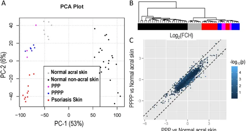
Fig 1Acompares gene expression from PPPP, PPP, normal acral skin, normal non-acral skin
and non-acral psoriasis vulgaris using a principal component analysis (PCA) plot. There is dis-tinct separation between normal acral, normal non-acral skin and psoriasis vulgaris. However gene expression in PPPP and PPP patients cluster together and cannot be differentiated. Unsu-pervised clustering of the expression profiles for all samples shows no clear distinction between PPP and PPPP patients (Fig 1B, dendrogram on the right y-axis) as they appear in the same cluster. Gene-wise comparison between the two groups found no gene to be differentially expressed at a FDR (False Discovery Rate) lower than 0.05.
Comparison of FCH of PPP vs Normal acral skin and PPPP vs Normal acral skin shows a strong correlation (r = 0.89) suggesting that transcriptomes of PPP and PPPP share most of their features (Fig 1C).
To study the association between the differences in the PPP and PPPP expression versus acral skin, we estimated the correlation coefficient and its distribution under the assumption of independence via simulation. We generated 1000 random samples of 3 independent variables, correspondent to PPP, PPPP and normal acral skin from a Normal distribution of mean mij
and sdiwhere i is the number of probe sets and j = 1,2,3 representing the mean for each gene
Difference in gene expression between normal palms/soles and normal
non-acral skin
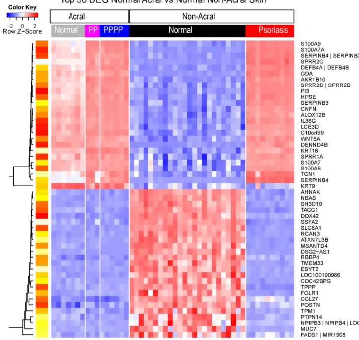
PCA plot shows striking differences in gene expression between normal palm/soles and non-acral skin (Fig 1A). A total of 4489 probe sets showed a significant difference (FDR<0.05) of
at least 2 FCH in expression between normal palm/soles and non-acral skin. Among those, there was an increase in keratin-6 (5-fold), keratin-16 (10-fold) and keratin-9 (16-fold), an increase in b-defensin4 (97 fold), lipocalin-2 (7 fold), IL-36-gamma (27 fold) and IL-36 recep-tor antagonist (8 fold). There was also a decrease (18 fold) in CCL27. Heat maps of representa-tive samples confirm that gene expression in normal palms/soles and non-acral skin is different (Fig 2).
Difference in gene expression between PPPP/PPP and normal acral
skin
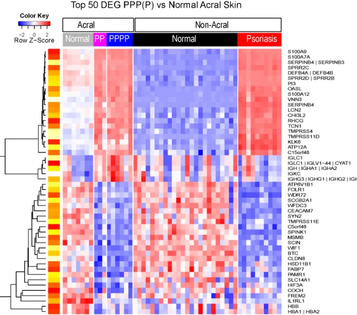
Important differences in gene expression between PPPP/PPP and normal acral skin can be seen on a PCA plot (Fig 1A). Since no differences were found between the two groups (PPP and PPPP), gene expression data from both groups of patients were combined and compared with normal non-acral skin. There was a significant (FDR<0.05) difference of at least 2 FCH
in gene expression for a total of 550 probe sets between PPPP/PPP and normal acral skin (the PPPP/PPP transcriptome). Among those genes there was an increase in b-defensin4 (3-fold) and lipocalin2 (3-fold), 2'-5'-oligoadenylate synthetase-like (OASL; 7 fold) and IL-36-gamma Fig 1. (A) Principal component analysis comparing gene expression from PPPP, PPP normal acral skin, normal non-acral skin and non-acral psoriasis vulgaris; PPP and cluster together PPPP. (B) Dendogram with unsupervised clustering of the expression profiles for all samples shows no clear distinction between PPP and PPPP as they appear in the same cluster. (C) There is a strong correlation between comparison of Fold Change (FCH) for PPP vs Normal acral skin and PPPP vs Normal acral skin.
(2-fold). Heat maps of representative samples confirm that gene expression of PPPP/PPP and normal acral skin is different (Fig 3).
Difference in gene expression between PPPP/PPP and psoriasis
vulgaris
PCA plot shows differences in terms of gene expression between PPPP/P and psoriasis vulgaris
(Fig 1A). A total of 403 probe sets showed at least a 2-fold change in gene expression with a
Fig 2. Heat map representing the expression profiles of the top 50 differentially expressed genes (DEG) of normal acral versus normal non-acral skin.Gene expression patterns from normal acral and non-acral skin are strikingly different. For DEG (FDR<0.05, FCH>2), the top 25 up and 25 down-regulated genes in terms of the fold change are presented according to an unsupervised cluster analysis. Yellow-red scale: red represents low gene expression and yellow high gene expression.
FDR of<0.05 between PPPP/PP and psoriasis vulgaris. However, we had already highlighted
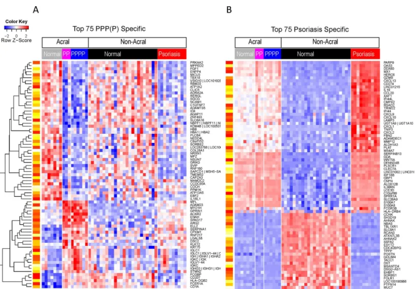
the large differences between acral and non-acral skin. To account for this we compared the PPPP/PP transcriptome (DEG between PPPP/PP and normal acral skin) with those that are DEG in the psoriasis vs normal non-acral skin. Heat maps of top genes show that gene expres-sion of PPPP/PPP and psoriasis vulgaris is different (Fig 4). One can observe that the top psori-asis genes, including IFNg genes OASL, MX1; IL17-regulated CCL20 and IL19 are not different in the PPPP/PPP as compared with normal acral skin. For genes in the PPPP/PP transcriptome however, some dysregulation is observed in psoriasis but not above the FCH cut-off.
Fig 3. Heat map representing the expression profiles of the top 50 differentially expressed genes (DEG) of PPP/PPPP versus normal acral skin.Gene expression patterns from PPP/PPPP and normal acral skin are strikingly different. For DEG (FDR<0.05, FCH>2), the top 25 up and 25 down-regulated genes in terms of the fold change are presented according to an unsupervised cluster
analysis. Yellow-red scale: red represents low gene expression and yellow high gene expression.
Increase in expression in PPPP/PPP of proteins usually expressed in
neural tissues
The upper right portion ofFig 4(PPP/PPPP specific) shows genes that are more highly expressed in PPPP/PPP as compared to psoriasis vulgaris and normal acral skin. Among these are genes coding for proteins usually found in the brain and/or associated with neurons such as G Protein Regulated Inducer of Neurite Outgrowth 1 (GPRIN1), and Disintegrin and Metallo-proteinase Domain-Containing Protein 23 (ADAM23). Using immunohistochemistry increased protein levels of GPRIN1 and and ADAM23 observed (Fig 5). All 3 proteins were mainly localized to keratinocytes (Fig 5). The increase in GPRIN1 and in ADAM23 expression was confirmed by RT-PCR (p<0.05 and p = 0.08 (trend with a two fold increase)) respectively;
not shown).
Fig 4. Heat map representing the expression profiles of the top 75 PPP/PPPP specific (A) and psoriasis specific (B) genes. The psoriasis PPP (P) specific genes are those that are differentially expressed in the PPP/PPPP versus normal acral skin and not differentially expressed in the psoriasis versus normal non-acral skin (FDR<0.05; FCH>2 in both comparisons). Likewise psoriasis specific genes are those that are differentially expressed in psoriasis as
compared with normal non-acral skin but are not differentially expressed in the PPP/PPPP versus normal acral skin comparisons. In both cases the top 75 are selected based on the largest differences observed between the log2FCH of the PPP/PPPP vs acral and the psoriasis versus non-acral skin comparison. The expression of several genes, including ADAM23 and GPRIN, is higher in PPP/PPPP (lower left portion ofFig 5A) Genes are presented according to an unsupervised cluster analysis. Yellow-red scale: red represents low gene expression and yellow high gene expression.
Discussion
To our knowledge this is one of the first study using transcriptomes to explore differences in patients with PPPP and PPP. We found that gene expression in PPPP and PPP clustered together and it was not possible to differentiate PPPP from PPP using micro-array analysis. Fig 5. Representative images of ADAM23 and GPRIN1 localization in normal acral skin, psoriasis vulgaris and PPPP as shown by immunohistochemistry.There is an increase in expression of ADAM23 and GPRIN1 in PPPP as compared to psoriasis vulgaris and normal acral skin. There is absence of staining with a non-specific isotype antibody (negative control).
Comparison of overall gene expression differences as well as comparisons between fold change expression of various genes did not show significant differences between PPPP and PPP. There was a strong correlation in FCH between PPP versus normal acral skin and PPPP versus nor-mal acral skin which further supports our conclusions that transcriptomes of both conditions are not significantly different. The difficulty in differentiating PPPP from PPP at the clinical, histological and molecular level has been previously reported [2,8,9]. Polymorphism in the genes coding for IL-19, IL-20 and LTA (lymphotoxin alpha) have been associated with both PPP and psoriasis [8,9]. However, polymorphism in TNF (tumor necrosis factor) promoters 238 and 308 have been associated with psoriasis vulgaris but not with PPP [10]. From a mor-phological point of view, there are no universally accepted criteria or descriptions to differenti-ate between PPP and PPPP. Patients presenting sterile pustules on an erythematous
palmoplantar skin without induration, scaling and without evidence of psoriasis are often labeled as having PPP. Patients with psoriasis on the scalp, trunk and limbs sometimes having typical well demarcated, indurated and scaly plaques on their palms and soles with occasional pustules on palms and soles are usually labeled as having PPPP. Unfortunately many patients have a palmoplantar morphology that is an intermediary between these two extremes. In addi-tion, the presence of psoriasis outside palms and soles has been reported in up to 24% of patients with a typical presentation of PPP [11,12].
In the current study microarray analysis was also used to compare gene expression in PPP/ PPPP to psoriasis vulgaris and normal acral skin in order to explore the pathophysiology of PPP/PPPP. Comparison of gene expression of normal acral skin and normal non-acral skin revealed an increase in expression of keratin 9, 6 and 16 on palms and soles which was expected given the hyperproliferative phenotype of acral skin and the relative specificity of keratin 9 for acral skin [13]. The increase in expression of antibacterial peptides b-defensin-4 and lipocalin-2 in normal acral skin as compared to normal non-acral skin is probably caused by the higher bacterial load present on acral skin. The increased in expression of b-defensin-4 and lipocalin-2 combined with the hyperproliferative phenotype of acral skin indicate that from morphologi-cal and functional point of view, the skin of palms and soles is intermediate between normal non-acral skin and psoriasis vulgaris. The decrease in CCL27, which is involved in homing T lymphocytes to the skin is intriguing and suggests that there might be mechanisms to limit adaptive immunity inflammatory reactions on palms and soles. From an evolutionary perspec-tive intense inflammation on palms and soles could limit the ability to run, walk and use vari-ous tools, which in turn could limit survival.
CCL20, CXCL2, CXCL10 and CXCL13 in PPPP/PPP suggests that acral keratinocytes may respond differently to cytokines such as IL-17A which regulates the production of many che-mokines including CCL20. These differences also suggest a more limited inflammatory reac-tion on palm and soles following activareac-tion of innate or adaptive immunity to preserve the ability to walk, run and use hands for survival.
We observed an increased expression of GPRIN1 and ADAM23 at the gene and protein level in PPP/PPPP as compared to psoriasis and/or normal acral skin. The localization of these proteins to keratinocytes was unexpected. Further studies should explore the role of these pro-teins in PPPP/PPP as they could be new therapeutic targets. Limitations of the present study include the absence of clearly established clinical criteria to differentiate PPP and PPPP, the small number of patients biopsied. Larger studies including female-only and male-only patients are needed to confirm these findings as they will be more adequately powered to iden-tify significant differences between PPP and PPPP subgroups.
In conclusion, transcriptome analysis of patients diagnosed clinically as having PPP and PPPP were similar but strikingly different from normal palms/soles and psoriasis vulgaris. This suggests that PPP and PPPP cannot be differentiated based on gene expression.
Author Contributions
Conceived and designed the experiments: RB JGK. Performed the experiments: CB KMB JFD RB XL. Analyzed the data: RB MSF CB JGK. Contributed reagents/materials/analysis tools: RB JGK CB. Wrote the paper: RB JGK.
References
1. Mrowietz U, van de Kerkhof PC. Management of palmoplantar pustulosis: do we need to change? Brit-ish Journal of Dermatology. 2011; 164(5):942–6. Epub 2011/02/01. doi:10.1111/j.1365-2133.2011. 10233.xPMID:21275942.
2. Brunasso AM, Puntoni M, Aberer W, Delfino C, Fancelli L, Massone C. Clinical and epidemiological comparison of patients affected by palmoplantar plaque psoriasis and palmoplantar pustulosis: a case series study. British Journal of Dermatology. 2013; 168(6):1243–51. Epub 2013/01/11. doi:10.1111/ bjd.12223PMID:23301847.
3. de Waal AC, van de Kerkhof PC. Pustulosis palmoplantaris is a disease distinct from psoriasis. J Der-matolog Treat. 2011; 22(2):102–5. doi:10.3109/09546631003636817PMID:20687846.
4. Ammoury A, El Sayed F, Dhaybi R, Bazex J. Palmoplantar pustulosis should not be considered as a variant of psoriasis. Journal of the European Academy of Dermatology and Venereology: JEADV. 2008; 22(3):392–3. doi:10.1111/j.1468-3083.2007.02344.xPMID:18269621.
5. Bissonnette R, Nigen S, Langley RG, Lynde CW, Tan J, Fuentes-Duculan J, et al. Increased expres-sion of IL-17A and limited involvement of IL-23 in patients with palmo-plantar (PP) pustular psoriasis or PP pustulosis; results from a randomised controlled trial. Journal of the European Academy of Derma-tology and Venereology: JEADV. 2013. Epub 2013/10/12. doi:10.1111/jdv.12272PMID:24112799. 6. Suarez-Farinas M, Li K, Fuentes-Duculan J, Hayden K, Brodmerkel C, Krueger JG. Expanding the
pso-riasis disease profile: interrogation of the skin and serum of patients with moderate-to-severe psopso-riasis. The Journal of investigative dermatology. 2012; 132(11):2552–64. doi:10.1038/jid.2012.184PMID: 22763790; PubMed Central PMCID: PMC3472561.
7. Johnson WE, Li C, Rabinovic A. Adjusting batch effects in microarray expression data using empirical Bayes methods. Biostatistics. 2007; 8(1):118–27. doi:10.1093/biostatistics/kxj037PMID:16632515. 8. Kingo K, Mossner R, Koks S, Ratsep R, Kruger U, Vasar E, et al. Association analysis of IL19, IL20 and
IL24 genes in palmoplantar pustulosis. British Journal of Dermatology. 2007; 156(4):646–52. Epub 2007/02/01. doi: BJD7731 [pii] doi:10.1111/j.1365-2133.2006.07731.xPMID:17263806.
9. Hashigucci K, Yokoyama M, Niizeki H, Yamasaki Y, Akiya K, Tojo T, et al. Polymorphism in the tumor necrosis factor B gene is associated with Palmoplantar pustulosis. Tissue antigens. 1999; 54(3):288– 90. Epub 1999/10/16. PMID:10519368.
palmoplantaris. Journal of Investigative Dermatology. 2005; 124(1):282–4. Epub 2005/01/19. doi: JID23556 [pii] doi:10.1111/j.0022-202X.2004.23556.xPMID:15654990.
11. Enfors W, Molin L. Pustulosis palmaris et plantaris. A follow-up study of a ten-year material. Acta der-mato-venereologica. 1971; 51(4):289–94. Epub 1971/01/01. PMID:4105778.
12. Ashurst PJ. Relapsing Pustular Eruptions of the Hands and Feet. British Journal of Dermatology. 1964; 76:169–80. Epub 1964/04/01. PMID:14140741.
13. Fu DJ, Thomson C, Lunny DP, Dopping-Hepenstal PJ, McGrath JA, Smith FJ, et al. Keratin 9 is required for the structural integrity and terminal differentiation of the palmoplantar epidermis. Journal of Investigative Dermatology. 2014; 134(3):754–63. Epub 2013/08/22. doi: jid2013356 [pii] doi:10.1038/ jid.2013.356PMID:23962810; PubMed Central PMCID: PMC3923277.
14. Kim DY, Kim JY, Kim TG, Kwon JE, Sohn H, Park J, et al. A comparison of inflammatory mediator expression between palmoplantar pustulosis and pompholyx. Journal of the European Academy of Dermatology and Venereology: JEADV. 2013; 27(12):1559–65. doi:10.1111/jdv.12203PMID: 23802874.
15. Murakami M, Hagforsen E, Morhenn V, Ishida-Yamamoto A, Iizuka H. Patients with palmoplantar pus-tulosis have increased IL-17 and IL-22 levels both in the lesion and serum. Experimental dermatology. 2011; 20(10):845–7. doi:10.1111/j.1600-0625.2011.01325.xPMID:21732985.
16. Hagforsen E, Hedstrand H, Nyberg F, Michaelsson G. Novel findings of Langerhans cells and interleu-kin-17 expression in relation to the acrosyringium and pustule in palmoplantar pustulosis. Br J Derma-tol. 2010; 163(3):572–9. doi:10.1111/j.1365-2133.2010.09819.xPMID:20426778.
17. Kanazawa N, Nakamura T, Mikita N, Furukawa F. Novel IL36RN mutation in a Japanese case of early onset generalized pustular psoriasis. Journal of Dermatology. 2013; 40(9):749–51. Epub 2013/07/10. doi:10.1111/1346-8138.12227PMID:23834760.