Transferases from Amoeba
Dictyostelium discoideum
Divya R. Nair1, Ratna Ghosh1, Alzu Manocha1, Debasisa Mohanty1, Shweta Saran2, Rajesh S. Gokhale1,3,4*
1National Institute of Immunology, New Delhi, India,2School of Life Sciences, Jawaharlal Nehru University, New Delhi, India,3Institute of Genomics and Integrative Biology, Council of Scientific and Industrial Research, Delhi, India,4Jawaharlal Nehru Centre for Advanced Scientific Research, Bangalore, India
Abstract
The life cycle of Dictyostelium discoideum is proposed to be regulated by expression of small metabolites. Genome sequencing studies have revealed a remarkable array of genes homologous to polyketide synthases (PKSs) that are known to synthesize secondary metabolites in bacteria and fungi. A crucial step in functional activation of PKSs involves their post-translational modification catalyzed by phosphopantetheinyl transferases (PPTases). PPTases have been recently characterized from several bacteria; however, their relevance in complex life cycle of protozoa remains largely unexplored. Here we have identified and characterized two phosphopantetheinyl transferases fromD. discoideumthat exhibit distinct functional specificity. DiAcpS specifically modifies a stand-alone acyl carrier protein (ACP) that possesses a mitochondrial import signal. DiSfp in contrast is specific to Type I multifunctional PKS/fatty acid synthase proteins and cannot modify the stand-alone ACP. The mRNA of two PPTases can be detected during the vegetative as well as starvation–induced developmental pathway and the disruption of either of these genes results in non-viable amoebae. Our studies show that both PPTases play an important role inDictyosteliumbiology and provide insight into the importance of PPTases in lower eukaryotes.
Citation:Nair DR, Ghosh R, Manocha A, Mohanty D, Saran S, et al. (2011) Two Functionally Distinctive Phosphopantetheinyl Transferases from Amoeba Dictyostelium discoideum. PLoS ONE 6(9): e24262. doi:10.1371/journal.pone.0024262
Editor:Vladimir N. Uversky, University of South Florida College of Medicine, United States of America
ReceivedJune 27, 2011;AcceptedAugust 3, 2011;PublishedSeptember 12, 2011
Copyright:ß2011 Nair et al. This is an open-access article distributed under the terms of the Creative Commons Attribution License, which permits unrestricted use, distribution, and reproduction in any medium, provided the original author and source are credited.
Funding:DBT’s National Bioscience Award for Career Development, India. Council of Scientific and Industrial Research, India (http://rdpp.csir.res.in/csir_acsir/ Home.aspx). The funders had no role in study design, data collection and analysis, decision to publish, or preparation of the manuscript.
Competing Interests:The authors have declared that no competing interests exist.
* E-mail: [email protected]
Introduction
Dictyostelium discoideum (Dicty) is a unicellular amoeba that undergoes multicellular differentiation when faced with starvation. This development process results in the formation of a fruiting body, which contains viable spores that germinate on return of favourable conditions. Various small metabolites are known to play a crucial role during this morphogenesis [1–6]. Two of the important developmental regulating factors (DRFs) – Differenti-ation Inducing Factor (DIF) and 4-methyl-5-pentylbenzene-1,3-diol (MPBD) are synthesized by large multifunctional polyketide synthases (PKSs) [7,8]. The genome of this organism has revealed an astoundingly large number of Type I PKSs [9], which are known to utilize a thio-template-based mechanism of biosynthesis. In this mechanism both starter and intermediate moieties are covalently acylated as thioesters either on the cysteine residue of ketosynthase (KS) domain or on the phosphopantetheine arm of acyl carrier protein (ACP). Phosphopantetheinyl modification of the ACP domains is a post-translational event and is essential for activity of PKSs. This modification is catalyzed by a group of enzymes known as phosphopantetheinyl transferases (PPTases) [10,11]. Several studies in recent years have revealed interesting insights into these enzymes; however, significance of PPTases in developmentally complex organisms is still obscure [12–22].
PKSs consist of three core catalytic domains – acyltransferase (AT), ACP and KS that act in a concerted manner with auxilliary
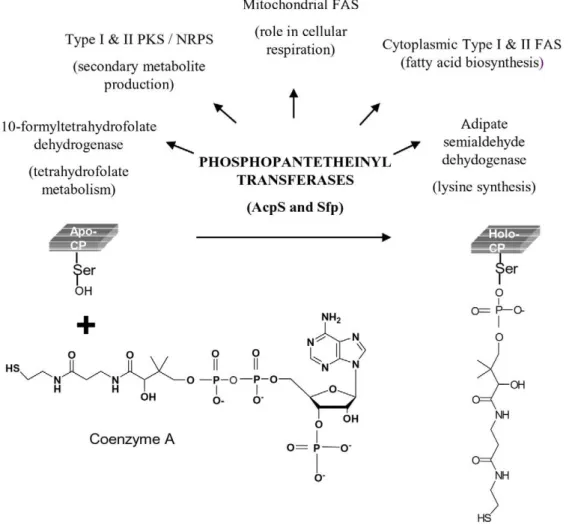
domains to yield the final product. In Type I PKSs various domains are present on a single polypeptide chain. In Type II PKSs these domains are present as discrete proteins that form a non-covalent complex. Type III PKSs are simple homodimeric proteins, and until recently were not known to require any carrier domains during repetitive condensations. In recent years, carrier domains have also been shown to provide starter units for Type III PKSs [7,8]. The modification of carrier domains involves the transfer of a 340 Da phosphopantetheine group derived from coenzyme A (CoA) on to the hydroxyl group of serine residue of ACP domains (Fig. 1). PPTases are also required for activating carrier proteins (CPs) of fatty acid synthases (FASs), non-ribosomal peptide synthases (NRPSs), adipate semialdehyde dehydrogenase [23] and 109-formyl tetrahydrofolate dehydrogenase [24].
Functions of PPTases in different organisms have revealed interesting themes. The genome ofB. subtilisencodes for AcpS and Sfp, former is involved in fatty acid synthesis and the latter in surfactin production [12]. However, in the absence of AcpS, Sfp has been shown to take over its function. This is in contrast to Mycobacterium tuberculosis where the roles of AcpS and Sfp have been reported to be non-redundant in nature [16]. The three PPTases from Myxococcus xanthus are categorized into -AcpS that is involved in fatty acid biosynthesis and two redundant Sfp proteins, modifying enzymes of secondary metabolic pathway [13]. In certain examples PPTases are known to be associated specifically with enzyme systems [18]. In parasites, the function of two PPTases is suggested to be dictated by the architecture of the proteins [17]. InToxoplasma gondii, the two PPTases have been proposed to cater to the Type I and Type II FAS systems. Interestingly, in Cryptosporidium parvuma single PPTase, Sfp is capable of modifying multifunc-tional Type I PKS and FAS and it does not contain AcpS. Plasmodium, which possesses only Type II FAS, has AcpS for mediating activation of CP. In fungi such asAspergillus nidulans
one of the three PPTases is an integral component of the FAS a-subunits and another one is suggested to be involved in activation of a mitochondrial FAS [14]. The third PPTase is demonstrated to be Sfp-like protein that is responsible for the activation of all its 27 PKSs and 14 NRPSs.
These studies clearly highlight that despite characterization of several different Sfp and AcpS-like PPTases, even now it is not possible to predict their selectivity for different protein types. It is therefore important to characterize them and delineate their functional specificities in a case-specific manner.
In this study, we have investigated the functional role of two PPTase homologues inDictyosteliumbiology. Here, we demonstrate that the two PPTases, DiAcpS and DiSfp are functionally discrete and non-redundant in nature. Furthermore, biochemical studies unambiguously show that DiSfp is required for the activation of multifunctional PKS/FAS, whereas, DiAcpS can modify only the stand-alone ACP. We also show that both PPTases are expressed during all the stages of its life cycle. Through our studies we demonstrate the importance of PPTases in the life cycle of Dictyostelium.
Figure 1. Schematic representation of phosphopantetheinyl transferase (PPTase) catalyzed conversion ofapo-ACP toholo-ACP.
Phosphopantetheine group of coenzyme A is transferred by the PPTases to a conserved serine group of ACP. This post-translational modification of carrier proteins is necessary for their function in various metabolic pathways.
Results and Discussion
Identification of Dictyostelium PPTases
The presence of 45 PKSs inDictyosteliumis quite unprecedented and to understand their relevance in multicellular development, we first decided to investigate into the modes by which PKSs are post-translationally modified by PPTases. We performed BLAST searches with Streptococcus pneumoniae AcpS and B. subtilisSfp on Dictybase (www.dictybase.org) to identify PPTase-like sequence(s) in the Dicty genome. Our analysis revealed the presence of two protein sequences with considerable homology. DDB0217726 (DiAcpS) shows 45% similarity withS. pneumoniaeAcpS, whereas, DDB0186752 (DiSfp) exhibits 53% similarity withB. subtilisSfp. Both these proteins show motifs characteristic of PPTases -[IV]G[ITV]D[ILV][VE] and W[CA][AL]KEAxxK. In addition, DiSfp also contains the (FNxSH) motif that is characteristic of Sfp type PPTases [17]. Comparative analysis of these protein sequences based on the crystal structures of B. subtilisAcpS and Sfp [28,29] also showed conservation of key residues. This includes R16 and R23 residues in DiAcpS involved in recognition of ACP and so also the amino acids D8, F25, R28, E58 and F74 (numbering corresponds toBacillusAcpS) reported to be involved in binding of CoA and Mg2+
. Careful analysis of DiSfp sequence also revealed the presence of residues – H118, S117, K52 and K193 which have been shown inB. subtilisSfp to be involved in CoA binding. Dendrogram-based phylogenetic analyses of Dicty PPTase sequences with other lower eukaryotes, including fungi and protozoan parasites readily provided their classification in two distinct groups of AcpS-like and Sfp-like sequences (Fig. 2A). Putative Sfp homologue from Neurospora does not group itself in either of the two clades. In order to understand the relevance of the two putative PPTases from Dicty, we decided to carry out genetic and biochemical investigations.
Gene knockout and expression studies of PPTases In order to generate genetic-knockouts of the individual genes, we adopted standard homologous recombination techniques. After transformation of the linear cassette (Figure S1), the Dicty amoeboid cells were aliquoted into 9664 wells. On systematic increase of antibiotic selection, ,50% wells showed growth of Dicty cells in the case ofdiacps. Fordisfpknockout transformants, survival rate was ,20%. PCR-based analysis was performed on 124 diacps clones and 96 disfp clones to confirm homologous recombination. However, in all the cases clones were found to be non-homologous recombinants (Figure S2). In order to validate experimental strategy,dipks37knockout was also attempted. Five positive clones fordipks37knockout could be confirmed and these mutants showed similar phenotypic characteristics as described earlier in the literature [7] (Figure S3). We attempted few different primer sets for PPTases, however, all our attempts provided only non-homologous recombinants. Our genetic studies could not provide any further information and therefore we proceeded to examine the presence of gene transcripts through different stages of this organism. RT-PCR analysis indicated presence of mRNA levels of both PPTases during all the stages of the life cycle of Dicty (Fig. 2B). Since the genetic tools for making conditional mutants or for RNAi studies are not robust in Dicty, we decided to biochemically delineate the functional implications of these proteins.
Cloning and expression of Phosphopantetheinyl Transferases
Both the proteins were heterologously expressed inE. coli. Diacps gene has 2 exons, which were amplified using nested primers from
genomic DNA. Similarly disfp also has 2 exons and these were PCR amplified individually and cloned into expression vector. Proteins were expressed by using a T7-based expression system and both the proteins could be readily purified using affinity chromatography. DiAcpS showed a band at around 27 kDa on SDS-PAGE rather than the expected molecular mass of 20 kDa. Meanwhile, DiSfp showed the theoretical molecular mass of 34 kDa. Identities of both the proteins were confirmed using mass spectrometric analysis (Figure S4).
Cell-free Assay with Mycobacterial Type I PKS
Preliminary analysis of the enzymatic activity of the PPTases was performed with multifunctional Type I mycobacterial PKSs. Several of these PKSs have been previously cloned and Figure 2. Phylogenetic analysis and expression profile of PPTases.A, Sequences of PPTases fromDictyosteliumand other lower eukaryotes were analyzed for their evolutionary relatedness. The sequences branch into two distinct groups, one group constituting AcpS-like sequences and the other formed by Sfp-like sequences. B, Expression profiles of DiAcpS and DiSfp were studied by RT-PCR and both were found to be expressing at all developmental stages. Stages analyzed were amoeboid (1), 0 hrs. after starvation (2), streaming (3), loose aggregate (4), mound (5), slug (6), early culminant (7) and fruiting body (8). IG7 (mitochondrial large rRNA) was used as the RT-PCR control.
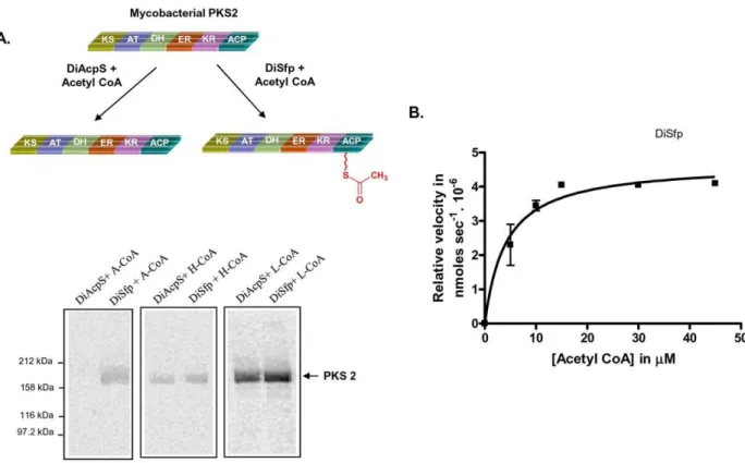
functionally characterized in our laboratory. Radiolabeled [1-3H] CoA was used as substrate and the transfer of radiolabel was detected by autoradiography. Whereas weak signal could be obtained with DiSfp, DiAcpS did not show any radioactive band. The profile remained unaltered even after increasing the concentration of DiAcpS and/or long exposure times. Since PPTases are also known to use acyl CoA as substrates [26,27,30], we used [1-14C] acetyl CoA for our further studies. Assays set up with PKS2, PKS12 and MAS (mycocersoic acid synthase) along with labeled acetyl CoA clearly indicated phosphopantetheinyla-tion only by DiSfp and no activity could be observed for DiAcpS (Fig. 3A). Catalytic efficiency of phosphopantetheine transfer with respect to acetyl CoA was estimated by Michaelis-Menten fit and kcat/Kmwas calculated to be 0.353 mM21min21(Fig. 3B). The
ACP concentration was fixed to be approximately 2.5-fold higher than the PPTase enzymes. We further probed if DiSfp was capable of transferring longer chain acyl-CoAs (hexanoyl and lauroyl CoA) on PKSs. Surprisingly, not only DiSfp but also DiAcpS showed ability to transfer both hexanoyl CoA and lauroyl CoA with comparable efficiencies (Fig. 3A). This unusual ability to only transfer longer chain acyl-CoA by DiAcpS on type I PKS was perplexing.
Comparative analysis of PPTase activity with type II stand-alone ACP
To ascertain the activity of PPTases with type II ACP, mycobacterial stand-alone ACP (Rv1344) was chosen. The purified protein shows two bands on SDS-PAGE, a 13 kDa band
corresponding to the intact protein size and an 11 kDa band, which possesses truncation at the N-terminus. Gel binding assays with this stand-alone carrier protein showed a reverse trend for the function of two PPTases in comparison to studies with Type I PKSs (described above). While DiAcpS could efficiently catalyze phosphopantetheinylation of Rv1344, DiSfp exhibited weak activity with acetyl-CoA (Fig. 4A). Kinetics of DiAcpS with the ACP was determined by varying the concentrations of acetyl CoA and the kcat/Kmwas estimated to be 0.162 mM21min21(Fig. 4B).
In order to unambiguously confirm phosphopantetheinylation of ACP, these proteins were separated on reverse-phase column and subjected to mass spectrometric analysis. DiAcpS mediated conversion of apo- to holo-ACP resulted in shift in the retention time of the protein from 20.7 minutes to 19.4 minutes (Fig. 5A). The peak at 19.4 minutes showed an increment of 340 Da, confirming modification with phosphopantetheine group. The enzymatic assays ofapo-ACP (13,474 Da) with hexanoyl CoA and lauroyl CoA exhibited higher molecular masses of 13,913 Da and 14,003 Da respectively, consistent with attachment of correspond-ing acyl-phosphopantetheine group. DiSfp could not efficiently catalyze conversion of apo-type II ACP to the holo-form, but showed reasonable activity with hexanoyl and lauroyl-CoAs (Fig. 5B).
Analysis of DiSfp and DiAcpS with ACP fragment of Mycobacterial PKS12
The specificity of DiAcpS and DiSfp to phosphopantetheinylate type II ACP and type I PKS ACP respectively prompted us to
Figure 3. Differential specificity ofDictyosteliumSfp (DiSfp) towards mycobacterial PKS2 and kinetic analysis.Gel-binding assays were set up with DiAcpS and DiSfp, taking [1-14C] acetyl-CoA (A-CoA), [1-14C] hexanoyl-CoA (H-CoA) and [1-14C] lauroyl-CoA (L-CoA) as co-substrates. A,
Panels show the autoradiogram of SDS-PAGE in which mycobacterial PKS2 was labeled by different acyl CoA substrates and PPTases as indicated. B, Gel-based kinetic analysis of DiSfp with respect to acetyl CoA. Concentration of acetyl CoA was varied from 5mM to 45mM.
examine whether this selectivity is due to architectural differences of the PKS proteins or is an inherent property of ACP domain. To resolve this issue, we cloned and expressed the PKS12 (Type I PKS) ACP fragment and then studied its phosphopantetheinyla-tion by the two PPTases. Boundaries of the module 1 ACP were defined using the PKS-NRPS database [31]. Gel-binding assay with the purified protein showed that the transfer of phospho-pantetheine group on to the isolated PKS12 ACP domain could be catalyzed solely by DiSfp. This is in concordance with the data wherein this ACP as part of the larger polypeptide could not be modified with DiAcpS (Fig. 6). Clearly, our work shows that the specificity of phosphopantetheinylation is not dictated by the modular architecture, but in fact involves specific recognition of the carrier domains. An earlier study with rat FAS ACP domain however, had come to reverse conclusion [32]. In that case an independently expressed ACP domain from multifunctional FAS system could be modified by bacterial AcpS that is known to modify Type II FAS ACP proteins. This apparent contradiction could be a manifestation of a broader specificity of AcpS protein or could also be argued in terms of the projected evolutionary relationship of two PPTases with primary and secondary metabolism [12,16,23,26].
Studies on Dictyostelium Type I PKS/FAS
The activity of Dicty PPTases was then evaluated by using cognate proteins from the organism. Since experiments with
mycobacterial proteins suggested that modular arrangement does not determine the selectivity, we used a smaller di-domain protein to understand phosphopantetheinylation specificity. ACP-TypeIII PKS di-domain of DiPKS1 (DDB0234164) was used for the assays with both PPTases [8]. Figure 7A illustrates that DiSfp is able to phosphopantetheinylate ACP-TypeIII PKS but DiAcpS fails to convert thisapo-protein to itsholoform. This reinforces our view that DiAcpS cannot function with type I PKS.
AcpS in several studies is suggested to be involved in primary metabolism of fatty acids [12,16,23,26]. We therefore wanted to investigate if similar scenario exists in Dicty. DiPKS16 (DDB0230068) was chosen as the candidate protein to test this hypothesis because this protein has been implicated as putative type I FAS [33]. DiPKS16 ACP domain boundaries were determined using the PKS-NRPS database [31]. The 258 bp gene was induced to over-express the C-terminus His6 tagged
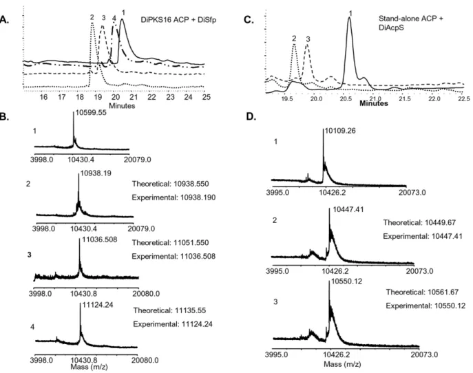
protein inE. coli. The purified ACP showed anomalous mobility on SDS-PAGE, indicating a molecular mass of,27 kDa rather than the calculated mass of 10.6 kDa. Similar anomalous migration of ACPs has been reported in literature and is attributed to their highly acidic nature [34]. Mass spectrometric analysis established the protein to be DiPKS16 ACP (Figure S5) Phosphopantetheinylation of this ACP was followed by autoradi-ography with radiolabeled acetyl CoA and here again only DiSfp showed enzymatic activity (Fig. 7B). HPLC-MALDI-TOF based assays provided further unambiguous analysis. As is evident from Figure 4. Differential specificity ofDictyosteliumAcpS (DiAcpS) towards mycobacterial stand-alone ACP and kinetic analysis.A, Panels show the autoradiogram of SDS-PAGE in which mycobacterial Rv1344 was labeled by different acyl CoA substrates and PPTases as indicated. Rv1344 shows the presence of two protein bands, lower one being the N-terminus truncated form. The truncated form is also seen to be incorporating radioactivity, suggestive of PPTase mediated modification. B, Michaelis-Menten kinetic analysis of DiAcpS was performed in a similar way as that for DiSfp.
figure 8A, DiSfp resulted in a shift of ACP retention time on reverse-phase column from 20.5 minutes to 19.2 minutes. These peaks on MALDI-TOF revealed molecular masses of 10,599 Da and 10,938 Da respectively (Fig. 8B). The increment of 340 Da is consistent with attachment of a phosphopantetheine arm. Similar activity was also observed with hexanoyl and lauroyl CoA, with expected increments in the molecular weight of ACP. ACP modified with hexanoyl CoA showed a retention time of 19.7 minutes and molecular mass of 11036 Da. Lauroyl CoA treated protein eluted at 20.1 minutes and revealed a molecular mass of 11124 Da. As opposed to this, DiAcpS lacked any activity towards this ACP. Intriguingly, DiAcpS failed to modify the type I ACP even with hexanoyl and lauroyl CoA. This is in contrast to our earlier observation with mycobacterial PKSs.
Role of DiAcpS in Dictyostelium
Our data strongly suggests that multifunctional PKSs are modified by DiSfp. This however raised an issue on the essential requirement of DiAcpS that was reflected in our genetic studies. A
BLAST search by using Saccharomyces stand-alone ACP with Dictybase identified DDB0184099 as a putative type II ACP, annotated as component of NADPH ubiquinone reductase. The gene was cloned intoE. coliexpression vector and over-expressed protein was purified using affinity chromatography. The protein showed a mobility of ,10 kDa on SDS-PAGE, less than the expected molecular weight of 13 kDa. The extraction of trypsinized fragments of DiACP on analysis with MALDI-TOF confirmed the identity of protein; however peptides from N-terminus could not be detected (Figure S6). HPLC-purified intact protein analysis with MALDI-TOF showed the molecular weight of this protein as 10109 Da. This deviation from the predicted molecular weight (13195 Da) could be attributed to N-terminus truncation of the protein (approximately 24 amino acids). Interestingly, sequence analysis based on MitoProt software predicted a 0.99 probability of the protein being localized to mitochondria. It is possible that this N-terminus could in fact be a signal for mitochondrial import. A similar scenario has also been observed inSaccharomycesACP where N-terminal leader sequence of about 35 amino acids and is proposed to be toxic for its expression inE. coli [35]. Saccharomyces, mtACP is known to be involved in octanoate biosynthesis which is a precursor to lipoic acid [36]. MtACP inNeurospora crassahas been demonstrated to be essential for the structural integrity of complex I of respiratory Figure 5. Rv1344 phosphopantetheinylation by DiAcpS.A, HPLC
chromatogram of assays with DiAcpS. B, HPLC chromatogram for DiSfp reactions. Reactions were carried out with coenzyme A (peak 2), hexanoyl CoA (peak 3) and lauroyl CoA (peak 4). Peak 1 representsapo-form of the ACP in each case. Each peak was subjected to MALDI-TOF analysis and the molecular masses obtained for each peak have been indicated. doi:10.1371/journal.pone.0024262.g005
chain [37]. It is likely that DiACP may have an analogous role to play in Dicty biology. RT-PCR analysis indeed confirmed presence of DiACP transcript throughout the developmental stages.
Similar to earlier studies with mycobacterial type II ACP, Dictyosteliumstand-alone ACP (DiACP) also showed activity with DiAcpS and not with DiSfp (Fig. 7C). To unambiguously confirm the phosphopantetheinylation by DiAcpS, modified protein was subjected to HPLC and the eluted protein was analyzed on MALDI-TOF. The protein peak was obtained at a retention time of 19.7 minutes as against 20.7 minutes ofapo-ACP, and showed an increase in molecular mass from 10109 Da to 10449 Da (Fig. 8C). Similar reaction with hexanoyl CoA also led to a shift in the retention time to 20.4 minutes. As expected, HPLC peak analysis showed an increase of 441 Da in the molecular mass of the protein (Fig. 8D). DiSfp showed no activity with this protein.
roles of two PPTases in Dictyostelium. Through their action on PKS/FAS systems, these enzymes are expected to be vital in the initiation and progress of developmental pathway.
Materials and Methods
Materials
Genomic DNA was isolated from D. discoideum AX2 strain. [1-14C] acyl CoAs (55 mCi/mmol) were obtained from American Radiolabeled Chemicals. Non-radioactive acyl-CoA substrates were purchased from Sigma.
Gene-knockouts and RT-PCR studies
Knockouts were constructed as described by Kuwayamaet al. [38]. Blasticidin resistance (Bsr) cassette was cloned between the XbaI-HindIIIsites in pBS from pUC-Bsr vector. Upstream region of the respective PPTases were then cloned as 59flanks between NotI-XbaIsites. Similarly, downstream regions were cloned as 39 flanks between HindIII-KpnI sites. For constructing dipks37 knockout cassette, regions around its ACP domain were selected.
This construct was then digested withNotIandKpnIto release the entire knockout cassette (Figure S1). The digested product was column-purified and precipitated. Linear cassette was then transformed intoDictyosteliumamoeboid cells. Following transfor-mation, cells were aliquoted into 96-wells plates and supplement-ed with 5mg/mL blasticidin after 24 hrs. Antibiotic dose was increased to 10mg/mL after live cells started re-appearing and cells which grew to confluence were analyzed by PCR (Figure S2).
RNA for RT-PCR was extracted from amoeboid cells and developmental stages using TRI reagent and was subsequently treated with DNAse and column-purified using QIAgen kit. RT-PCR was performed using Invitrogen kit with oligodT primer and gene expression checked using gene-specific PCR primers. RT-PCR conditions used were as follows: One cycle of 60 min at 50uC for reverse transcription and one cycle of 30 sec at 98uC, followed by 30 cycles of 15 sec at 98uC, 30 sec at 45uC annealing, and 30 sec at 68uC for extension. The details of the primer sets used are included in Table S1. IG7 (mitochondrial large rRNA) was used as the RT-PCR control.
Cloning, Expression and Purification of Proteins
The D. discoideum PPTases, diacps (DDB0217726) and disfp (DDB0186752) were amplified from genomic DNA using gene-specific primers and cloned into TOPO cloning vector (Invitro-gen). Sequences of all the primers used in this study have been enlisted in Table S1. For expression of N-terminally His6-tagged
protein, the genes were cloned into pET28(a) (Novagen) and protein expression was checked in BL21(DE3) strain ofEscherichia coli. For DiAcpS expression, culture was grown at 37uC till O.D. reached 0.6 and was induced with 0.5 mM IPTG at 18uC. DiSfp culture was grown at 37uC till O.D. reached 0.6 and was induced with 0.5 mM IPTG at 30uC. Mycobacterial PKS12 ACP domain of module 1 was PCR amplified from pTC5 [39] and cloned into pBS (Stratagene). The gene was then cloned into pET21(c) (Novagen) and expressed in BL21 cells at 22uC by inducing with 0.5 mM IPTG. DiPKS16 ACP and stand-alone ACP were amplified fromDictyosteliumgenomic DNA and cloned into pBS. Subsequent cloning was done in pET21(c) for expression of C-terminal His6-tagged protein. Expression conditions were same as
that for the PKS12 ACP. All proteins were purified to homogeneity using Ni2+
-NTA affinity chromatography.
HPLC-Mass spectrometry coupled assays
Phosphopantetheinylation assay was set up with 54mM
apo-ACP, 34mM DiAcpS/DiSfp, 12.5 mM MgCl2, 250mM CoA and
50 mM Tris-Cl (pH 8.0), in a final volume of 75mL [18]. Reaction was quenched with 50 mM EDTA after 60 minutes of incubation at 37uC. 50mL of this reaction mix was then loaded onto Phenomenex, C18 reverse-phase HPLC column and eluted with 20 minutes linear gradient from 12% ACN to 90% ACN in H2O with 0.1% CF3CO2H. The eluted products were
concen-trated, resuspended in 50% ACN and 0.1% CF3CO2H and then
analyzed on 4800 MALDI TOF/TOF Analyzer.
Gel-Binding Assays
Radioactive assays were set up with 57mM ofapo-ACP, 20mM PPTase, 12.5 mM MgCl2, 45mM [1-14C] Acetyl-CoA and
50 mM Tris-Cl (pH 8.0), in a final volume of 20mL. Reaction was quenched directly by adding non-reducing SDS dye after incubation of 20 minutes at 30uC. Samples were loaded on SDS-PAGE, gel was dried and analyzed using a phosphorimager (BAS5001).
Kinetic Analysis
Kinetic parameters for DiAcpS and DiSfp were determined with 20mM of either of the PPTase, and 5–45mM [1-14C] acetyl-CoA. Concentration of mycobacterial PKS2 and Rv1344 was fixed at 54mM. Radiolabeled ACP was quantified using
phosphorimager (BAS5001). All the experiments were carried out in triplicates and standard deviations were estimated by using GraphPad.
Supporting Information
Figure S1 Generation of diacps, disfp and dipks37
knockouts inDictyosteliumby homologous recombina-tion.Blasticidin resistance cassette (Bsr) was cloned between the XbaI and HindIII sites of pBluescript vector, named as PBS-Bsr
vector. Sequences in the 59region and 39region ofdiacpsanddisfp genes were PCR amplified from genomic DNA and cloned in the NotI/XbaI and HindIII/KpnI of the PBS-Bsr vector, so as to flank the Bsr cassette.Dipks37knockout was prepared by cloning the 59and 39regions of its ACP domain. The vector was digested with NotI and KpnI enzymes to release the knockout cassette. The digested DNA was column-purified, precipitated and transformed into amoeboid cells.
(PDF)
Figure S2 PCR analysis to checking homologous recom-bination of knockout cassettes in Dictyostelium amoe-boid cells. A, strategy for confirming homologous recombina-tion. Black shaded blocks represent regions upstream and downstream to the 59and 39flanks respectively. Numbers above the lanes in panels B, C and D depict the primer numbers used. B, confirmatory PCR fordiacpsknockout. No amplification was seen with primer set 1+2, whereas, expected amplifications were observed for primer sets 1+3 and 2+4. This suggests non-homologous recombination. C,disfpknockout clone also shows a similar pattern, indicating non-homologous recombination. D, dipks37 knockout clone shows expected fragments with all the primer sets, confirming homologous recombination.
(PDF)
Figure S3 Phenotypic defects observed in dipks37
knockout mutants. Dipks37 knockout mutant generated by homologous recombination exhibit a similar phenotype as reported by Noel and co-workers. As compared to the wild type slug (A.), mutants (C.) are slender and break apart. Mutant fruiting bodies (D.) also show abnormal phenotype by slopping down on their stalks and giving a messy appearance. Whereas, wild type fruiting bodies (B.) remain erect on their stalks.
(PDF)
Figure S4 Mass spectrometric identification of DiAcpS and DiSfp –MALDI-TOF spectra of both DiAcpS and DiSfp is represented along with the list of peptides that were identified. (PDF)
Figure S5 Mass spectrometric identification of myco-bacterial PKS12 ACP domain and DiPKS16 ACP domain –MALDI-TOF spectra of both proteins is represented along with the list of peptides that were identified.
(PDF)
Figure S6 Mass spectrometric identification of Dicty stand-alone ACP (DDB0184099) – MALDI-TOF spectra of both proteins is represented along with the list of peptides that were identified.
(PDF)
Table S1 List of primers used. Gene names are indicated against the primer sequences. FP and RP refer to forward primer and reverse primer respectively.
(PDF)
Author Contributions
Conceived and designed the experiments: DRN RSG. Performed the experiments: DRN. Analyzed the data: DRN RSG. Contributed reagents/ materials/analysis tools: RG AM DM SS. Wrote the paper: DRN RSG.
References
1. Anjard C, Chang WT, Gross J, Nellen W (1998) Production and activity of spore differentiation factors (SDFs) inDictyostelium. Development 125: 4067–4075. 2. Sternfeld J, David CN (1979) Ammonia plus another factor are necessary for
differentiation in submerged clumps ofDictyostelium. J Cell Sci 38: 181–191.
3. Saran S (1999) Calcium levels during cell cycle correlate with cell fate of
Dictyostelium discoideum. Cell Biol Int 23: 399–405.
4. Serafimidis I, Kay RR (2005) New prestalk and prespore inducing signals in
5. Saito T, Taylor GW, Yang JC, Neuhaus D, Stetsenko D, et al. (2006) Identification of new differentiation inducing factors fromDictyostelium discoideum. Biochim Biophys Acta 1760: 754–761.
6. Saito T, Kato A, Kay RR (2008) DIF-1 induces the basal disc of theDictyostelium
fruiting body. Dev Biol 317: 444–453.
7. Austin MB, Saito T, Bowman ME, Haydock S, Kato A, et al. (2006) Biosynthesis ofDictyostelium discoideumdifferentiation-inducing factor by a hybrid type I fatty acid-type III polyketide synthase. Nat Chem Biol 2: 494–502.
8. Ghosh R, Chhabra A, Phatale PA, Samrat SK, Sharma J, et al. (2008) Dissecting the functional role of polyketide synthases inDictyostelium discoideum: biosynthesis of the differentiation regulating factor 4-methyl-5-pentylbenzene-1,3-diol. J Biol Chem 283: 11348–11354.
9. Eichinger L, et al. (2005) The genome of the social amoeba Dictyostelium discoideum. Nature 435: 43–57.
10. Walsh CT, Gehring AM, Weinreb PH, Quadri LE, Flugel RS (1997) Post-translational modification of polyketide and nonribosomal peptide synthases. Curr Opin Chem Biol 1: 309–315.
11. Copp JN, Neilan BA (2006) The phosphopantetheinyl transferase superfamily: phylogenetic analysis and functional implications in cyanobacteria. Appl Environ Microbiol 72: 2298–2305.
12. Mootz HD, Finking R, Marahiel MA (2001) 49-phosphopantetheine transfer in primary and secondary metabolism of Bacillus subtilis. J Biol Chem 276: 37289–37298.
13. Meiser P, Muller R (2008) Two functionally redundant Sfp-type 49 -phosphopantetheinyl transferases differentially activate biosynthetic pathways inMyxococcus xanthus. Chembiochem 9: 1549–1553.
14. Marquez-Fernandez O, Trigos A, Ramos-Balderas JL, Viniegra-Gonzalez G, Deising HB, et al. (2007) Phosphopantetheinyl transferase CfwA/NpgA is required forAspergillus nidulanssecondary metabolism and asexual development. Eukaryot Cell 6: 710–720.
15. Copp JN, Roberts AA, Marahiel MA, Neilan BA (2007) Characterization of PPTNs, a cyanobacterial phosphopantetheinyl transferase from Nodularia spumigenaNSOR10. J Bacteriol 189: 3133–3139.
16. Chalut C, Botella L, de Sousa-D’Auria C, Houssin C, Guilhot C (2006) The nonredundant roles of two 49-phosphopantetheinyl transferases in vital processes of Mycobacteria. Proc Natl Acad Sci 103: 8511–8516.
17. Cai X, Herschap D, Zhu G (2005) Functional characterization of an evolutionarily distinct phosphopantetheinyl transferase in the apicomplexan
Cryptosporidium parvum. Eukaryot Cell 4: 1211–1220.
18. Huang Y, Wendt-Pienkowski E, Shen B (2006) A dedicated phosphopantethei-nyl transferase for the fredericamycin polyketide synthase fromStreptomyces griseus. J Biol Chem 281: 29660–29668.
19. Finking R, Solsbacher J, Konz D, Schobert M, Schafer A, et al. (2002) Characterization of a new type of phosphopantetheinyl transferase for fatty acid and siderophore synthesis in Pseudomonas aeruginosa. J Biol Chem 277: 50293–50302.
20. McAllister KA, Peery RB, Zhao G (2006) Acyl carrier protein synthases from gram-negative, gram-positive, and atypical bacterial species: Biochemical and structural properties and physiological implications. J Bacteriol 188: 4737–4748. 21. Barekzi N, Joshi S, Irwin S, Ontl T, Schweizer HP (2004) Genetic characterization of pcpS, encoding the multifunctional phosphopantetheinyl transferase ofPseudomonas aeruginosa. Microbiology 150: 795–803.
22. Sanchez C, Du L, Edwards DJ, Toney MD, Shen B (2001) Cloning and characterization of a phosphopantetheinyl transferase fromStreptomyces verticillus
ATCC15003, the producer of the hybrid peptide-polyketide antitumor drug bleomycin. Chem Biol 8: 725–738.
23. Lambalot RH, Gehring AM, Flugel RS, Zuber P, LaCelle M, et al. (1996) A new enzyme superfamily - the phosphopantetheinyl transferases. Chem Biol 3: 923–936.
24. Donato H, Krupenko NI, Tsybovsky Y, Krupenko SA (2007) 10-formyltetrahy-drofolate dehydrogenase requires a 49-phosphopantetheine prosthetic group for catalysis. J Biol Chem 282: 34159–34166.
25. Quadri LE, Weinreb PH, Lei M, Nakano MM, Zuber P, et al. (1998) Characterization of Sfp, aBacillus subtilisphosphopantetheinyl transferase for peptidyl carrier protein domains in peptide synthetases. Biochemistry 37: 1585–1595.
26. Gehring AM, Lambalot RH, Vogel KW, Drueckhammer DG, Walsh CT (1997) Ability ofStreptomycesspp. acyl carrier proteins and coenzyme A analogs to serve as substrates in vitro forE. coliholo-ACP synthase. Chem Biol 4: 17–24. 27. Carreras CW, Gehring AM, Walsh CT, Khosla C (1997) Utilization of
enzymatically phosphopantetheinylated acyl carrier proteins and acetyl-acyl carrier proteins by the actinorhodin polyketide synthase. Biochemistry 36: 11757–11761.
28. Parris KD, Lin L, Tam A, Mathew R, Hixon J, et al. (2000) Crystal structures of substrate binding toBacillus subtilisholo-(acyl carrier protein) synthase reveal a novel trimeric arrangement of molecules resulting in three active sites. Structure 8: 883–895.
29. Reuter K, Mofid MR, Marahiel MA, Ficner R (1999) Crystal structure of the surfactin synthetase-activating enzyme sfp: a prototype of the 49 -phosphopan-tetheinyl transferase superfamily. Embo J 18: 6823–6831.
30. McAllister KA, Peery RB, Meier TI, Fischl AS, Zhao G (2000) Biochemical and molecular analyses of theStreptococcus pneumoniaeacyl carrier protein synthase, an enzyme essential for fatty acid biosynthesis. J Biol Chem 275: 30864–30872. 31. Ansari MZ, Yadav G, Gokhale RS, Mohanty D (2004) NRPS-PKS: a
knowledge-based resource for analysis of NRPS/PKS megasynthases. Nucleic Acids Res 32: 405–413.
32. Reed MA, Schweizer M, Szafranska AE, Arthur C, Nicholson TP, et al. (2003) The type I rat fatty acid synthase ACP shows structural homology and analogous biochemical properties to type II ACPs. Org Biomol Chem 1: 463–471. 33. Zucko J, Skunca N, Curk T, Zupan B, Long PF, et al. (2007) Polyketide synthase
genes and the natural products potential ofDictyostelium discoideum. Bioinformatics 23: 2543–2549.
34. Byers DM, Gong H (2007) Acyl carrier protein: structure-function relationships in a conserved multifunctional protein family. Biochem Cell Biol 85: 649–662. 35. Stuible HP, Meier S, Wagner C, Hannappel E, Schweizer E (1998) A novel phosphopantetheine:protein transferase activating yeast mitochondrial acyl carrier protein. J Biol Chem 273: 22334–22339.
36. Brody S, Oh C, Hoja U, Schweizer E (1997) Mitochondrial acyl carrier protein is involved in lipoic acid synthesis inSaccharomyces cerevisiae. FEBS Lett 408: 217–220.
37. Sackmann U, Zensen R, Rohlen D, Jahnke U, Weiss H (1991) The acyl-carrier protein in Neurospora crassamitochondria is a subunit of NADH:ubiquinone reductase (complex I). Eur J Biochem 200: 463–469.
38. Kuwayama H, Obara S, Morio T, Katoh M, Urushihara H, et al. (2002) PCR-mediated generation of a gene disruption construct without the use of DNA ligase and plasmid vectors. Nucleic Acids Res 30: E2.