the Lichen Fungus,
Umbilicaria muehlenbergii
Sook-Young Park1,2., Min-Hye Jeong1,3., Hai-Ying Wang4
, Jung A. Kim1, Nan-Hee Yu1,3, Sungbeom Kim2, Yong Hwa Cheong1, Seogchan Kang5, Yong-Hwan Lee2*, Jae-Seoun Hur1*
1Korean Lichen Research Institute, Sunchon National University, Sunchon, Korea,2Dept. of Agricultural Biotechnology, Fungal Bioinformatics Laboratory, Center for Fungal Genetic Resources, and Center for Fungal Pathogenesis, Seoul National University, Seoul, Korea,3Dept. of Biology, Sunchon National University, Sunchon, Korea, 4College of Life Sciences, Shandong Normal University, Jinan, China,5Dept. of Plant Pathology & Environmental Microbiology, The Pennsylvania State University, University Park, Pennsylvania, United States of America
Abstract
Transformation-mediated mutagenesis in both targeted and random manners has been widely applied to decipher gene function in diverse fungi. However, a transformation system has not yet been established for lichen fungi, severely limiting our ability to study their biology and mechanism underpinning symbiosis via gene manipulation. Here, we report the first successful transformation of the lichen fungus,Umbilicaria muehlenbergii, via the use of Agrobacterium tumefaciens. We generated a total of 918 transformants employing a binary vector that carries the hygromycin B phosphotransferase gene as a selection marker and the enhanced green fluorescent protein gene for labeling transformants. Randomly selected transformants appeared mitotically stable, based on their maintenance of hygromycin B resistance after five generations of growth without selection. Genomic Southern blot showed that 88% of 784 transformants contained a single T-DNA insert in their genome. A number of putative mutants affected in colony color, size, and/or morphology were found among these transformants, supporting the utility ofAgrobacterium tumefaciens-mediated transformation (ATMT) for random insertional mutagenesis ofU.muehlenbergii. This ATMT approach potentially offers a systematic gene functional study with genome sequences ofU. muehlenbergiithat is currently underway.
Citation:Park S-Y, Jeong M-H, Wang H-Y, Kim JA, Yu N-H, et al. (2013)Agrobacterium tumefaciens-Mediated Transformation of the Lichen Fungus,Umbilicaria muehlenbergii. PLoS ONE 8(12): e83896. doi:10.1371/journal.pone.0083896
Editor:Sung-Hwan Yun, Soonchunhyang University, Republic of Korea
ReceivedSeptember 16, 2013;AcceptedNovember 9, 2013;PublishedDecember 30, 2013
Copyright:ß2013 Park et al. This is an open-access article distributed under the terms of the Creative Commons Attribution License, which permits unrestricted use, distribution, and reproduction in any medium, provided the original author and source are credited.
Funding:This work was supported by grants from the Korea National Research Resource Center Program through the National Research Foundation of Korea (NRF, 2012M3A9B8021726), the National Institute of Biological Resources (The Genetic Evaluation of Important Biological Resources) of Korea, the National Research Foundation of Korea grant funded by the Korea government (2008-0061897, 2013-003196, 2013-009744, and 2013R1A1A2013062), and the Next-Generation BioGreen 21 Program of Rural Development Administration in Korea (PJ00821201). The funders had no role in study design, data collection and analysis, decision to publish, or preparation of the manuscript.
Competing Interests:Co-corresponding author Dr. Yong-Hwan Lee is a PLOS ONE Editorial Board member. This does not alter the authors’ adherence to all the PLOS ONE policies on sharing data and materials.
* E-mail: [email protected] (JSH); [email protected] (Y-HL)
.These authors contributed equally to this work.
Introduction
Availability of an efficient transformation system is crucial for the experimental study of gene function. Polyethylene glycol (PEG)-mediated introduction of DNA to protoplasts has been widely utilized for fungal transformation. Generation of good protoplasts through efficient digestion of the fungal cell wall using a mixture of hydrolytic enzymes is critical for high transformation efficiency [1–3]. Accordingly, this approach is not readily applicable to fungi that are recalcitrant to cell wall degrading enzymes.
Agrobacterium tumefaciens has been found to introduce and integrate engineered T-DNA efficiently into the genome of
Saccharomyces cerevisiae [4] and several filamentous fungi [5]. A. tumefaciens-mediated transformation (ATMT) works well with several intact fungal tissues (e.g., spores, mycelia, gill tissues from mushroom), offering an alternative means for transforming fungi that do not readily produce protoplasts. In addition, the transformation efficiency ofAspergillus awamorivia ATMT was up to 600-fold higher than by PEG-mediated transformation [5].
Since these initial reports of fungal ATMT, this method has been successfully applied to transform phylogenetically diverse fungi. Many binary vectors with different features and utilities have been developed to support molecular genetic studies of fungi via ATMT [6–9]. Random insertional mutagenesis of the fungal genome via ATMT has been successfully applied to several fungi to identify many genes essential for their life cycle and pathogenicity [8,10– 13]. ATMT also facilitates efficient targeted gene manipulation via homologous recombination [4,14,15].
heterologous expression offers an alternative approach to explore lichen polyketide biosynthesis. However, this approach still remains to be validated the production of identical substances that generated by lichen thallus.
With efficient transformation methods, direct mutagenesis of candidate genes in lichen fungi via random insertional mutagenesis
or targeted deletion offers a more direct approach to discover and study such genes. Unfortunately, however, no such method has been established in lichen fungi. This is mainly because axenically cultured lichen fungi typically grow extremely slowly and have a tough cell well [46,47]. Recently, we found a relatively fast growing and dimorphic lichen fungus, Umbilicaria muehlenbergii
(Figure 1). Relative to other lichen fungi,U.muehlenbergiicould be grown easily and quickly in liquid nutrient media. Moreover, polysaccharides from Umbilicaria species showed antitumor and anti-HIV activities [48–50]. Establishment of a transformation system forU.muehlenbergiiwill offer a valuable tool for studying its genes as well as those in other lichen fungi. We therefore tested whether ATMT could be applied toU. muehlenbergii, this has led to the generation of mitotically stable transformants. To our knowledge, this is the first time transformation of a lichen fungus is reported. This method may be applicable to other lichen fungi, potentially enabling direct manipulations of their genes and genomes.
Results
Isolation ofU. muehelenbergii
Lichen thalli ofU. muehlenbergiiwere collected from a rock at Mt. Tulaopoding, Jilin province, China in 2012. The lichen was identified by Dr. Wang in Shandong Normal University (Jinan 250014, China). A voucher specimen was deposited in the herbarium of the university and duplicated in the Korean Lichen Research Institute (KoLRI) at Sunchon National University (Sunchon 540–950, Korea). Lichen-forming fungus (LFF) of U. muehlenbergiiwas isolated by the tissue culture method [51] from the lichen thalli. Hyphal and yeast-like growth forms of the LFF were isolated. A number of conidia were produced both on solid PDA medium and in liquid PDB medium. Analysis of internal transcribed spacer (ITS) sequence indicated that the two growth
Figure 1. Morphologies ofU. muehlenbergii.Images of the hyphae phase (A) and the yeast phase (B) were obtained using a stereoscope (left) and a scanning electron microscope (right).
doi:10.1371/journal.pone.0083896.g001
Figure 2. Map of binary vector pSK1044.pBHt2 contains the hygromycin B resistance gene under the control of theAspergillus nidulans trpC promoter [8] was used to construct pSK1044 by insertion of theeGFPgene cassette between theEcoRI andHindIII sites in the multi-cloning site of pBHt2.
forms of LFF were genetically identical to the originally isolatedU. muehlenbergiistrain.
Agrobacterium Tumefaciens-mediated Transformation of
U. muehlenbergii
In order to test whetherA. tumefacienscan transfer T-DNA toU. muehlenbergii (Figure 1), we used the binary vector pSK1044 (Figure 2 and Information S1). First, we evaluated the sensitivity of
U. muehlenbergii to hygromycin B to determine an optimal concentration for selecting transformants. The fungus was highly sensitive to hygromycin B, with complete growth inhibition at 20 mg/ml (Figure 3A). Therefore this concentration was used for selecting transformants.
Co-cultivation ofA. tumefacienscells with yeast-like conidia ofU. muehlenbergii in the presence of acetosyringone (AS), a phenolic compound that is secreted from wounded plants, led to the appearance of hygromycin B-resistant colonies approximately one month after transfer to selection medium. To test the effect of co-cultivation time on transformation efficiency, we evaluated 24, 36, and 48 hrs of co-cultivation. The longer co-cultivation at 48 hrs led to.64-fold increase in the number of transformants, resulting in 27 to 98 transformants per 16106conidia (Figure 3B and 3C). However, increasing the co-cultivation time decreased the frequency of transformants that contained a single copy of T-DNA (Figure 3D). Therefore, we choose 36 hrs of co-cultivation time to generate transformants forU. muehlenbergii.
Assessment of the mitotic stability of 24 transformants showed that all maintained hygromycin B resistance after being cultured for five generations in the absence of hygromycin B (data not shown).
Generation of a Pool of T-DNA Tagged Mutants and
Expression of Green Fluorescent Protein inU.
muehlenbergii
We conducted insertional mutagenesis of U. muehlenbergii via ATMT, resulting in 918 hygromycin B-resistant mutants. South-ern analysis of 784 transformants revealed that 88.0% (690 transformants) appeared to have a single copy T-DNA insertion (Figure 3E and 3F). Small fractions of transformants showed two (9.9%), three (1.8%), and four or more (0.3%) copies of T-DNA inserted in their genome (Figure 3F).
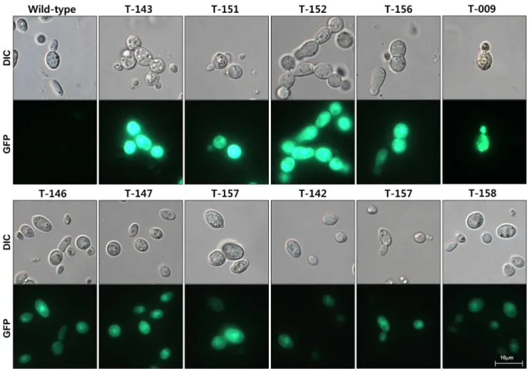
We also tested the transformants for green fluorescence, as pSK1044 contains theeGFPgene under the control ofCochliobolus heterostrophus GAPD gene promoter in its T-DNA. Thirteen randomly selected transformants were screened using fluorescence microscopy (Table 1). All of them consistently displayed green fluorescence, although fluorescence in five transformants, includ-ing UmT-009, UmT-143, UmT-151, UmT-152 and UmT-156, was stronger than in the others (Figure 4). Differential expression of eGFP among these transformants suggested that the level of
eGFP expression might vary depending on the sites of T-DNA integration (i.e., positional effect).
Identification of Genomic Sequences Flanking Inserted T-DNA
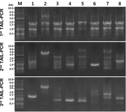
Thermal asymmetry interlaced-PCR (TAIL-PCR) and subse-quent sequencing were used to identify the genomic locations of T-DNA insertions and the context of flanking sequences in selected transformants. To increase the efficiency of TAIL-PCR in amplifying the flanking regions, we employed a mix of arbitrary degenerate primers (AD-1,2,3,4, and 6 = 1:1:1:1:1) with one specific primer for the primary (RB1), secondary (RB2-1) and tertiary (RB3) PCR reactions. This approach led to successful amplification of genomic sequences flanking the right border of the inserted T-DNA from all 50 transformants analyzed (Figure 5, Table 1). The size of the PCR products ranged from 0.2 kb to 4 kb. Products from primary PCR reactions typically displayed similar band patterns, whereas more specific bands appeared in secondary and tertiary PCR reactions (Figure 5). Sequence comparison among 50 flanking regions (Information S2) suggests that insertion of T-DNA appears to be without preferential sequence contexts (Figure 6 and Information S2). Unlike in plants or other fungi tested [52–55], we did not find any truncated T-DNA in these 50 transformants (Figure 6 and Information S2).
Since the genome ofU. muehlenbergiihas not yet been sequenced, sequences of individual flanking regions were used to search the GenBank protein database using BLASTX. No significant match was found with sequences from 30 transformants, whereas flanking sequences from the remaining 20 transformants matched sequenc-es corrsequenc-esponding to known or hypothetical gene products (Table 1).
Phenotypic Characterization of Transformants
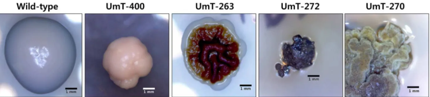
Phenotypic screening of transformants led to the identification of putative mutants that differ from the wild-type strain in colony color, size, and/or morphology (Figure 7). Compared with the round, white colonies of the wild-type U. muehlenbergii typically formed on PDA, colonies of some transformants displayed differential morphological features including heavily pigmented (263 and 272), reduced size (400 and UmT-272), and/or different morphologies (UmT-263, UmT-272 and UmT-270) (Figure 7). One putative mutant, UmT-270, exhibited pseudohyphal growth with yellow pigmentation (Figure 7). How-ever, sequences flanking T-DNA in these putative mutants did not exhibit significant similarities to any previously known gene products in the GenBank database.
Discussion
Lichens have colonized a wide range of ecosystems, including extreme environments such as deserts and arctic regions. As summarized in the Introduction, many lichens are known to produce pharmacologically active metabolites [18–40]. Lack of a transformation system for lichen fungi has hampered efforts to study the genetic mechanisms underpinning important biological and ecological processes associated with lichens and their mycobionts. Success in ATMT of diverse fungi in both the Ascomycota and the Basidiomycota [5,8,56–58] motivated us to
Figure 3. Growth, transformation efficiency, and Southern analysis.(A) Sensitivity of a wild-typeU. muehlenbergiistrain to hygromycin B was evaluated by culturing it on PDA amended with 0, 5, 10 and 20mg/ml of hygromycin B. (B) Membranes overlaid with only fungal cells (1ston the left), and a mix ofA. tumefaciensand fungal cells (2nd, 3rdand 4th) were placed on PDA containing 20mg/ml hygromycin B after culturing on co-cultivation medium for 24, 36 and 48 hrs (2nd, 3rdand 4th). The pictures were taken after 28 days of culture on this selection medium. (C) Effect on the transformation efficiency by increasing the co-cultivation time. Data presented as the average of eight plates per treatments. Error bars indicates standard error. (D) Distribution of T-DNA copy number among transformants of different time interval of co-cultivation. Genomic DNAs were digested withHindIII, a restriction enzyme that does not cut thehphcassette (see Figure 2). The digested DNAs, fractionated using 0.7% agarose gel and transferred to a nylon membrane, were probed with a labeledhphcassette. (E) Representative result of Southern blot analysis from 15 randomly selected transformants and a wild-type strain. (F) Distribution of T-DNA copy numbers among 784 transformants.
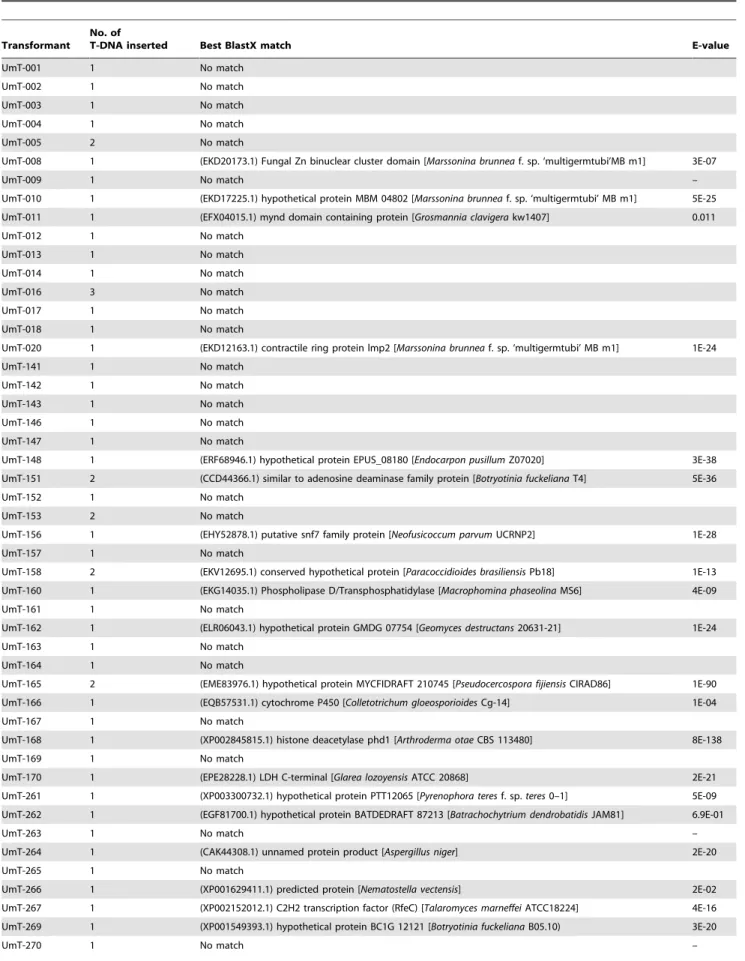
Table 1.Results from BlastX searches of the NCBI database withU.muehlenbergiigenomic sequences flanking inserted T-DNA as queries.
Transformant No. of
T-DNA inserted Best BlastX match E-value
UmT-001 1 No match
UmT-002 1 No match
UmT-003 1 No match
UmT-004 1 No match
UmT-005 2 No match
UmT-008 1 (EKD20173.1) Fungal Zn binuclear cluster domain [Marssonina brunneaf. sp. ‘multigermtubi’MB m1] 3E-07
UmT-009 1 No match –
UmT-010 1 (EKD17225.1) hypothetical protein MBM 04802 [Marssonina brunneaf. sp. ‘multigermtubi’ MB m1] 5E-25
UmT-011 1 (EFX04015.1) mynd domain containing protein [Grosmannia clavigerakw1407] 0.011
UmT-012 1 No match
UmT-013 1 No match
UmT-014 1 No match
UmT-016 3 No match
UmT-017 1 No match
UmT-018 1 No match
UmT-020 1 (EKD12163.1) contractile ring protein lmp2 [Marssonina brunneaf. sp. ‘multigermtubi’ MB m1] 1E-24
UmT-141 1 No match
UmT-142 1 No match
UmT-143 1 No match
UmT-146 1 No match
UmT-147 1 No match
UmT-148 1 (ERF68946.1) hypothetical protein EPUS_08180 [Endocarpon pusillumZ07020] 3E-38
UmT-151 2 (CCD44366.1) similar to adenosine deaminase family protein [Botryotinia fuckelianaT4] 5E-36
UmT-152 1 No match
UmT-153 2 No match
UmT-156 1 (EHY52878.1) putative snf7 family protein [Neofusicoccum parvumUCRNP2] 1E-28
UmT-157 1 No match
UmT-158 2 (EKV12695.1) conserved hypothetical protein [Paracoccidioides brasiliensisPb18] 1E-13
UmT-160 1 (EKG14035.1) Phospholipase D/Transphosphatidylase [Macrophomina phaseolinaMS6] 4E-09
UmT-161 1 No match
UmT-162 1 (ELR06043.1) hypothetical protein GMDG 07754 [Geomyces destructans20631-21] 1E-24
UmT-163 1 No match
UmT-164 1 No match
UmT-165 2 (EME83976.1) hypothetical protein MYCFIDRAFT 210745 [Pseudocercospora fijiensisCIRAD86] 1E-90
UmT-166 1 (EQB57531.1) cytochrome P450 [Colletotrichum gloeosporioidesCg-14] 1E-04
UmT-167 1 No match
UmT-168 1 (XP002845815.1) histone deacetylase phd1 [Arthroderma otaeCBS 113480] 8E-138
UmT-169 1 No match
UmT-170 1 (EPE28228.1) LDH C-terminal [Glarea lozoyensisATCC 20868] 2E-21
UmT-261 1 (XP003300732.1) hypothetical protein PTT12065 [Pyrenophora teresf. sp.teres0–1] 5E-09
UmT-262 1 (EGF81700.1) hypothetical protein BATDEDRAFT 87213 [Batrachochytrium dendrobatidisJAM81] 6.9E-01
UmT-263 1 No match –
UmT-264 1 (CAK44308.1) unnamed protein product [Aspergillus niger] 2E-20
UmT-265 1 No match
UmT-266 1 (XP001629411.1) predicted protein [Nematostella vectensis] 2E-02
UmT-267 1 (XP002152012.1) C2H2 transcription factor (RfeC) [Talaromyces marneffeiATCC18224] 4E-16
UmT-269 1 (XP001549393.1) hypothetical protein BC1G 12121 [Botryotinia fuckelianaB05.10) 3E-20
apply ATMT to U. muehlenbergii. Successful transformation of U. muehlenbergiinow makes it possible to manipulate its genes and also opens the possibility of using this fungus as a surrogate (i.e., heterologous expression system) for studying the function of genes from other fungi.
The observed efficiency of transformation, 27 to 98 transfor-mants per 16106conidia, is not very high compared to those of other fungi [5,8,48]. Several studies showed that the efficiency of fungal ATMT is affected by several factors, including a) the concentration of acetosyringone (AS), b)A. tumefaciensstrain and its growth conditions, c) numbers of fungal and bacterial cells mixed for co-cultivation [4,5,8,10,57]. Since AS functions as a signal for activating a set of virulence (vir) genes in A. tumefaciens that are required for T-DNA transfer [59], the presence of AS is essential for ATMT of fungi [5,8,57]. Adjustments of AS concentration during the induction and co-cultivation stages may help improve
the efficiency. The choice of an A. tumefaciens strain is another factor that can be explored, as some strains ofA. tumefaciensworked better than others in transforming fungi (S. Kang, unpublished result). In addition, the growth conditions of A. tumefaciens cells prior to co-cultivation also affected transformation efficiency [8,48], potentially offering another control point that can be improved and the ratio of bacterial to fungal cells can also be adjusted optimally.
Since many of these factors may also affect the number of T-DNA copies inserted in individual transformants [8,13,57], simply employing conditions that provide the greatest number of transformants may not be desirable for certain applications, such as insertional mutagenesis and targeted gene disruption. A key advantage of insertional mutagenesis via ATMT over chemical or radiation mutagenesis is that inserted T-DNA serves as a convenient tag for cloning and identifying mutated genes (e.g.,
Figure 4. Expression of eGFP in 11 transformants. DIC, differential interference contrast images; GFP, fluorescence image.
doi:10.1371/journal.pone.0083896.g004 Table 1.Cont.
Transformant No. of
T-DNA inserted Best BlastX match E-value
UmT-272 1 No match –
UmT-400 1 No match –
Figure 6 and Table 1). Insertion of T-DNA at multiple loci in a mutant, therefore, can cause a substantial nuisance when it comes to identifying the gene causing a phenotype of interest. For such mutants from fungi with a sexual stage, a genetic cross can be performed to determine which T-DNA segregates with the mutant phenotype before cloning the mutated gene. However, for fungi likeU. muehlenbergiithat require a relatively long time to complete
the sexual cycle, genomic sequences flanking all T-DNA inserts need to be functionally validated (e.g., complementation, targeted mutation or both) to characterize the gene of interest. Thus, for efficient insertional mutagenesis and subsequent functional anal-yses of mutated genes, employing transformation conditions that ensure a high percentage of transformants possessing a single copy of T-DNA is highly desirable. Fortunately, with 36 hrs of
co-Figure 5. Agarose gel analysis of products resulted from TAIL-PCR of eight randomly selected transformants.The gels from the top to the bottom show products from the primary, secondary, and tertiary reactions, respectively.
doi:10.1371/journal.pone.0083896.g005
Figure 6. Genomic sequences ofU. muehlenbergiiflanking the right border of inserted T-DNA.All sequences are shown 59R39. Bold letters correspond to the 30 bp border sequences.
cultivation, only one copy of T-DNA was inserted in the genome of 88% of theU. muehlenbergiitransformants (Figure 3E), which is higher than in other fungi [5,57,60].
Isolation of three different types of putative morphological mutants from a pool of 918 transformants supports the utility of ATMT for mutagenizingU. muehlenbergiivia random insertion of T-DNA and subsequently identifying the mutated genes (Figure 7). Pseudohyphal growth of UmT-270 (Figure 7) suggests that a gene required for the regulation of a dimorphic switch may be disrupted. Identification of this gene as well as those tagged in other mutants will help understand how growth is controlled inU. muehlenbergii. Unfortunately, amplified genomic sequences that flank the right border of T-DNA in these mutants did not exhibit significant similarity to any known or hypothetical gene products (Table 1). It is possible that the genes tagged in these mutants may be unique toU. muehlenbergii. Alternatively, T-DNA insertion may be more indirectly, e.g. by inactivating a promoter or destabilizing mRNA.
In addition, to examine homologous recombination, we conducted amplification of four conserved MAP kinase genes in eukaryotes [61], Ste7, Fus3, Hog1, and Ste12, with degenerated primer sets to acquire sequence information of the genes. However, no amplified fragment was obtained from Ste7 and
Ste12and no sequence matches were obtained forFus3andHog1, suggesting that the orthologs inU. muehlenbergiimay have unique start and/or end sequences. Therefore, genome sequence of U. muehlenbergiiwill be needed to carry out homologous recombina-tion.
Since genome sequencing of U. muehlenbergii is currently underway, we will integrate T-DNA insertion mutants and phenotype data into U. muehlenbergii genome data. In addition, we will apply this system for homologous recombination of theU. muehlenbergiiand other lichen-forming fungi. This ATMT system will open an era of systematic gene functional studies for lichen fungi.
Materials and Methods
Fungal Cultures and Growth Conditions
A culture ofU. muehlenbergii KoLRI_LF000956, obtained from the Korean Lichen Center at Sunchon National University (Sunchon 540–742, Korea), was stored in the form of a conidial suspension in 15% glycerol at –80uC. It was revitalized by shake culturing in 100 ml of potato dextrose broth (PDB, Difco Laboratories) at 150 rpm and 15uC in the dark. For transforma-tion, after harvesting yeast-like cells in 14 days old culture were harvested by centrifugation at 5,000 rpm for 5 min and washed
three times with sterilized distilled water to remove polysaccha-rides.
Amplification of ITS Region and Sequencing
The ITS region was amplified from the two growth forms of LFF and the thallus ofU. muehlenbergiiwith the primers ITS4 and ITS5 (Table 2) using i-StarMAXII PCR master mix system (iNtRON Biotechnology, Sungnam city, Korea). A Takara PCR thermal cycler MP (Takara, Tokyo, Japan) was employed for 30 cycles of PCR reaction. The amplified PCR products were purified using MEGAquick-spin Total Fragment DNA Purifica-tion Kit (iNtRON Biotechnology) and sequenced on both strands with the same primers used for the PCR amplification. ITS
Figure 7. Colonies of four putative mutants ofU. muehlenbergiigenerated via ATMT.From left to right, colonies of a wild-type strain and four mutants are shown. These strains were grown for 28 days on PDA.
doi:10.1371/journal.pone.0083896.g007
Table 2.Primers used in this study.
Target Name Sequence (59–39)a
ITS region ITS4 TCCTCCGCTTATTGATATGC
ITS5 GGAAGTAAAAGTCGTAACAAGG
GAPD promoter
GAPD promoter-F
GAATTCGAATTCGAATTGGGTACTC
GAPD promoter-R
GGATCCGGATCCTTTGAAGATTGGG
eGFP ORF EGFP-F GGATCCATGGTGAGCAAGGGCG
EGFP-R GCATGCTTACTTGTACAGCTCGTC
TrpC terminator
Ncterm-F GCATGCATCATTCCACTCAACATTCAGGC
Ncterm-R AAGCTTATCATCATGCAACATGCATGTACTG
hph gene HYG_5F GGCTTGGCTGGAGCTAGTGGAGG
HYG_3R CTCCGGAGCTGACATCGACACCAAC
TAIL-PCR RB1 GGCACTGGCCGTCGTTTTACAAC
RB2-1 CTGGCGTAATAGCGAAGAGG
RB3 CCCTTCCCAACAGTTGCGCA
AD1 AGWGNAGWANCAWAGG
AD2 WAGTGNAGWANCANAGA
AD3 WAGTGNAGWANCANGTT
AD4 WAGTGNAGWANCANGAA
AD6 WGTGNAGWANCANAGA
aUnderlined sequences correspond to restriction enzyme sites introduced for
cloning purpose:EcoRI (GAATTC),BamHI (GGATCC),SphI (GCATGC), andHindIII (AAGCTT).
sequences were analyzed through NCBI (http://www.ncbi.nlm. nih.gov/).
Plasmid Construction
Fluorescent protein constructs were made by using eGFP, enhanced green fluorescent protein gene. The primers used to amplify the promoter,eGFPcoding sequences and terminator are shown in Table 2. Each primer contains a restriction enzyme site at the 59 end to facilitate subsequent construction. PCR amplifications were performed using the FailSafe PCR system (Epicentre, Madison, WI, USA). The PCR products were isolated from gels, purified by QIA quick columns (Qiagen) and then cloned in pGEM-T Easy (Promega Corp.). All clones were verified by sequencing.BamHI andSphI fragment ofeGFPcoding sequence (720 bp) and a SphI and HindIII fragment of terminator from
Neurospora crassa trpC (260 bp) under the control of the EcoR
I-BamHI fragment of Cochlioborus heterostrophus GAPD promoter (466bp) was ligated into EcoRI and HindIII site from the multi-cloning site of pGEM-3ZF (Promega Corp.). Subsequently, the
EcoRI andHindIII fragments were cloned between theEcoRI and
HindIII sites of binary vector pBHt2 [8], named pSK1044 (Fig. 2 and Information S1).
ATMT ofU. muehlenbergii
Initially, A. tumefaciens strain AGL-1 was transformed with binary vector pSK1044 (Figure 2). ATMT was carried out as previously described [57] with a few minor modifications. Cells of
A. tumefaciens were grown at 28uC for 2 days in 1 ml minimal medium (MM) [62] supplemented with kanamycin (50mg/ml). After transferring 100ml bacterial cells into 900ml of induction medium (IM) [4] containing 200mM acetosyringone (AS), they were grown for 6 hrs at 28uC. For co-cultivation, freshly harvested cells ofU. muehlenbergiiwere used. After mixing equal volumes ofU. muehlenbergii(16107conidia/ml) and A. tumefacienscells, 200ml of the mix was spread on sterilized 0.45mm pore cellulose membrane (cellulose nitrate, 47 mm diameter, Whatman Ltd, Maidstone, UK) overlaid on co-cultivation medium amended with 200mM AS. Following co-cultivation at 23uC for 24 hrs or 36 hrs, the membranes were transferred to PDA amended with 20mg/ml hygromycin B and 250mg/ml cefatoxim to select transformants. Approximately one month later, individual colonies were trans-ferred to 24-well plates (SPL, Korea) containing PDA. For isolation of pure culture, method for isolation of bacterial colonies was applied. All transformants were cultured in PDB, mixed with glycerol to make a 15% solution, and stored at –80uC for long-term storage. Colony color, size, and morphology of individual transformants were examined by culturing them on PDA.
Genomic DNA Isolation and Southern Analysis
Transformants and the wild-type strain were grown in 30 ml of PD broth at 15uC in an orbital shaker (200 rpm) for 7 days. Genomic DNA was extracted using DNAeasy mini kit. One microgram of DNA from each strain was digested withHindIII, a restriction enzyme that can be used to determine single copy integration of T-DNA, and fractionated in 0.7% agarose gels at 40 V for 6 hrs in 0.5% Tris-Acetate-EDTA buffer. Fractionated DNAs were transferred to Hybond N+ membrane (Amersham International, Little Chalfont, England) using 106SSC (16SSC is 0.15 M NaCl plus 0.015 M sodium citrate).
Hybridization of Southern blots was conducted using the hygromycin B resistance gene as a probe. The probe was prepared by PCR amplification of a 1.4 kb fragment from pBHt2 [8] using a primer pair, HYG_5F and HYG_3R (Table 2). The resulting
PCR product was isolated from a gel using QIAquick spin columns (Qiagen, Valencia, CA, U.S.A.) to produce a probe, which was labeled with32P-dCTP by random priming (Rediprime II DNA labeling system, GE Heathcare Life Sciences). Hybrid-ization was performed at 65uC overnight in 66SSPE (16SSPE: 0.18 M NaCl, 1 mM EDTA, and 10 mM sodium phosphate (pH 7.4)) containing 1% sodium dodecyl sulfate (SDS) and 100mg of denatured salmon sperm DNA per ml. After hybridization, blots were washed twice in 26SSPE and 0.1% SDS for 5 min at 65uC. Signals were detected by autoradiography using BAS-MS imaging plate (Fuji Film).
Microscopy
To screen for expression of eGFP in transformants, conidial suspensions from randomly chosen transformants were dropped onto a slide glass and observed using a Zeiss Axio Imager A1 fluorescence microscope (Carl Zeiss, Oberkochen, Germany). A filter set with excitation at 470/40 nm and emission at 525/50 nm was used.
Scanning electron microscope observations of U. muehlenbergii
hyphae and yeast-like cells (Figure 1) were carried out by fixing cultures with 2.5% glutaraldehyde in 0.1 M sodium phosphate buffer (PB, pH7.2) for overnight at 4uC and then treating them with 1% OsO4 in PB for 1 hr at 4uC. The fixed specimens were dehydrated in an ascending series of ethyl alcohol and subse-quently sputter-coated with OsO4. The specimens were imaged using a field emission scanning electron microscope (FE-SEM; S-4800; Hitachi High-Technologies, Tokyo, Japan) operating at 15– 20 kV.
TAIL-PCR and Sequencing
TAIL-PCR was performed to isolate genomic DNA sequences flanking inserted T-DNA as described previously [52]. The secondary or tertiary PCR products were treated with using ExoSAP-ITH (USB, Cleveland, OH, USA) according to the manufacturer’s instruction and sequenced using primer RB3. Resulting sequences were analyzed through NCBI (http://www. ncbi.nlm.nih.gov/) to search for protein homology at the insertion site of T-DNA in each transformant.
Mitotic Stability of Transformants
To determine the mitotic stability of transformants, 24 randomly selected transformants were cultured on PDA without hygromycin B. Following five successive transfers, colonies were tested for growth on fresh PDA amended with hygromycin B (20mg/ml).
Supporting Information
Information S1 pSK1044.
(PDF)
Information S2 Fifty full genome sequences with
RB-border sequences are generated by TAIL-PCR.
(PDF)
Author Contributions
References
1. Daboussi MJ, Djeballi A, Gerlinger C, Blaiseau PL, Bouvier I, et al. (1989) Transformation of seven species of filamentous fungi using the nitrate reductase gene ofAspergillus nidulans. Curr Genet 15: 453–456.
2. Leung H, Lehtinen U, Karjalainen R, Skinner D, Tooley P, et al. (1990) Transformation of the rice blast fungusMagnaporthe grisea to hygromycin B resistance. Curr Genet 17: 409–411.
3. Marmeisse R, Gay G, Debaud JC, Casselton LA (1992) Genetic transformation of the symbiotic basidiomycete fungusHebeloma cylindrosporum. Curr Genet 22: 41–45.
4. Bundock P, den Dulk-Ras A, Beijersbergen A, Hooykaas PJ (1995) Trans-kingdom T-DNA transfer fromAgrobacterium tumefacienstoSaccharomyces cerevisiae. EMBO J 14: 3206–3214.
5. de Groot MJ, Bundock P, Hooykaas PJ, Beijersbergen AG (1998)Agrobacterium tumefaciens-mediated transformation of filamentous fungi. Nat Biotechnol 16: 839–842.
6. Frandsen RJN (2011) A guide to binary vectors and strategies for targeted genome modification in fungi usingAgrobacterium tumefaciens-mediated transfor-mation. J of Microbiol Methods 87: 247–262.
7. Khang C, Park S, Lee Y, Kang S (2005) A dual selection based, targeted gene replacement tool forMagnaporthe griseaandFusarium oxysporum. Fungal Genet Biol 42: 483–492.
8. Mullins E, Romaine CP, Chen X, Geiser D, Raina R, et al. (2001)Agrobacterium tumefaciens-mediated transformation ofFusarium oxysporum: An efficient tool for insertional mutagenesis and gene transfer. Phytopathology 91: 173–180. 9. Paz Z, Garcia-Pedrajas MD, Andrews DL, Klosterman SJ, Baeza-Montanez L,
et al. (2011) One step construction of Agrobacterium-recombination-ready-plasmids (OSCAR), an efficient and robust tool for ATMT based gene deletion construction in fungi. Fungal Genet Biol 48: 677–684.
10. Islam MN, Nizam S, Verma PK (2012) A highly efficientAgrobacteriummediated transformation system for chickpea wilt pathogenFusarium oxysporumf. sp.ciceri using DsRed-Express to follow root colonisation. Microbiol Res 167: 332–338. 11. Jeon J, Park SY, Chi MH, Choi J, Park J, et al. (2007) Genome-wide functional analysis of pathogenicity genes in the rice blast fungus. Nat Genet 39: 561–565. 12. Liu H, Bao X (2009) Overexpression of the chitosanase gene inFusarium solani viaAgrobacterium tumefaciens-mediated transformation. Curr Microbiol 58: 279– 282.
13. Munch S, Ludwig N, Floss DS, Sugui JA, Koszucka AM, et al. (2011) Identification of virulence genes in the corn pathogenColletotrichum graminicolaby Agrobacterium tumefaciens-mediated transformation. Mol Plant Pathol 12: 43–55. 14. Kim HS, Park SY, Lee S, Adams EL, Czymmek K, et al. (2011) Loss of
cAMP-dependent protein kinase A affects multiple traits important for root pathogenesis byFusarium oxysporum. Mol Plant-Microbe Interact 24: 719–732. 15. Gouka RJ, Gerk C, Hooykaas PJ, Bundock P, Musters W, et al. (1999)
Transformation of Aspergillus awamori by Agrobacterium tumefaciens-mediated homologous recombination. Nat Biotechnol 17: 598–601.
16. Hawksworth DL, Kirk PM, Sutton BC, Pegler DN (1995) Dictionary of the Fungi. Wallingford: CAB.
17. Lutzoni F, Pagel M, Reeb V (2001) Major fungal lineages are derived from lichen symbiotic ancestors. Nature 411: 937–940.
18. Huneck S (1999) The significance of lichens and their metabolites. Naturwis-senschaften 86: 559–570.
19. Nishikawa Y, Ohno H (1981) Studies on the water-soluble constituents of lichens. IV. Effect of antitumor lichen-glucans and related derivatives on the phagocytic activity of the reticuloendothelial system in mice. Chem Pharm Bull (Tokyo) 29: 3407–3410.
20. Rezanka T, Dembitsky VM (2006) The colleflaccinosides, two chiral bianthraquinone glycosides with antitumor activity from the lichen Collema flaccidumcollected in Israel and Russia. Nat Prod Res 20: 969–980. 21. Watanabe M, Iwai K, Shibata S, Takahashi K, Narui T, et al. (1986)
Purification and characterization of mouse alpha 1-acid glycoprotein and its possible role in the antitumor activity of some lichen polysaccharides. Chem Pharm Bull (Tokyo) 34: 2532–2541.
22. Candan M, Yilmaz M, Tay T, Erdem M, Turk AO (2007) Antimicrobial activity of extracts of the lichen Parmelia sulcata and its salazinic acid constituent. Z Naturforsch C 62: 619–621.
23. Candan M, Yilmaz M, Tay T, Kivanc M, Turk H (2006) Antimicrobial activity of extracts of the lichenXanthoparmelia pokornyiand its gyrophoric and stenosporic acid constituents. Z Naturforsch C 61: 319–323.
24. Cansaran D, Kahya D, Yurdakulola E, Atakol O (2006) Identification and quantitation of usnic acid from the lichen Usnea species of Anatolia and antimicrobial activity. Z Naturforsch C 61: 773–776.
25. Gambichler T, Skrygan M, Tigges C, Kobus S, Glaser R, et al. (2009) Significant upregulation of antimicrobial peptides and proteins in lichen sclerosus. Br J Dermatol 161: 1136–1142.
26. Garcia Rowe J, Garcia Gimenez MD, Saenz Rodriguez MT (1999) Some lichen products have antimicrobial activity. Z Naturforsch C 54: 605–609. 27. Gollapudi SR, Telikepalli H, Jampani HB, Mirhom YW, Drake SD, et al. (1994)
Alectosarmentin, a new antimicrobial dibenzofuranoid lactol from the lichen, Alectoria sarmentosa. J Nat Prod 57: 934–938.
28. Ingolfsdottir K, Bloomfield SF, Hylands PJ (1985) In vitro evaluation of the antimicrobial activity of lichen metabolites as potential preservatives. Antimicrob Agents Chemother 28: 289–292.
29. Ivanova V, Backor M, Dahse HM, Graefe U (2010) Molecular structural studies of lichen substances with antimicrobial, antiproliferative, and cytotoxic effects fromParmelia subrudecta. Prep Biochem Biotechnol 40: 377–388.
30. Manojlovic NT, Vasiljevic PJ, Maskovic PZ, Juskovic M, Bogdanovic-Dusanovic G (2012) Chemical Composition, Antioxidant, and Antimicrobial Activities of LichenUmbilicaria cylindrica(L.) Delise (Umbilicariaceae). Evid Based Comple-ment Alternat Med 2012: 452431.
31. Mitrovic T, Stamenkovic S, Cvetkovic V, Tosic S, Stankovic M, et al. (2011) Antioxidant, antimicrobial and antiproliferative activities of five lichen species. Int J Mol Sci 12: 5428–5448.
32. Rankovic B, Rankovic D, Maric D (2010) Antioxidant and antimicrobial activity of some lichen species. Mikrobiologiia 79: 812–818.
33. Tay T, Turk AO, Yilmaz M, Turk H, Kivanc M (2004) Evaluation of the antimicrobial activity of the acetone extract of the lichenRamalina farinaceaand its (+)-usnic acid, norstictic acid, and protocetraric acid constituents. Z Naturforsch C 59: 384–388.
34. Turk AO, Yilmaz M, Kivanc M, Turk H (2003) The antimicrobial activity of extracts of the lichenCetraria aculeataand its protolichesterinic acid constituent. Z Naturforsch C 58: 850–854.
35. Turk H, Yilmaz M, Tay T, Turk AO, Kivanc M (2006) Antimicrobial activity of extracts of chemical races of the lichenPseudevernia furfuraceaand their physodic acid, chloroatranorin, atranorin, and olivetoric acid constituents. Z Naturforsch C 61: 499–507.
36. Yilmaz M, Tay T, Kivanc M, Turk H, Turk AO (2005) The antimicrobial activity of extracts of the lichenHypogymnia tubulosaand its 3-hydroxyphysodic acid constituent. Z Naturforsch C 60: 35–38.
37. Yilmaz M, Turk AO, Tay T, Kivanc M (2004) The antimicrobial activity of extracts of the lichen Cladonia foliacea and its (-)-usnic acid, atranorin, and fumarprotocetraric acid constituents. Z Naturforsch C 59: 249–254. 38. Harmala P, Hiltunen R, Oksman-Caldentey KM, Laakso T, Kauppinen V
(1992) Isolation and in vitro cultivation of lichen algae and their antimicrobial properties. Fitoterapia 58: 217.
39. Bugni TS, Andjelic CD, Pole AR, Rai P, Ireland CM, et al. (2009) Biologically active components of a Papua New Guinea analgesic and anti-inflammatory lichen preparation. Fitoterapia 80: 270–273.
40. Tanas S, Odabasoglu F, Halici Z, Cakir A, Aygun H, et al. (2010) Evaluation of anti-inflammatory and antioxidant activities ofPeltigera rufescenslichen species in acute and chronic inflammation models. J Nat Med 64: 42–49.
41. Nash ITH (1996) Lichen Biology; Nash THI, editor. Cambridge: Cambridge University Press.
42. Culberson CF, Armaleo D (1992) Induction of a complete secondary-product pathway in a cultured lichen fungus. Exp Mycol 16: 52–63.
43. Huneck S, Yaoshimura I (1996) Identification of Lichen Substances: Springer-Verlaq.
44. Gagunashvili AN, Davidsson SP, Jonsson ZO, Andresson OS (2009) Cloning and heterologous transcription of a polyketide synthase gene from the lichen Solorina crocea. Mycol Res 113: 354–363.
45. Simmenmann SJ, Andresson OS, Brown DW, Miao VPW (2000) Cloning and heterologous expression ofSolorina crocea pyrG. Curr Genet 2000: 333–338. 46. Molina MC, Crespo A (2000) Comparison of development of axenic cultures of
five species of lichen-forming fungi. Mycol Res 104: 595–602.
47. McDonald TR, Mueller O, Dietrich FS, Lutzoni F (2013) High-throughput genome sequencing of lichenizing fungi to assess gene loss in the ammonium transporter/ammonia permease gene family. BMC Genomics 14: 225. 48. Hirabayashi K, Iwata S, Ito M, Shigeta S, Narui T, et al. (1989) Inhibitory effect
of a lichen polysaccharide sulfate, GE-3-S, on the replication of human immunodeficiency virus (HIV) in vitro. Chem Pharm Bull (Tokyo) 37: 2410– 2412.
49. Nishikawa Y, Tanaka M, Shibata S, Fukuoka F (1970) Polysaccharides of lichens and fungi. IV. Antitumour active O-acetylated pustulan-type glucans from the lichens ofUmbilicariaspecies. Chem Pharm Bull (Tokyo) 18: 1431–1434. 50. Stepanenko LS, Maksimov OB, Fedoreev SA, Miller GG (1998) Polysaccharides
of lichens and their sulfated derivatives: antivial activity. Chem Nat Compd 34: 337–338.
51. Yamammoto Y, Kinoshita Y, Yoshimura I (2002) Culture of thallus fragments and redifferentiation of lichen; Kranner I, Beckett RP, Varma AK, editors. Berlin: Springer. 34–46 p.
52. Choi J, Park J, Jeon J, Chi MH, Goh J, et al. (2007) Genome-wide analysis of T-DNA integration into the chromosomes ofMagnaporthe oryzae. Mol Microbiol 66: 371–382.
53. Bundock P, Hooykaas PJ (1996) Integration ofAgrobacterium tumefaciensT-DNA in theSaccharomyces cerevisiaegenome by illegitimate recombination. Proc Natl Acad Sci U S A 93: 15272–15275.
55. Sha Y, Li S, Pei Z, Luo L, Tian Y, et al. (2004) Generation and flanking sequence analysis of a rice T-DNA tagged population. Theor Appl Genet 108: 306–314.
56. Marchand G, Fortier E, Neveu B, Bolduc S, Belzile F, et al. (2007) Alternative methods for genetic transformation ofPseudozyma antarctica, a basidiomycetous yeast-like fungus. J Microbiol Methods 70: 519–527.
57. Rho HS, Kang S, Lee YH (2001)Agrobacterium tumefaciens-mediated transforma-tion of the plant pathogenic fungus,Magnaporthe grisea. Mol Cells 12: 407–411. 58. Chen EC, Su YH, Kanagarajan S, Agrawal DC, Tsay HS (2009) Development
of an activation tagging system for the basidiomycetous medicinal fungusAntrodia cinnamomea. Mycol Res 113: 290–297.
59. Winans SC (1992) Two-way chemical signaling inAgrobacterium-plant interac-tions. Microbiol Rev 56: 12–31.
60. Maruthachalam K, Klosterman SJ, Kang S, Hayes RJ, Subbarao KV (2011) Identification of pathogenicity-related genes in the vascular wilt fungus Verticillium dahliae by Agrobacterium tumefaciens-mediated T-DNA insertional mutagenesis. Mol Biotechnol 49: 209–221.
61. Gustin MC, Albertyn J, Alexander M, Davenport K (1998) MAP kinase pathways in the yeastSaccharomyces cerevisiae. Microbiol Mol Biol Rev 62: 1264– 1300.
![Figure 2. Map of binary vector pSK1044. pBHt2 contains the hygromycin B resistance gene under the control of the Aspergillus nidulans trpC promoter [8] was used to construct pSK1044 by insertion of the eGFP gene cassette between the EcoRI and HindIII sites](https://thumb-eu.123doks.com/thumbv2/123dok_br/18392772.357758/2.918.91.445.92.438/contains-hygromycin-resistance-aspergillus-nidulans-promoter-construct-insertion.webp)