Role of Pectinolytic Enzymes Identified in
Clostridium thermocellum
Cellulosome
Soumyadeep Chakraborty1‡, Vania O. Fernandes2‡, Fernando M. V. Dias2, Jose A. M. Prates2, Luis M. A. Ferreira2, Carlos M. G. A. Fontes2, Arun Goyal1*, Maria S. J. Centeno2*
1Department of Biotechnology, Indian Institute of Technology Guwahati, Guwahati, Assam, India,2 CIISA-Faculdade de Medicina Veterinária, Universidade de Lisboa, Lisboa, Portugal
‡These authors contributed equally to this work.
*arungoyl@iitg.ernet.in(AG);zecenteno@fmv.ulisboa.pt(MSJC)
Abstract
The cloning, expression and characterization of three cellulosomal pectinolytic enzymes viz., two variants of PL1 (PL1A and PL1B) and PL9 fromClostridium thermocellumwas car-ried out. The comparison of the primary sequences of PL1A, PL1B and PL9 revealed that these proteins displayed considerable sequence similarities with family 1 and 9 polysaccha-ride lyases, respectively. PL1A, PL1B and PL9 are the putative catalytic domains of protein sequence ABN54148.1 and ABN53381.1 respectively. These two protein sequences also contain putative carbohydrate binding module (CBM) and type-I dockerin. The associated putative CBM of PL1A showed strong homology with family 6 CBMs while those of PL1B and PL9 showed homology with family 35 CBMs. Recombinant derivatives of these three enzymes showed molecular masses of approximately 34 kDa, 40 kDa and 32 kDa for PL1A, PL1B and PL9, respectively. PL1A, PL1B and PL9 displayed high activity toward polygalacturonic acid and pectin (up to 55% methyl-esterified) from citrus fruits. However, PL1B showed relatively higher activity towards 55% and 85% methyl-esterified pectin (cit-rus). PL1A and PL9 showed higher activity on rhamnogalacturonan than PL1B. Both PL1A and PL9 displayed maximum activity at pH 8.5 with optimum temperature of 50°C and 60°C respectively. PL1B achieved highest activity at pH 9.8, under an optimum temperature of 50°C. PL1A, PL1B and PL9 all produced two or more unsaturated galacturonates from pec-tic substrates as displayed by TLC analysis confirming that they are endo-pectate lyase be-longing to family 1 and 9, respectively. This report reveals that pectinolytic activity displayed byClostridium thermocellumcellulosome is coordinated by a sub-set of at least three multi-modular enzymes.
Introduction
Plant cell wall degradation carried out by saprophytic and phytopathogenic microbes is essen-tial for the recycling of carbon stored in plant biomass and of intrinsic biotechnological impor-tance. Plant cell walls are composed of a complex network of polysaccharides, primarily
OPEN ACCESS
Citation:Chakraborty S, Fernandes VO, Dias FMV, Prates JAM, Ferreira LMA, Fontes CMGA, et al. (2015) Role of Pectinolytic Enzymes Identified in Clostridium thermocellumCellulosome. PLoS ONE
10(2): e0116787. doi:10.1371/journal.pone.0116787
Academic Editor:Ligia O. Martins, Universidade Nova de Lisboa, PORTUGAL
Received:July 28, 2014
Accepted:December 9, 2014
Published:February 6, 2015
Copyright:© 2015 Chakraborty et al. This is an open access article distributed under the terms of the
Creative Commons Attribution License, which permits unrestricted use, distribution, and reproduction in any medium, provided the original author and source are credited.
Data Availability Statement:All relevant data are within the paper.
cellulose, hemicelluloses and pectic substances [1]. Pectins are a highly heterogeneous group of polymers, containing a high quantity of galacturonic acid, which contribute to the firmness and structure of plant tissues, and is predominantly found in the primary cell wall and middle la-mella. This recalcitrant carbohydrate is more soluble in water than cellulose and hemicelluloses, suggesting that it constitutes the initial target for plant associated microbes attack [2]. Pectins are divided into three polysaccharides i.e., homogalacturonan (HG), rhamnogalacturonan type-I (RG-I), and rhamnogalacturonan type-II (RG-II). HG is present as a linear backbone, while RG-I and RG-II are branched carbohydrates [3]. The backbone of RG-I pectin is composed of alternating rhamnose and galacturonic acid residues with a disaccharide repeated unit consisting of [(1,2)-α-L-rhamnose-(1,4)-α-D-galacturonic acid] where galacturonic acid residues may be acetylated at the O2 or O3 positions [4,5]. It is often referred as“hairy”because of the presence of multiple side chains of neutral polymers like arabinans, galactans and arabinogalactans which are attached to C4 of the rhamnose residues [6,7]. RG-II consists of a polygalacturonan backbone with side chains complexes of about 30 monosaccharides including rare molecules such as apiose and aceric acid [8].
Due to its structural complexity, pectin degradation requires the concerted action of several enzymes. The enzymatic degradation of polygalacturonan involves two well-known enzymatic mechanisms: i) hydrolysis by glycoside hydrolases (GH) that cleave glycoside bonds in the polysaccharide and ii)β-elimination reactions carried out by polysaccharide lyases resulting in oligomers withΔ4,5 unsaturated residues at the non-reducing end [9,10]. Polysaccharide ly-ases (EC 4.2.2.-) belong to a large group of enzymes defined as carbohydrate-active-enzymes and have been classified into 23 families (May 2014), according to CAZy database [11]. Pectate lyases of families 1, 2, 3, 9, and 10 catalyse theβ-eliminative cleavage ofα(1! 4)-glycosidic bond between D-galactopyranosyluronic acid (GalpA) residue in pectate (a low methylesterified form of pectin), and generateΔ4,5 unsaturated GalpA as the product [12], which exhibits a maximum absorbance at around 235 nm [13].
Pectate lyases are widely distributed among microbial plant pathogens likeErwinia[14,15,
16] although they have also been found in saprophytic bacteria including the genusBacillus
[17,2,18] andClostridium[12].Clostridium thermocellumis an anaerobic, saccharolytic and thermophilic bacterium that organizes a consortium of plant cell wall degrading enzymes in a large multienzymatic complex termed the cellulosome [19,20]. The cellulosome is assembled via the interaction of individual type-I dockerins located at the C-terminus of enzymes into one of the nine cohesins of the scaffoldin subunit, CipA. CipA also bears a family 3 carbohy-drate-binding module (CBM) which accounts for its cellulose-targeting function and a dock-erin type-II that mediates the attachment of the entire complex into the bacterial cell surface [19,21]. Despite its specialization in the hydrolysis of crystalline cellulose, the cellulosome con-tains in addition to several cellulases, an extensive group of hemicellulases, which have been ex-tensively characterized [22,23,24,25,26], and are believed to increase the accessibility of the bacterium into its primary substrate.
The majority of glycoside hydrolases that attack cellulose and hemicelluloses are modular enzymes consisting of catalytic modules appended to non-catalytic carbohydrate-binding modules (CBMs) [10]. Pectinases on the contrary generally have a relatively simple structure lacking CBMs, which is possibly explained by the accessibility of pectins to soluble biocatalysts [27]. CBMs are described to date into 69 families (May 2014), according to CAZy database (http://www.cazy.org/Carbohydrate-Binding-Modules.html) and continue to expand. The present study provides data indicating thatC. thermocellumcellulosome secretes modular polysaccharides lyases belonging to PL families 1 and 9. The role of this subset of enzymes in the anaerobic conversion of biomass by cellulosomes was investigated.
Materials and Methods
Bacterial strains, plasmids and culture conditions
TheEscherichia colistrains used in this study were NZYStar (NZYTech Ltd.), BL21 (DE3) and BL21(DE3) pLysS (Novagen). The plasmid vectors used were pNZY28 (NZYTech Ltd.), pGEM-T Easy vector (Promega), pET21a and pET28a (Novagen).E. colistrains containing re-combinant plasmids were cultured in LB broth medium supplemented with 100μg/mL
ampi-cillin or 50μg/mL kanamycin. To generate the recombinant proteins encoded by pET21a or
pET28a expression vectors,E. coliBL21 (DE3) were cultured at 37°C to mid-exponential phase (A550= 0.6) and at this point isopropyl-β-D-thiogalactoside (IPTG) was added to a final
con-centration of 1 mM. Incubation conditions after induction with IPTG were 16h at 19°C for PL1A and PL9, 12h at 24°C for PL1B.
Substrates used in enzyme assays
Polygalacturonic acid (PGA) from citrus fruits, rhamnogalacturonan from soyabean (RGAS) and potato (RGAP), pectic galactans from potato (PGP) and lupin (PGL), were purchased from Megazyme. Pectins from citrus fruits (with varying degrees of methyl-esterification, PC) and apple (PA) were purchased from Sigma Chemical Co., USA.
General recombinant DNA procedures
Bacterial transformation, agarose gel electrophoresis, plasmid DNA preparation, restriction en-donuclease digestion and ligation of DNA sequences were followed as described elsewhere [28].
Construction of recombinant plasmids
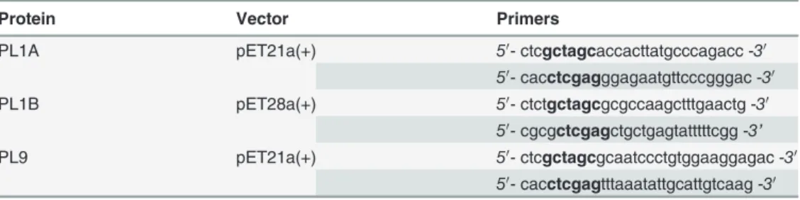
Genes encoding PL1A, PL1B and PL9 were amplified fromC. thermocellumgenomic DNA, using one IU of thermostable DNA polymerase NZYSpeedy Proof (NZYTech Ltd.) and primer pairs, described inTable 1.NheI/XhoI restriction sites were used for directional cloning of the respective amplified DNA sequences into the expression vectors pET21a and pET28a. The re-actions, in a final volume of 50μl, were subjected to 30 cycles at the following temperatures:
95°C for 1 min, 55°C for 1 min and 72°C for 2.5 min. The amplified genes after running on aga-rose gel were purified by gel extraction kit (Qiagen), and were cloned into pNZY28 (NZYTech Ltd) or pGEMT-Easy (Promega) and sequenced to ensure that no mutation occurred during PCR. Recombinant pNZY28 and pGEMT-Easy derivatives were digested withNheI/XhoI re-striction enzymes (NZYTech Ltd. or Promega).pL1a andpL9 genes were cloned into similarly digested expression vector pET21a(+) whereaspL1b gene was cloned into pET28a(+) vector. Recombinant PL1A and PL9 proteins contained a C-terminal His6-tag, whereas PL1B
con-tained an N-terminal His6-tag.
Table 1. Primers used in PCR ofpl1A, pl1Bandpl9genes.
Protein Vector Primers
PL1A pET21a(+) 50- ctcgctagcaccacttatgcccagacc -30
50- cacctcgagggagaatgttcccgggac -30
PL1B pET28a(+) 50- ctctgctagcgcgccaagctttgaactg -30
50- cgcgctcgagctgctgagtatttttcgg -3’
PL9 pET21a(+) 50- ctcgctagcgcaatccctgtggaaggagac -30
50- cacctcgagtttaaatattgcattgtcaag -30
The nucleotides shown in bold are the restriction enzyme sites, which were used to clone amplified genes into the expression vectors pET21a(+) and pET28a(+).
Expression and purification of PL1A, PL1B and PL9
PL encoding genes were expressed usingE. coliBL21 (DE3) where the cells were induced with 1mM IPTG, only when the culture ODA600reaches 0.4–0.6. Induced cells were grown
over-night at 24°C and 180 rpm for protein production. Cells were harvested by centrifugation at 12000 g, 4°C for 20 min and the bacterial pellets were resuspended in 50 mM sodium HEPES buffer (pH 7.5) containing 1 M NaCl, 5 mM CaCl2and 10 mM imidazole for PL1A and PL9.
PL1B containing cells were resuspended in 50 mM Tris-HCl buffer (pH 8.6) containing only 100 mM NaCl. These three recombinant proteins containing His-tags were purified by immo-bilized nickel ion affinity chromatography as described previously [29]. For PL1A and PL9 the buffer was exchanged to 50 mM Tris-HCl, pH 8.5, containing 5 mM CaCl2and 100 mM NaCl
and for PL1B the buffer was exchanged to 50 mM Tris-HCl (pH 8.6) containing 100 mM NaCl. The purity and size of recombinant enzymes were evaluated by SDS-PAGE [30].
Enzyme assays
The enzyme activity of PL1A, PL1B and PL9 was determined against different pectic substrates. 30μg of PL1A or PL9 was incubated with 0.5% (w/v) of substrate dissolved in 50 mM Tris-HCl
buffer pH 8.5 containing 5 mM CaCl2and 100 mM NaCl at 60°C for 20 min. The assay of
PL1B was carried out by incubating 7μg of enzyme with 0.1% (w/v) of substrate in 50 mM
Glycine-NaOH buffer (pH 9.8) containing 0.6 mM CaCl2for 15 min at 50°C. The reactions
were stopped by incubation on ice for 10 min and centrifuged at 13,000gfor 5 min. The supernatant containing the released unsaturated products was measured by spectrophotometer (Ultrospec III Pharmacia and Cary 100 Bio Varian). The molar extinction coefficient used for the unsaturated product released at A232nm, was 5,200 M−1cm−1[31] and at A235nm, was
4,600 M−1cm−1[32]. 1 Unit of enzyme was defined as the amount of enzyme that forms 1
μmol
of 4,5-unsaturated product per minute, under the described assay conditions.
To determine the maximum activity of PL1A and PL9 at different pH values, all enzymes were incubated with appropriate substrates at 50°C in the following buffers: 50 mM MES (pH 6.5); 50 mM Tris-HCl (pH 7.0 to 8.5) and 50 mM NaHCO3(pH 9.0 to 12.0), and the activity
was determined at A232nm as described above. Activity of PL1B at different pH values was
de-termined by incubating with PGA at 50°C using following buffers: 50 mM Tris-HCl (pH 7.6– 8.8), 50 mM Glycine-NaOH (pH 9.0–10.6) and 50 mM Na2HPO4-NaOH (10.8–12), and the
activity was determined at A235nm as mentioned earlier. The optimal activities of PL1A and
PL9 at a range of temperature from 10 to 100°C, were determined spectrophotometrically at A232nm, by incubating the enzymes in 50 mM Tris-buffer pH 8.5, for 20 min. The optimal
ac-tivity of PL1B in the temperatures range from 10 to 100°C was spectrophotometrically deter-mined at A235nm in 50 mM Glycine-NaOH buffer pH 9.8, after 15 min of incubation. The
thermostability of PL1A and PL9 was evaluated, by incubating the enzyme at different temper-atures (30 to 100°C) in 50 mM Tris-HCl pH 8.5, and for PL1B the enzyme was incubated at same temperature range in 50 mM Tris-HCl (pH 8.6) for 30 min [33], and then the residual ac-tivity was measured by assay methods described earlier.
Kinetic parameters of these three Clostridial enzymes were measured against PGA (citrus) having an average molecular weight approximately, 25000 g/mol [34]. 20μl (1.5 mg/ml) of
en-zyme (PL1A or PL9) was used in 1 ml reaction mixture containing 50 mM Tris-HCl (pH 8.5), 5 mM CaCl2with varying concentrations (0.01 to 0.5% w/v) of PGA were incubated at 50°C.
The unsaturated product formation was monitored spectrophotometrically at A232nm.
Simi-larly, 20μl (1.4 mg/ml) of PL1B was used in 1 mL of reaction volume containing 50 mM
Gly-cine-NaOH (pH 9.8), 0.6 mM CaCl2and varying concentration of PGA (0.01 to 0.5% w/v).
spectophotometrically at A235nm. Kcatand Kmwere determined using the Michaelis-Menten
equation. All the reactions were carried out in triplicate and results were reported as mean±SD.
Analysis of enzyme degradation products
PL1A (6μg), PL1B (7μg) and PL9 (6μg) were separately incubated in 1 ml reaction volume
containing 0.1% (w/v) PGA or citrus pectin (25% methyl-esterified). The reaction was carried out under optimized conditions of pH and temperature for different time intervals from 0 to 60 min. After the reaction enzyme was deactivated by keeping on ice for 5 min and the sample was treated with equal volumes of ethanol to precipitate un-hydrolyzed polysaccharides and protein. Ethanol was removed and samples were concentrated to 500μl by heating at 50°C. 1μl
of sample was then loaded on the TLC plate (readymade silica coated aluminum TLC plates obtained from Merck, Germany) for running the degradation products under a solvent system containing butan-1-ol/water/acetic acid in the ratio of 5:3:2 [35]. The spots on TLC plates were visualized by a solution containing 0.5% (w/v)α-naphthol and 5% (v/v) sulphuric acid in etha-nol [36], after heating at 95°C for 10 min in hot air oven. Standard oligogalacturonides like D-galacturonic acid (S1), di-D-galacturonic acid (S2) and tri-D-galacturonic acid (S3) (procured from Sigma Chem. Co., USA) were used to analyze the degradation product formed from different substrates upon enzymatic treatment.
Results
Molecular architecture of three pectin degrading enzymes from
Clostridium thermocellum
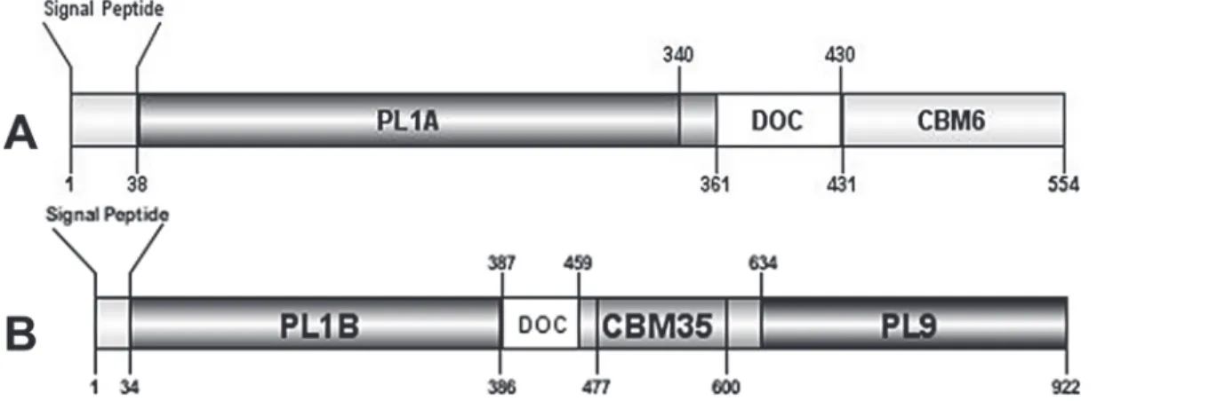
Inspection of two protein sequences fromC. thermocellumrevealed that they contain one gene (pL1a) in the sequence ABN53381.1 (Fig. 1A) and two genes (pL1b andpL9) in the sequence ABN53381.1 (Fig. 1B) all putatively expressing lyase activities. Both the protein sequences are associated with type I dockerin, which is the signature module of the cellulosomal proteins. Analysis of the deduced amino acid sequence of the three enzymes revealed characteristic N-terminal signal sequences with putative cleavage sites located between Ala-37/Thr-38
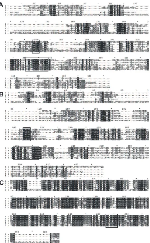
(ABN54148.1) and between Ala-33/Ala-34 (ABN53381.1) suggesting that the proteins are ex-ported into the extracellular space. Homology searches using Blast (www.ncbi.nlm.nih.gov/ BLAST), revealed downstream the signal peptide of ABN54148.1 a putative 302-aa family 1 PL (PL1A) followed by a 70-aa dockerin domain and a 124-aa C-terminal family 6 CBM (CBM6) (Fig. 1A). ABN53381.1 contains a 353-aa N-terminal family 1 PL (PL1B) and a 289-aa C-ter-minal family 9 PL (PL9). Sandwiched between these two catalytic domains is a 73-aa dockerin domain and 124-aa family 35 CBM (CBM35) (Fig. 1B). Alignment of PL1A (Fig. 2A) and PL1B (Fig. 2B) domain with other PL1 homologues revealed two consensus sequence patterns in the enzymes catalytic domains,“VWIDH”and“VxxRxPxxRxGxxHxxxN”, which are signa-ture regions of pectate lyases (Pel) [37,38,39]. The conserved arginine residue observed in the second region, identified as R-218 in the superfamily pectate lyase C (PelC fromErwinia chysanthemi) is the catalytic residue involved in proton abstraction [40,41]. Cleavage of glycosidic bonds in which the aglycone sugar is galacturonic acid can be acid-base-assisted catalysis, mediated by glycoside hydrolases [42] or viaβ-elimination reaction, which is initiated by proton abstraction from C-5 of the galacturonosyl residue on the reducing end of the glycosidic bond [43]. As R-218 belongs to a potential group or groups involved in the proton abstraction in PelC and as it is highly conserved in catalytic domains of PL1A (position 190 in
mechanism as expected for pectate lyases. The“VWIDH”region is highly conserved in PL1A and PL1B (Fig. 2A and 2B) and it is involved in the membrane transport and in the protein fold [44]. Structural motifs (parallelβ-helix) are also identified in pectate lyases fromErwinia chysanthemiandBacillus subtilis[40,45,46].
PL9 catalytic domain bears profound similarity with Pel9A fromErwinia chryanthemi. Pel9A showed an endolytic cleavage pattern where it cleaves the polysaccharide by anti-β -elim-ination mechanism, where a base catalyzed abstraction of proton is carried out from the C5 carbon [47]. In case of Pel9A the putative base is Lysine rather than Arginine found in other endo-pectate lyases [48]. Such a consensus sequence was found in the catalytic domain of PL9, whose translated amino acid sequence is highly conserved in all the aligned sequences marked within a box inFig. 2C. The lysine residue involved in proton abstraction duringβ-elimination is located in position 269 of the PL9 sequence (Fig. 2C).
Cloning, expression and purification of recombinant PL1A, PL1B and
PL9
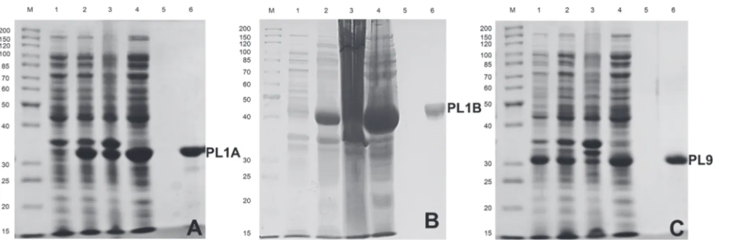
DNA sequences of 906, 1059 and 867 bp, encoding PL1A, PL1B and PL9 respectively, were am-plified by PCR and cloned into pET21a and pET28a expression vector as described in the method section. The recombinant proteins containing the His6-tags were purified by
immobi-lized metal ion affinity chomatography. The expression and purification of PL1A, PL1B and PL9 proteins was analyzed by SDS-PAGE as shown inFig. 3A, B and Cdisplaying molecular size of approximately 34, 40 and 32 kDa, respectively.
Biochemical properties of PL1A, PL1B and PL9
The biochemical role ofC. thermocellumcellulosomal PL1A, PL1B and PL9 enzymes was in-vestigated by analyzing their activity against different substrates. All the three enzymes PL1A, PL1B and PL9 were predominantly active towards polygalacturonic acid (PGA) and pectin both from citrus (Fig. 4). PL1B displayed relatively higher activity with 55% and 85% methyl-esterified pectins from citrus than PL1A and PL9. On the other hand PL1A and PL9 showed 30–40% relative activity with rhamnogalacturonan from potato (RGAP) and soyabean (RGAS), whereas PL1B showed only 8% relative activity.
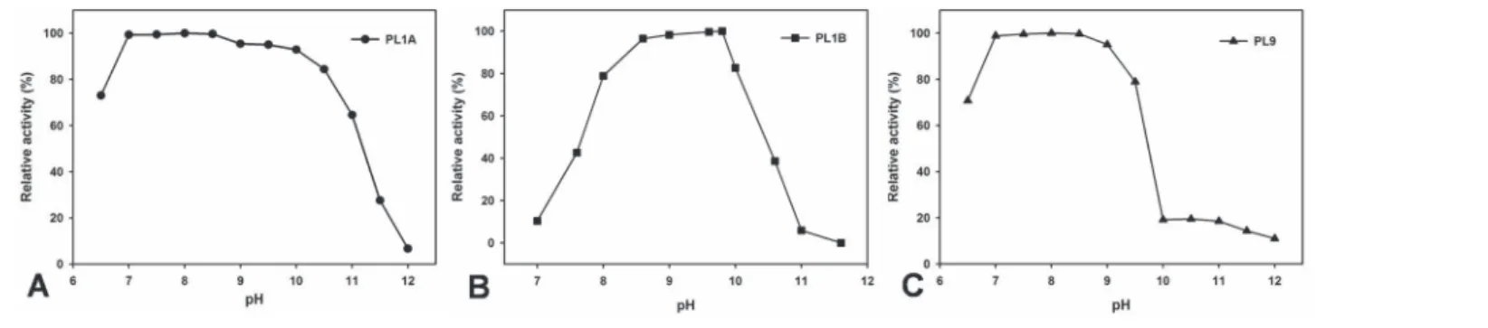
The effect of pH and the temperature on the activity of the recombinant PL1A, PL1B and PL9 enzymes against PGA was determined. The results showed that PL1A, PL1B and PL9 were
Figure 1. Molecular architecture of modular (A) protein sequence with accession no. ABN54148.1 that comprises of N-terminal PL1A catalytic domain, followed by DOC, type-I dockerin and C terminal CBM6 binding domain.(B) protein sequence with ac no. ABN53381.1 that comprises of N terminal PL1B catalytic domain, followed by DOC, type-I dockerin and CBM35 binding domain, with a C-terminal PL9 catalytic domain.
active under alkaline conditions. PL1A and PL9 were active within pH range (6.5–9.5) showing highest activity at pH 8.5 (Fig. 5A and C). PL1B was active within pH range (8–10) displaying highest activity at pH 9.8 (Fig. 5B).
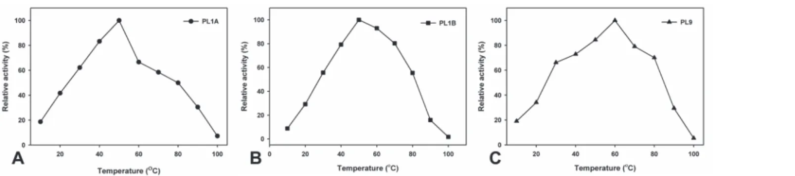
The optimum temperature was found at 50°C for both PL1A and PL1B and 60°C for PL9 (Fig. 6A, B and C) which were expected because these enzymes originated from a thermophilic bacterium. Nevertheless all the three recombinant enzymes, PL1A, PL1B and PL9 displayed thermostability within the temperature range of 30 to 70°C for 30 min (Fig. 7A, B and C).
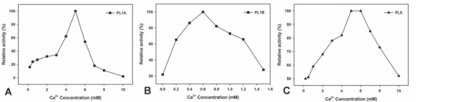
All the three enzymes showed an exclusive requirement of Ca2+ions to achieve their maxi-mum activity. PL1A and PL1B showed only 20% of their maximaxi-mum activity in the absence of 5 mM and 0.6 mM Ca2+ions, respectively, whereas PL9 showed 50% of its maximum activity in absence of 5 mM Ca2+ions (Fig. 8A, B and C). The optimum Ca2+ion concentration re-quired to achieve 100% pectinolytic relative activity were 5 mM for both PL1A and PL9, whereas 0.6 mM for PL1B.
Kinetic parameters of these three enzymes were determined against PGA and are presented inTable 2. The data revealed that PL1A, PL1B and PL9, showed turnover number values (Kcat)
of 1.3, 1.76, and 1.32 min−1respectively (Table 2). The catalytic efficiency (K
cat/Km) values
ex-hibited by PL1A, PL1B and PL9 were 41, 62 and 35 mM−1min−1respectively, revealing that
PL1B exhibit higher catalytic efficiency on PGA, than PL1A and PL9.
Functional properties of recombinant PL1A, PL1B and PL9
Products released by the enzymatic cleavage of PL1A, PL1B and PL9 of PGA and pectin (citrus) were determined. The reactions were carried out under optimum conditions of pH and temperature for each individual enzyme as mentioned in Methods section. The samples from enzymatic reaction were collected at different time intervals of 0, 5, 10, 15, 20, 30, 45 and 60 min and separated through TLC. PL1A produced unsaturated di- and tri-galacturonates
PDB: 3KRG) and 5 (Acidovorax Avenae Subsp Citrulli, PDB: 4HWV). (B) PL1B was aligned with following proteins: 1 (PL1B,Clostridium thermocellum ATCC 27405); 2 (Bacillus Sp. N16–5, PDB: 3VMV); 3
(Thermotoga Maritima, PDB: 3ZSC); 4 (Xanthomonas CampestrisATCC 33913, PDB: 2QX3); 5 (Bacillus Sp. TS-47, PDB: 1VBL). (C) PL9 was aligned with following proteins: 1 (PL9,Clostridium thermocellum ATCC 27405); 2 (Clostridium straminisolvensJCM 21531, GAE89695.1); 3 (Clostridium cellulovorans 743B, YP_003842407.1); 4 (Acetivibrio cellulolyticus, WP_010245176.1); 5 (Caldicellulosiruptor kristjanssonii I77R1B, YP_004026944.1).
doi:10.1371/journal.pone.0116787.g002
Figure 3. Hyper-expression and purification of PL1A, PL1B and PL9 usingE. coliBL21 (DE3) cells.The purity of the proteins was analysed by SDS-PAGE using 10% (w/v) gel showing(A)PL1A (34 kDa);(B)PL1B (40 kDa);(C)PL9 (32 kDa); Lane M: Fermentas high range protein molecular weight marker; Lane 1: Uninduced BL21 cells; Lane 2: IPTG induced BL21 cells; Lane 3: Cell pellet after sonication; Lane 4: Cell free extract, Lane 5: Last wash from column and Lane 6: Purified recombinat enzyme.
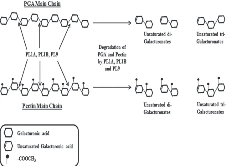
along with other oligosaccharides of higher size. The accumulation of unsaturated tri-galactur-onates and higher size oligosaccharides was predominant after 5 min of the start of reaction (Fig. 9A). It was evident from the TLC results that an increase in the amount of unsaturated di-and tri-galacturonates was found with increase in time di-and found to be highest at 60 min of re-action time (Fig. 9A). PL1B from the beginning of the reaction started producing unsaturated di and tri-galacturonates, and no higher size oligosaccharides were observed. Accumulation of unsaturated di- and tri-galacturonates increased with time and found to be highest at 60 min of the reaction (Fig. 9B). PL9 produced unsaturated tri-galacturonates and oligosaccharides of higher size, and the accumulation of this two products were found to be constant from 5 to 60 min of the reaction time (Fig. 9C). The cleavage pattern of these three enzymes suggests that they cleave within the poly-galacturonan main chain of PGA and pectin (citrus) thus following an endo cleaving pattern. The mechanism by which these three enzymes fromClostridium thermocellumcleaves theα-1,4 linkages in pectic polysaccharides thus resulting in enzymatic degradation is explained schematically inFig. 10.
Discussion
C. thermocellumis known to hydrolyze not only cellulose but also hemicelluloses [22,23,24,
25,26,49]. It was also shown thatC. thermocellumcould utilize polygalacturonic acid and
Figure 4. Substrate specificity of PL1A, PL1B and PL9 towards pectic polysaccharides, where PGA: Polygalacturonic acid, PC 25: Pectin (Citrus) (25% methyl-esterified), PC 55: Pectin (Citrus) (55% methyl-esterified), PC 85: Pectin (Citrus) (85% methyl-esterified), PA: Pectin (Apple), RGAP: Rahmnogalacturonan (Potato), RGAS: Rahmnogalacturonan (Soyabean), PGL: Pectic galactan (Lupin), PGP: Pectic galactan (Potato).
doi:10.1371/journal.pone.0116787.g004
Figure 5. Effect of pH on the activity of (A) PL1A; (B) PL1B; (C) PL9 towards PGA as substrate.
pectins as carbon sources [50]. The results described in this paper demonstrate thatC. thermocellumcellulosome is composed of enzymes that are able to attack pectin and can hydrolyze these complex polysaccharides. We have identified and characterized for the first time three cellulosomal pectinolytic enzymes PL1A, PL1B and PL9 from this microorganism. The data revealed PL1A, PL1B and PL9 catalytic activity on polygalacturonic acid (PGA) and pectin (citrus). Sequence similarity studies with proteins in biological databanks placed PL1A, PL1B and PL9 in families 1 and 9 of pectate lyases (PL), respectively. Till date there are 22 fam-ilies of polysaccharide lyases, of which PL famfam-ilies 1, 2, 3, 9 and 10 contain pectate lyases (http://www.cazy.org/search?page=recherche&recherche=4.2.2.2&tag=9).
Analysis of primary sequences of the cellulosomal enzymes under analysis here revealed a modular organization with the presence of a CBM, which is rare in pectinases [12]. It is gener-ally believed that pectins are more accessible to enzyme attack than cellulose and hemicellu-loses, and as such it has been assumed that there has been less evolutionary pressure for pectinases to contain CBMs. However, Rgl11A and Pel10A fromP. cellulosaand Pel4A from
Clostridium cellulovoransare examples of prokaryotic pectinases that contain a cellulose-binding domain [12,27,51]. This report showed that CBMs are prevalent within cellulosomal pectinases and might be involved in potentiating the degradation of less recalcitrant substrates. Previous studies showed that functionally active family 6 and 35 CBM’s bind strongly to cellu-lose [52,53]. Family 6 CBMs display considerable promiscuity in ligand binding with different modules showing affinity for amorphous cellulose, xylans andβ-glucans [54]. In addition, CBM family 35 also reveals considerable plasticity in ligand recognition which is not surprising considering that this family shares sequence similarities with CBM 6. Both these families, CBM 6 and CBM 35, are structurally related to theβ-jelly-roll CBM superfamily [55] and can be viewed as a subfamily of the largeβ-jelly-roll CBM superfamily [56]. CBMs are prevalent in
Figure 6. Effect of temperature on the activity of (A) PL1A; (B) PL1B; (C) PL9 towards PGA as substrate.
doi:10.1371/journal.pone.0116787.g006
Figure 7. Thermostability of (A) PL1A; (B) PL1B; (C) PL9 towards PGA, after 30 min of incubation of the enzyme at different temperatures.
plant cell wall degrading enzymes and as a general function promote the interaction of the en-zyme with their target substrate [55]. PL1A contains a CBM 6, while PL1B and PL9 exhibit a CBM 35. The presence of CBMs in the structure of PL1A, PL1B and PL9 suggests that they are important in increasing their catalytic efficiency by bringing the enzymes into close proximity to their target substrates. However, in a recent work Montanier and colleagues [57] while ana-lysing the biological role of 4 members of family CBM35, it was revealed that the biological role of CBM35s is not dictated solely by the substrate specificity of their appended catalytic do-mains as members of these CBM family may recognize the products of pectin hydrolysis. Struc-turally, PL1A, PL1B and PL9 consist of an individual dockerin-containing enzyme integrated into theC. thermocellumcellulosome by CipA cohesin-dockerin interaction with a non-catalyt-ic module CBM-like.
PL1A, PL1B and PL9 are characteristic pectate lyases and preferentially hydrolyze polygalacturonic acid, though they also act on pectins. Moreover, all the three enzymes displayed significantly higher activity with 55% and 85% methyl-esterified pectin (citrus). Simi-lar high activity of pectate lyase on pectins with high degree of methyl-esterification has been previously reported only fromBacillus subtillis[17]. PL1A and PL9 showed significant activity with rhamnogalacturonan from potato (RGAP) and soyabean (RGAS), as compared with PL1B. The enzymes that degrade the backbones of pectic substances utilize two distinct cleavage mechanisms, the hydrolysis or theβ-elimination. The method used to evaluate the PL1A, PL1B and PL9 activities provide evidence that these enzymes are lyases, as they catalyse theβ-eliminative cleavage of glycosidic bonds with the production ofΔ4,5 unsaturated galac-turonates, which can be followed spectrophotometrically at absorbances of 232 to 235 nm. PL1A and PL9 was active within the pH range of 6.5–9.5 with highest activity at pH 8.5, which is similar to those of Rgl11Y fromC. cellulolyticumcellulosome (pH 8.5) and fromC. cellulovoranscellulosome Pel4A (pH 8.0) [12,58], but PL1B was active within the pH range of 8–10 and showed highest activity at pH 9.8 similar to PelA fromBacillus sp. which showed pH optima of 10 [59]. All four enzymes have a requirement of Ca2+ions to achieve their maximum
Figure 8. Effect of concentration of Ca2+ions on the activity of (A) PL1A; (B) PL1B; (C) PL9 against PGA as substrate.
doi:10.1371/journal.pone.0116787.g008
Table 2. Kinetic parameters of PL1A, PL1B and PL9 with Polygalacturonic Acid (PGA) from citrus.
Enzyme Substrate Kcat(min−1) Km(mM) Kcat/Km(mM−1min−1)
PL1A PGA(citrus) 1.3±0.03 0.0313±0.0005 41±0.23
PL1B PGA(citrus) 1.76±0.05 0.0286±0.0002 62±0.43
PL9 PGA(citrus) 1.32±0.05 0.0378±0.0004 35±0.4
One unit of enzymatic activity (U) was defined as the amount of enzyme in mg that produces 1 mmol/L of unsaturated product per minute.
activity. It has been suggested that the pH value of some plant tissues changes during microbial attack which possibly indicate that the degradation of plant cell wall polysaccharides occurs se-quentially according to the pH of plant tissues [12]. It is also known that pectate lyases require Ca2+forin vitroactivity and presumably utilize the abundant Ca2+in the plant cell wall forin vivoactivity [38,60].
Analysis of the degradation products of cellulosomal PLs by TLC conclusively inferred that PL1A, PL1B and PL9 followed an endo cleavage pattern on PGA and pectin (citrus), cleaving these substrates endolytically as was previously reported for PelA fromClostridium cellulovorans[58]. These enzymes produced unsaturated di, tri and higher oligogalacturonates from PGA and pectin (citrus). PelC fromB. subtilis[17], also an endo pectate lyase, showed a similar cleavage pattern producing mixtures of different degradation products, whereas PelX fromErwinia chysanthemian exo-pectate lyase always produced a single degradation product either unsaturated di or tri-galacturonates [61]. Hence, PL1A, PL1B and PL9 under
Figure 9. Thin layer chomatography (TLC) showing the enzymatic degradation products of PGA (citrus) and pectin (citrus) (25% methyl-esterified). Chromatogram displaying hydrolysis by (A) PL1A (B) PL1B and (C) PL9 at 0, 5, 10, 15, 20, 30, 45 and 60 min. Standard oligosaccharides used were S1: D-galacturonic acid; S2: Di-D-galacturonic acid; S3: Tri-D-galacturonic acid.
doi:10.1371/journal.pone.0116787.g009
Figure 10. Schematic presentation of mode of action of PL1A, PL1B and PL9 against PGA and pectin (citrus) hydrolysis leading to production of corresponding unsaturated oligo-galacturonates.
investigation are conclusively endo pectate lyases. Pectic substrates are highly heterogeneous which may require many enzymes with different specificities and catalytic mechanisms for their complete breakdown. Therefore, the ability of these cellulosomal enzymes to degrade pec-tic substances suggests that cellulosomes are designed for the degradation of an entire set of carbohydrates within plant cell walls, and not only cellulose and hemicellulose. It is clear that within cellulosomes other enzymes presently of unknown function may target the degradation of pectic polysaccharides.
Conclusion
Thermostable enzymes are important resources in various industrial processes that occur at higher temperatures. Hence enzymes described in this study will be competent enough for in-dustrial processes like fruit juice extraction, vegetable and fruit maceration or bioscouring of cotton fabric to increase the efficiency of dying at improved temperatures. These enzymes can be used as a cocktail for further efficient and complete degradation of pectic polysaccharides.
Author Contributions
Conceived and designed the experiments: SC VOF FMVD JAMP LMAF CMGAF AG MSJC. Performed the experiments: SC VOF MSJC. Analyzed the data: SC VOF CMGAF AG MSJC. Contributed reagents/materials/analysis tools: AG CMGAF. Wrote the paper: SC VOF FMVD CMGAF AG MSJC.
References
1. Carpita NC, Gibeaut DM (1993) Structural models of primary cell walls in flowering plants: consistency of molecular structure with the physical properties of the walls during growth. Plant J 3: 1–30. PMID: 8401598
2. Ochiai A, Itoh T, Maruyama Y, Kawamata A, Mikami B, et al. (2007) A novel structural fold in polysac-charide lyases:Bacillus subtilisfamily 11 rhamnogalacturonan lyase yesw with an eight-bladedβ -pro-peller. J Biol Chem 282: 37134–37145. PMID:17947240
3. Darvill AG, McNeil M, Albersheim P (1978) Structure of plant cell walls VIII. A new pectic polysaccha-ride. Plant Physiol 62: 418–422. PMID:16660529
4. McNeil M, Darvill AG, Albersheim P (1980) Structure of plant cell walls X. Rhamnogalacturonan I, a structurally complex pectic polysaccharide in the walls of suspension-cultured sycamore cells. Plant Physiol 66: 1128–1134. PMID:16661590
5. McDonough MA, Kadirvelraj R, Harris P, Poulsen JN, Larsen S (2004) Rhamnogalacturonan lyase re-veals a unique thee-domain modular structure for polysaccharide lyase family 4. FEBS Lett 565: 188– 194. PMID:15135077
6. Ridley BL, O Neil MA, Mohnen D (2001) Pectins: structure, biosyntesis and oligogalacturonic-related signaling. Phytochemistry 57: 929–967. PMID:11423142
7. De Vries RP, Visser J (2001) Aspergillus enzymes involved in degradation of plant cell wall polysaccha-rides. Microbiol Mol Biol Rev 65: 497–522. doi:10.1128/MMBR.65.4.497-522.2001PMID:11729262 8. O Neill MA, Warrenfeltz D, Kates K, Pellerin P, Doco T, et al. (1996) Rhamnogalacturonan-II, a pectic polysaccharide in the walls of growing plant cell, forms a dimer that is covalently cross-linked by a bo-rate ester. J Biol Chem 271: 22923–22930. PMID:8798473
9. Linhardt RJ, Galliher PM, Cooney CL (1986) Polysaccharide lyases. Appl Biochem Biotechnol 12: 135–176. PMID:3521491
10. Davis G, Henrissat B (1995) Structures and mechanisms of glycosyl hydrolases. Struct 3: 853–859. PMID:8535779
11. Cantarel BL, Coutinho PM, Rancurel C, Bernard T, Lombard V, et al. (1999) The Carbohydrate-Active EnZymes database (CAZy): an expert resource for Glycogenomics. Nucl Acids Res 37: D 233–238. doi:10.1093/nar/gkn663PMID:18838391
13. Jurnak F, Kita N, Garrett M, Heffron SE, Scavetta R, et al. (1996) Functional implications of the three-di-mensional structures of pectate lyases. In Pectin and Pectinases, vol. 14, pp. 295–308. Edited by Visser J. & Voragen A. G. J.. Amsterdam: Elsevier. PMID:7716248
14. Hugouvieux-Cotte-Pattat N, Condemine G, Nasser W, Reverchon S (1996) Regulation of pectinolysis inErwinia chrysanthemi. Annu Rev Microbiol 50: 213–257. PMID:8905080
15. Pissavin C, Robert-Baudouy J, Hugouvieux-Cotte-Pattat N ( 1996) Regulation of pelZ, a gene of the pelBC cluster encoding a new pectate lyase inErwinia chrysanthemi3937. J Bacteriol 178: 7187– 7196. PMID:8955401
16. Shevchik VE, Robert-Baudouy J, Hugouvieux-Cotte-Pattat N (1997) The pectate lyase PelI ofErwinia chrysanthemibelongs to a new family. J Bacteriol 179: 7321–7330. PMID:9393696
17. Soriano M, Diaz P, Pastor FI (2006) Pectate lyase C fromBacillus subtilis: a novel endo-cleaving en-zyme with activity on highly methylated pectin. Microbiology 152: 617–625. PMID:16514142
18. Sukhumsiirchart W, Kawanishi S, Deesukon W, Chansiri K, Kawasaki H, et al. (2009) Purification, char-acterization, and overexpression of thermophilic pectate lyase ofBacillussp. RN1 isolated from a hot spring in Thailand. Biosci Biotechnol Biochem 73(2): 268–73. PMID:19202269
19. Bayer EA, Kening R, Lamed R (1983) Adherance ofClostridium thermocellumto cellulose. J. Bacteriol. 156: 818–827. PMID:6630152
20. Lamed R, Setter E, Bayer EA (1983) Characterization of a cellulose-binding, cellulose-containing com-plex inClostridium thermocellum. J Bacteriol 156: 828–836. PMID:6195146
21. Bayer EA, Lamed R, White BA, Flint HJ (2008) From cellulosomes to cellulosomics. The Chemical Re-cord 8: 364–377. doi:10.1002/tcr.20160PMID:19107866
22. Zverlov VV, Fuchs KP, Schwarz WH, Velikodvorskays GA (1994) Purification and cellulosomal locali-zation ofClostridium thermocellummixed linkageβ-glucanase LicB (1,3–1,4-β-D-glucanase). Biotech-nol Lett 16: 29–34.
23. Halstead JR, Vercoe PE, Gilbert HJ, Davidson K, Hazlewood GP (1999) A family 26 mannanase pro-duced byClostridium thermocellumas a component of the cellulosome contains a domain which is conserved in mannanases from anaerobic fungi. Microbiology 145: 3101–3108. PMID:10589717 24. Fernandes AC, Fontes CMGA, Gilbert HJ, Hazlewood GP, Fernandes TH, et al. (1999) Homologous
xylanases fromClostridium thermocellum: evidence for bi-functional activity, synergism between xylanase catalytic modules and the presence of xylan-binding domains in enzyme complexes. Biochem J 342: 105–110. PMID:10432306
25. Blum DL, Kataeva IA, Li XL, Ljungdahl LG (2000) Feruloyl esterase activity of theClostridium thermocellumcellulosome can be attributed to previously unknown domains of XynY and XynZ. J Bacteriol 182: 1346–1351. PMID:10671457
26. Fontes CM, Gilbert HJ (2010) Cellulosomes: highly efficient nanomachines designed to deconstruct plant cell wall complex carbohydrates. Annual Reviews Biochem 79: 655–681. doi: 10.1146/annurev-biochem-091208-085603PMID:20373916
27. McKie VA, Vincken JP, Voragen AGJ, Van Den Broek L, Stimson E, et al. (2001) A new family of rhamnogalacturonan lyases contains an enzyme that binds to cellulose. Biochem J 355: 167–177. PMID:11256961
28. Sambrook J, Fritsch EF, Maniatis T (1989) Molecular Cloning; a Laboratory Manual, 2aedition, New York.: Cold Spring Harbor Laboratory.
29. Carvalho AL, Goyal A, Prates JAM, Bolam DM, Gilbert HJ, et al. (2004) Crystal structure and functional properties of the family 11 carbohydrate-binding module of cellulosomal cellulase. Lic26A-Cel5E of Clostridium thermocellum. J Biol Chem 279: 34785–34793.
30. Laemmli UK (1970) Cleavage of structural proteins during the assembly of the head of bacteriophage T4. Nature (London) 227: 680–685. PMID:5432063
31. Collmer A, Riad JL, Mount MS (1988) Assay methods for pectic enzymes. Meth Enzymol 161: 329– 335.
32. Hasegawa S, Nagel CW (1966) A New Pectic Acid Transeliminase produced exocellularly by a Bacil-lus. J Food Sc 31: 838–845.
33. Fontes CMGA, Hall J, Hirst BH, Hazlewood GP, Gilbert HJ (1995) The resistence of cellulases and xylanases to proteolytic inactivation. Appl Microbiol Biotechnol 43: 52–57. PMID:7766136
35. Lojkwoska E, Masclaux C, Boccara M, Robert-Baudouy J, Hugouvieux-Cotte-Pattat N (1995) Charac-terization ofpelLgene encoding a novel pectate lyase ofErwinia chysanthemi3937. Mol Microbiol 16 (6): 1183–1195. PMID:8577252
36. Cote GL, Leathers TD (2005) A method for surveying and classifyingLeuconostoc sp. Glucansucrases according to strain-dependent acceptor product patterns. J Ind Microbiol Biotechnol 32: 53–60. PMID: 15714308
37. Hinton JCD, Sidebotham JM, Gill DR, Salmond GPC (1989) Extracellular and periplasmatic isoen-zymes of pectate lyase fromErwinia caratovorasubspeciescarotovorabelong to different gene fami-lies. Mol Microbiol 3: 1785–1795. PMID:2695748
38. Barras F, Van Gigsegem F, Chatterjee AK (1994) Extracellular enzymes and pathogenesis of soft-rot Erwinia. Annu Rev Phytopathol 32: 201–234.
39. Henrissat B, Heffron SE, Yoder MD, Lietzke SE, Jurnak F (1996) Functional implication of structure-based sequence alignment of proteins in the extracellular pectate lyase superfamily. Plant Physiol 107: 963–976. PMID:7716248
40. Yoder MD, Keen NT, Jurnak F (1993) New domain motif: the structure of pectate lyase. Science 260: 1503–1507. PMID:8502994
41. Scavetta RD, Herron SR, Hotchkiss AT, Kita N, Keen NT, et al. (1999) Structure of plant cell wall frag-ment complexed to pectate lyase C. Plant Cell. 11: 1081–1092. PMID:10368179
42. Koshland DE (1953) Stereochemistry and the mechanism of enzymatic reactions. Biol Rev Camb Philos Soc 28: 416–436.
43. Moran FS, Nasuno S, Starr MP (1968) Extracellular and intracellular polygalacturonic acid trans eliminase of Erwinia carotovora. Arch Biochem Biophys 123: 298–306. PMID:5642600
44. Bruhlmann F, Keen NT (1997) Cloning, sequence and expression of thepelgene from anAmycolata sp. Gene 201: 45–51.
45. Lietzke SE, Yoder MD, Keen NT, Jurnak F (1994) The three dimensional structure of pectate lyase E, a plant virulence factor fromErwinia chysanthemi. Plant Physiol 106: 849–862. PMID:12232373 46. Pickersgill R, Jenkins J, Harris G, Nasser W, Robert-Baudouy J (1994) The structure ofBacillus subtilis
pectate lyase in complex with calcium. Nature Struct Biol 1: 717–723. PMID:7634076
47. Anderson VE (1998) Comprehensive Biological Catalysis: A Mechanistic Reference (Sinnnot, M., ed) Vol. 2, pp. 115–133, Academic Press, London.
48. Jenkins J, Shevchik VE, Hugouvieux-Cotte-Pattat N, Pickersgill RW (2004) The Crystal Structure of Pectate Lyase Pel9A from Erwinia chrysanthemi. J Biol Chem 279: 9139–9145. PMID:14670977 49. Zverlov VV, Fuchs KP, Schwarz WH (2002) Chi18A, the endochitinase in the cellulosome of the
thermoplylic, cellulolytic bacteriumClostridium thermocellum. Appl Environ Microbiol 68: 3176–3179. PMID:12039789
50. Spinnler HE, Lavigne B, Blachere H (1986) Pectinolytic activity ofClostridium thermocellum: its use for anaerobic fermentation of sugar beet pulp. Appl Microbiol Biotechnol 23: 434–437.
51. Brown IE, Mallen MH, Charnock SJ, Davies GJ, Black GW (2001) Pectate lyase 10A from Pseudomo-nas cellulosais a modular enzyme containing a family 2a carbohydrate-binding module. Biochem J 355: 155–165. PMID:11256960
52. Henshaw JL, Bolam DN, Pires VMR, Czjzek M, Henrissat B, et al. (2004) The family 6 carbohydrate binding module CmCBM6-2 contains two ligand-binding sites with distinct specificities. J Biol Chem 279: 21552–21559. PMID:15004011
53. Bolam DN, Xie H, Pell G, Hogg D, Galbraith G, et al. (2004) X4 Modules represent a new family of car-bohydrate-binding modules that display novel properties. J Biol Chem 279: 22953–22963. PMID: 15004012
54. Czjzek M, Bolam DN, Mosbah A, Allouch J, Fontes C, et al. (2004) The location of the ligand-binding site of carbohydrate-binding modules that have evolved from a common sequence is not conserved. J Biol Chem 276: 48580–48587. PMID:11673472
55. Boraston AB, Bolam DN, Gilbert HJ, Davies GJ (2004) Carbohydrate-binding-modules: fine-tuning polysaccharide recognition. Biochem J 382: 769–781. doi:10.1042/BJ20040892PMID:15214846 56. Tunnicliffe RB, Bolam DN, Pell G, Gilbert HJ, William MP (2005) Structure of a Mannan-specific Family
35 Carbohydrate-Binding Module: Evidence for Significant Conformational Changes upon Ligand Bind-ing. J Mol Biol 347(2): 287–296. PMID:15740741
58. Tamaru Y, Doi RH (2001) Pectate lyase A, an enzymatic subunit of theClostridium cellulovorans cellulosome. Proc Natl Acad Sci USA 98: 4125–4129. doi:10.1073/pnas.071045598PMID:11259664 59. Soriano M, Blanco A, Diaz P, Pastor FI (2000) An unusual pectate lyase from a Bacillus sp. with high
activity on pectin: cloning and characterization. Microbiology 146: 89–95. PMID:10658655 60. Herron SR, Scavetta RD, Garrett M, Legner M, Jurnak F (2003) Characterization and implications of
Ca2+binding to pectate lyase. J Biol Chem 278: 12271–12277. PMID:12540845