Universidade Nova de Lisboa
Instituto de Higiene e Medicina Tropical
Biological characterization of de-ubiquitylating enzymes
(UBPs/UCHs) in
Plasmodium spp
as potential drug targets
Zoraima Naymbi da Silva Neto
DISSERTAÇÃO PARA OBTENÇÃO DO GRAU DE DOUTOR EM
CIÊNCIAS BIOMÉDICAS
ESPECIALIDADE PARASITOLOGIA
Instituto de Higiene e Medicina Tropical
Biological characterization of de-ubiquitylating enzymes
(UBPs/UCHs) in
Plasmodium spp
as potential drug targets
Autor: Zoraima Naymbi da Silva Neto
Orientador: Doutora Dinora Maria da Silva Lopes
Comissão tutorial: Professor Doutor Virgilio Estólio do Rosário
Investigador Doutor João Alexandre Rodrigues
Doutora Dinora Maria da Silva Lopes
Dissertação apresentada para cumprimento dos requisitos necessários à obtenção do grau de Doutor em Ciências Biomédicas e especialidade de Parasitologia.
Publicações/Publications
1.Neto Z, Machado M, Lindeza A, Gazzarini M, do Rosário V, Lopes D. Treatment of
Plasmodium chabaudi parasites with curcumin: drug interaction and implications in the ubiquitin proteosome pathway (UPS). Journal of parasitology Research (2013). Article ID 429736.
2.Silva R J, Ramos S A, Machado M, Moura D F, Neto Z, Canto Cavalheiro M M, Figueiredo P, Rosário D V, Amaral F C A, Lopes, D. A review of anti-malarial plants used in traditional medicine in communities in Portuguese speaking countries: Brazil, Mozambique, Cape Verde, Guinea Bissau, São Tome and Principe and Angola.
Memórias doInstituto Oswaldo Cruz, (2011) 106: 142-158.
3.Fortes F, Dimbu R, Figuieredo P, Neto Z, Rosário D.V, Lopes D.Studies on pfdhfr and pfdhps mutations in Angola. Malaria Journal, 2011, 10 : 22.
4.Afonso A, Neto Z, Castro H, Lopes D, Alves C A, Tomas M A, Rosário D V.
Dedicatória
AGRADECIMENTOS/ACKNOWLEDGMENTS
Este trabalho foi realizado no Unidade de Parasitologia Médica (IHMT)/ Centro de Malária e Doenças Tropicais LA (IHMT) e foi financiado pelo projecto ” Estudos genéticos do
fenótipo de resistência acelerada a múltiplos fármacos (ARMD) no parasita Plasmodium: dinâmica de estudos de fitness com a referência: PTDC/BIA_MIC/6586172006. Tendo sido suportada pela bolsa de doutoramento da Fundação para a Ciência e Tecnologia (FCT) com referência: SFRH/BD/46203/2008 que teve uma duração de 4 anos (1 de Abril de 2009 a 1 de Abril de 2013). Para que a realização deste projecto fosse possível, foi fundamental poder contar com o apoio e colaboração de diversas pessoas, às quais quero aqui expressar os meus sinceros agradecimentos:
Ao Professor Doutor Virgílio E. do Rosário por me ter recebido como aluna estagiária em 2007, na antiga unidade de ensino e investigação UEI malária. Pelo apoio científico como bolseira de investigação (BIC) e posteriormente como estudante de doutoramento (BD). Obrigada por partilhar comigo todo o seu conhecimento científico ao longo destes anos.
À Doutora Ana Júlia Afonso da UEI de Parasitologia pelo período em que trabalhamos juntas de Janeiro 2008- Janeiro 2009 na antiga UEI malaria com quem tive a oportunidade de aprender imenso sobre a biologia do Plasmodium chabaudi e pelas discussões científicas que tivemos que resultaram na escrita deste projeto de doutoramento.
À Doutora Dinora Lopes pela orientação deste projeto, pela ajuda cientifica dada na elaboração e conclusão das experiencias cientificas ao longo deste doutoramento. Quero agradecer em particular pelos fins-de-semana em que tivemos que fazer culturas de
Plasmodium e pelas colheitas as 6h00 da manhã. Fora do âmbito científico gostaria de agradecer também pela amizade ao longo destes 7 anos no IHMT.
À Doutora Karine Le Roch da Universidade da California (UCR) pelo apoio prestado durante o decorrer das experiencias realizadas no seu laboratório em particular a transfeção em Plasmodium falciparum que foi um desafio muito grande mas também uma óptima
“curva” de aprendizagem.
Marta Machado toda a sua experiencia na área da bioquímica e ensaios enzimaticos. Os meus agradecimentos também pelo apoio na elaboração do artigo resultante do trabalho
publicado no “Journal of parasitology research.”
À Professora Ana Maria Tomás da Universidade do Porto IBMC (Instituto de Biologia molecular e celular) por me ter recebido no seu laboratório durante o período de 2009 como bolseira da investigação (BIC) para a transferência de conhecimento básico de transfecção em Plasmodium berghei e Plasmodium chabaudi no âmbito do projecto ARMD.
Ao Doutor Filomeno Fortes, Director do Programa Nacional de Controlo da Malária (PNCM) em Angola pelo incentivo e pelos debates científicos que temos tido sobre a possibilidade de se fazer investigação científica de boa qualidade em Angola.
À Marta Machado “Machada” pelo carinho e pela amizade prestada durante a elaboração deste projeto. Obrigada Marta pela tua ajuda que foi indispensável principalmente com as experiencias do biotério, por tratar das culturas de Plasmodium falciparum nos fins-de-semana, por me ajudares com as colheitas de Plasmodium falciparum em horários esquisitos como a meia-noite até as 3 da manhã. Não tenho palavras para agradecer ajuda que me deste. Não me esqueci do bilhete que fiquei de pagar para ires de férias para Angola e desfrutar do calor e das praias.
À Paula Figueiredo e Catarina Alves pelo apoio prestado durante a elaboração deste projeto.
RESUMO
A malária ainda constitui um grande problema de saúde pública e a resistência aos antimaláricos ameaça todos os esforços efectuados com vista ao combate e controle desta doença. Existe uma grande necessidade de se identificar novos compostos de preferência que actuem em novos alvos terapêuticos. A via da ubiquitinação/proteosoma já foi identificada como um alvo terapêutico interessante. Mutações nas enzimas de des-ubiquitilação (DUBs) que catalizam a remoção da ubiquitina estão associadas ao desenvolvimento de doenças infecciosas e não infecciosas.
Neste projecto quatro DUBs foram identificadas no genoma do parasita Plasmodium falciparum e foram caracterizadas. A expressão dos genes que codificam estas enzimas ao longo do ciclo de vida do parasita na presença e ausência de fármaco foi efectuada por RT-PCR.Anticorpos policlonais obtidos a partir de ratinhos foram utilizados para a deteção da abundancia das proteínas ao longo do ciclo de vida do parasita. Utilizou-se ainda a tecnica de transfeção com o objectivo de criar uma linha knockout para determinar se estas proteínas são essências para o parasita. Proteínas recombinantes foram expressas em células de E.coli e actividade enzimática das mesmas foi testada usando um substrato específico para as DUBs. O inibidor das DUBs com actividade antimalarica, curcumina foi usado quer in vitro para testar a sua actividade sobre as proteínas recombinantes, mas também in vivo no modelo de malaria roedora de Plasmodium chabaudi em associação com cloroquina e artemisinina.Um ensiao de proteomica foi também usado para ver que proteínas estão alteradas em resposta ao tratamento com curcumina.
Os resultados demonstram que em P. falciparum os genes pfuch-l1, pfuch-l3, pfuch-l54 e pfubp-8 são diferencialmente expressos ao longo do ciclo de vida do parasita e as respectivas proteínas são mais abundantes no estadio de trofozoito e esquizonte. O tratamento dos parasitas com artemisinina, cloroquina, curcumina induziu um aumento temporário na expressão dos genes seguido de um declínio. Não foi possível obter uma linha parasitária knockout pfuch-l1 e pfuch-l3 viável. As proteínas recombinantes foram expressas com sucesso em células de E. coli excepto a Pfuch-l54. As Pfuch-l1, Pfuch-l3, Pfubp-8 demonstraram actividade enzimática e interagiram com o susbstrato Ub-AMC. Os IC50 da curcumina nas proteínas recombinantes foram: Pfuch-l1 15µM, Pfuch-l3 25.4µM, Pfubp-8 10µM e para a proteina recombinante humana USP2, 5µM. A Curcumina quando testada nas células HepG2 apresenta alguma toxicidade in vitro, mas não apresenta uma alta toxicicidade em ratinhos e quando utilizada em associação com a cloroquina apresenta um efeito de sinergismo.Enquanto a associação da curcumina com artemisinina o resultado é antagónico.Os ensaios de proteomica em culturas de P. falciparum tratadas com curcumina revelaram 10 proteinas que se encontraram alteradas em resposta ao tratamento. Estas proteínas estão envolvidas no metabolismo do sulfato, tradução e degradação de proteínas, ciclo celular e organização celular.
ABSTRACT
Malaria continues to be a major public health concern. Drug resistance continues to threaten all efforts made to control the disease. Hence there is a race to identify new antimalarial drugs that act on newer targets, in order to minimize the spread of drug resistance. The ubiquitin/proteasome pathway has been idientified as a potential drug target. Mutations in de-ubiquitylating enzymes (DUBs), which catalyze the removal of ubiquitin, have been associated with the development of infectious and non infectious diseases. In this project four DUBs namely pfuch-l1, pfuch-l3, pfuch-l54 and pfubp-8 were identified in the Plasmodiumfalciparum genome and were characterized.
The expression profile of genes encoding DUBs throughout the parasite´s life cycle with and without drug treatment was carried out by RT-PCR. Polyclonal antibodies raised in mice were used to detect protein abundance in different stages of the parasite´s life cycle. An attempt was made to produce a DUB knockout line and determine whether they are essential for the parasite. Recombinant proteins were expressed in E. coli cells and their de-ubiquitylating activity was tested using a specific substrate for DUBs. The activity of curcumin (a Dub inhibitor) was evaluted in vitro on the recombinant proteins and its antimalarial activity was tested in association with chloroquine and artemisinin in an in vivo rodent malaria model, Plasmodium chabaudi. A proteomics approach was also used to determine what proteins were deregulated in response to curcumin treatment.
The results show that P. falciparum genes pfuch-l1, pfuch-l3, pfuch-l54 and pfubp-8 are differentially expressed throughout the parasite´s life cycle and those proteins are more abundant at the trophozoite and schizont stages of the parasite. Treatment of parasites with artemisinin, chloroquine, and curcumin induced a transient increase in the expression of those genes, followed by a steady decrease in the gene expression pattern. No viable pfuch-l1 and pfuch-l3 gene knockout lines were obtained. Recombinant proteins were successfully expressed in E. coli cells with the exception of Pfuch-l54.Pfuch-l1, Pfuch-l3, Pfubp-8 demonstrated de-ubiquitylating activity by cleaving the substrate Ub-AMC. In vitro IC50 of curcumin towards recombinant Pfuch-l1 was 15µM, for recombinant Pfuch-l3 was 25.4µM and for Pfubp-8 was 10µM and for human USP2 was 5µM.
Índice/Content
ACKNOWLEDGMENTS ... III RESUMO ... V ABSTRACT ... VII LIST OF FIGURES ... XIII LIST OF TABLES ... XIV
CHAPTER 1- INTRODUCTION ... 1
1.1 An introduction to malaria ... 2
1.1.1 A Historical overview on the discovery of malaria ... 2
1.1.2 An overview on the current malaria dissemination ... 3
1.1.3 The malaria parasite and its life cycle ... 5
1.2 Malaria control measures ... 8
1.2.1. Vector control measures ... 8
1.2.1.1. Chemical vector control measures ... 8
1.2.1.2. Non chemical vector control measures ... 8
1.2.2. Malaria vaccines ... 8
1.2.3. Antimalarial drugs ... 9
1.2.3.1. Chloroquine ... 9
1.2.3.2 Sulphadoxine/Pyrimethamine ... 10
1.2.3.3. Atovaquone/Proguanil ... 11
1.2.3.4. Antibiotics ... 11
1.2.3.5. Artemisinin and its derivatives ... 13
1.3 Plasmodium genome and its potential drug targets ... 14
1.3.1 Identification of potential drug targets ... 14
1.3.2. Discovery of the ubiquitin molecule in Plasmodium genome ... 15
1.3.4 Ubiquitin Ligases ... 19
1.3.5 Ubiquitin Ligases in the Plasmodium genome ... 20
1.3.6 The Proteosome ... 23
1.3.7 The proteasome in the Plasmodium genome ... 25
1.3.8 De-ubiquitylating enzymes ... 28
1.3.9 De-ubiquitylating (DUBs) enzymes in the Plasmodium genome ... 30
2.1. Biological Material ... 39
2.2. Methods ... 40
2.2.1. Expression profile study of genes encoding DUBs in Plasmodium falciparum strains 3D7 and Dd2, in the presence and absence of drug pressure ... 40
2.2.1.1 Culture of Plasmodium falciparum parasite strains 3D7 and Dd2 ... 40
2.2.1.2 Determination of in vitro IC50 of chloroquine, artemisinin and curcumin with SYBR GREEN based method ... 41
2.2.1.3 Cytotoxical evaluation of artemisinin, chloroquine and curcumin in Hepatocellular carcinoma cells (HepG2) ... 42
2.2.1.3.1. HepG2 culture ... 42
2.2.1.3.2. Cytotoxicity assay ... 42
2.2.1.4 Parasite culture for gene expression studies in the absence and presence of drugs . 43 2.2.1.5 Plasmodium falciparum RNA extraction and cDNA synthesis ... 43
2.2.1.6 Real time PCR conditions ... 44
2.2.1.7 Analysis of relative expression using the 2 - ∆∆ct method ... 46
2.2.1.8 Statistical analysis ... 46
2.2.2 Evaluating the importance of de-ubiquitylating enzymes in Plasmodium falciparum by generating a transgenic parasite line by homologous recombination ... 47
2.2.2.1 pHHpfuch-l1 and pHHpfuch-l3 knockout construction ... 48
2.2.2.2 PCR product purification ... 49
2.2.2.3 Transfection of parasites by electroporation ... 50
2.2.2.4 Selection of transfected parasites ... 51
2.2.3 Recombinant protein expression and in vitro activity of curcumin towards recombinant DUBs ... 53
2.2.3.1 Amplification of PCR products for production of recombinant proteins ... 54
2.2.3.2 PCR product purification ... 55
2.2.3.3 Cloning of PCR products into the protein expression vector pET28a+ ... 55
2.2.3.4 Transformation of BL21 DE3 RIL Codon Plus cells ... 58
2.2.3.5 Expression of the recombinant proteins in BL21 DE3 RIL Codon Plus cells ... 58
2.2.3.6 Purification of the recombinant proteins ... 60
2.2.3.7 Determination of the enzymatic activity of recombinant DUBs by cleavage of the fluoregenic substrate Ub-AMC ... 61
2.2.3.8 Screening and determination of the IC50 of curcumin on recombinant DUBs ... 62
2.2.3.9 Immunization procedure for the production of polyclonal antibodies ... 64
2.2.3.11 Pfuch-l1 protein quantification in response to drug treatment ... 65
2.2.4 In vivo efficacy of curcumin as antimalarial drug in Plasmodium chabaudi parasites ... 66
2.2.4.1 Selection of Plasmodium chabaudi parasite clones ... 66
2.2.4.2 Acute toxicity of curcumin ... 67
2.2.4.3 In vivo four day suppressive test of curcumin, curcumin/piperine, curcumin/piperine/chloroquine and curcumin/piperine/artemisinin ... 67
2.2.4.4 In vivo drug interaction studies and isobolograms ... 68
2.2.4.5 Statistical analysis ... 69
2.2.5 A proteomics (2DE) approach for the identification of Plasmodium falciparum schizont stage proteins altered in response to curcumin treatment ... 70
2.2.5.1 Preparation of Plasmodium falciparum parasites for proteomic analysis ... 70
2.2.5.2 2D-DIGE and Protein labeling ... 71
2.2.5.3 2DE Image acquisition and analysis ... 72
2.2.5.4 Trypsin digestion of spots of interest ... 74
2.2.5.5 Bioinformatics and protein database analysis ... 74
CHAPTER 3 - RESULTS & DISCUSSION ... 75
3.1 Expression profile of genes encoding DUBs in Plasmodium falciparum strains 3D7 and Dd2 and detection of protein abundance in different stages of the parasite´s life cycle ... 75
3.1.1 Determination of the in vitro IC50 of artemisinin, chloroquine and curcumin ... 76
3.1.2 Expression profile of gene pfuch-l1 in Plasmodium falciparum ... 77
3.3.3 Expression profile of gene pfuch-l3 in Plasmodium falciparum ... 83
3.3.4 Expression profile of gene pfuch-l54 in Plasmodium falciparum ... 85
3.3.5 Expression profile of gene pfubp-8 in Plasmodium falciparum ... 88
CHAPTER 4 - RESULTS & DISCUSSION ... 92
4.1 Evaluating the importance of de-ubiquitylating enzymes in Plasmodium falciparum by generating a transgenic parasite line by homologous recombination. ... 92
CHAPTER 5 - RESULTS AND DISCUSSION ... 98
5.1 Recombinant protein expression and in vitro protein activity... 98
5.1.1 Recombinant protein expression in E.coli cells BL21 DE3 codon Plus ... 99
5.1.2 Enzymatic activity of recombinant Plasmodium falciparum de-ubiquitylating enzymes (DUBs) ... 99
CHAPTER 6 - RESULTS & DISCUSSION ... 104
6.1 In vivo efficacy and acute toxicity test of curcumin in P.chabaudi parasites ... 104
6.1.1 In vivo efficacy of curcumin and chloroquine P. chabaudi parasites………..…..105
7.1 A proteomics (2DE) approach for the identification of Plasmodium falciparum schizont stage
proteins altered in response to curcumin treatment. ... 114
7.1.1 In gel protein identification ... 115
7.1.2 Plasmodium falciparum proteins deregulated in response to curcumin treatment ... 121
CHAPTER 8-General Conclusions ... 128
8.1.1 General Conclusions ... 129
8.1.2 Future studies ... 133
CHAPTER 9-Bibliographic References ... 135
CHAPTER10-APPENDIXES APPENDIX A - Gene sequences ... 160
APPENDIX B- CLUSTALW2 alignment…..………168
APPENDIX C - RT-PCR ... 174
APPENDIX D - Immunization protocol ... 179
APPDENDIX E - plate readings ... 182
APPENDIX F - Determination of Curcumin IC50 on recombinant proteins ... 183
APPENDIX G - Proteomics IEF run ... 185
APPENDIX H- Deubiquitylating enzymes interacting partners………187
LIST OF FIGURES
Figure 1. Malaria Distribution around the world 4
Figure 2. The life cycle of the malaria parasite 7
Figure 3.Antimalarial drug resistance around the world 12
Figure 4. A general structure of the ubiquitin molecule and its main features 18
Figure 5. A simplistic overview of the activation of the ubiquitin molecule 20
Figure 6. A general representation of the Ubiquitin/Proteosome system (UPS) 24
Figure 7. A general structure of the catalytic domain of ubiquitin carboxyl
hydrolase (UCH) and ubiquitin protease (UBP) 30
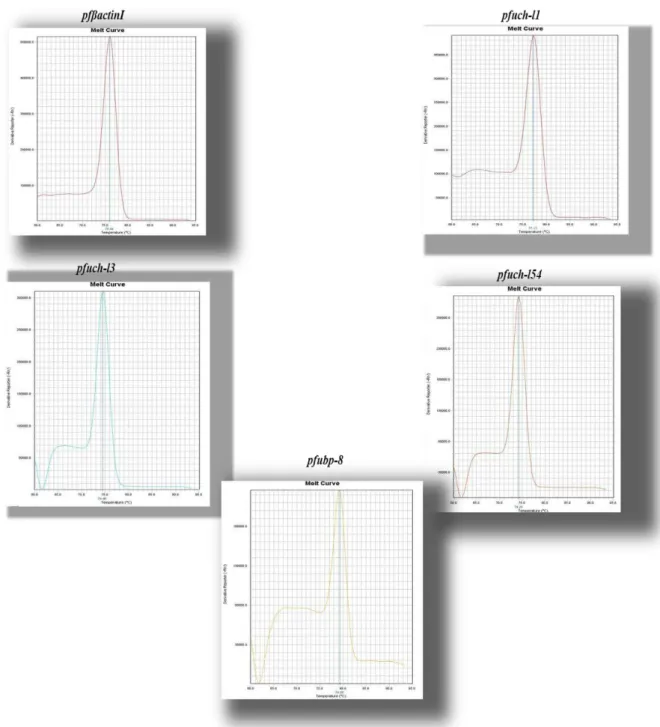
Figure 8. Illustration of the melting curves obtained by RT-PCR 45
Figure 9. A simple structure of the transfection vectors pHH and PARL-2 52
Figure 10. pET28a+ protein expression vector 57
Figure 11. Determination of the enzymatic activity of recombinant DUBs by
cleavage of the fluoregenic substrate Ub-AMC 63
Figure 12. A simple overview of the 2DE gel electrophoresis process 73
Figure 13. A simple representation of Plasmodium falciparum life cycle 80
Figure 14. Expression profile of gene pfuch-l1 in the absence and in the presence of drug treatment in Plasmodium falciparum clones 3D7 and Dd2 81 Figure 15. Confirmation of protein abundance at ring trophozoite and schizont
stage parasite lysates and parasite response to drug treatment 82 Figure 16. Expression profile of gene pfuch-l3 in the absence and in the presence of drug treatment in Plasmodium falciparum clones 3D7 and Dd2 84 Figure 17. Expression profile of gene pfuch-l54 in the absence and in the presence
of drug treatment in Plasmodium falciparum clones 3D7 and Dd2 87 Figure 18. Expression profile of gene pfubp-8 in the absence and in the presence of drug treatment in Plasmodium falciparum clones 3D7 and Dd2 89 Figure 19. Plasmodium falciparumpfuch-l1 gene knockout strategy and PARL-2
vector bearing the GFP tag. 96
Figure 20. Expression of recombinant proteins in E.coli cells BL21 DE3 Codon
Plus cells 101
Figure 21. Evaluation of recombinant protein activity 103
Figure 22. Parasitaemia evolution in mice infected with P. chabaudi clone AS-3CQ 107 Figure 23. Isobologram illustrating the in vivo interaction at the ED90 level
between drug A (curcumin) with drug B (chloroquine) 108 Figure 24. Parasitaemia evolution in mice infected with P. chabaudi clone AS-ART 110 Figure 25. Isobologram illustrating the in vivo interaction at the ED90 level
between drug A (Curcumin) with drug B (artemisinin) 111
Figure 26. Plasmodium falciparum protein samples labeled with cyanine dyes 117
Figure 27 Fluorescence intensity 3D images of spots deregulated in response to
curcumin treatment part I 118
Figure 28 Fluorescence intensity 3D images of spots deregulated in response to
curcumin treatment part II 119
Figure 29. Classification of human proteins according to the PANTHER database 120
Figure 30 Plasmodium falciparum proteins classified according to their protein
class by PANTHER database 124
Figure 31 Plasmodium falciparum proteins classified according to their biological
LIST OF TABLES
Table 1. Characterization of ubiquitin (Ub) and ubiquitin like proteins (UbLPs) in
Plasmodium falciparum 17
Table 2. Characterization of ubiquitin ligases in Plasmodium falciparum
22 Table 3. Characterization of the proteosome in Plasmodium falciparum
27
Table 4.Characterization of ubiquitylating enzymes (DUBs) and
de-ubiquitylating enzyme (DUBL) in Plasmodium falciparum 33
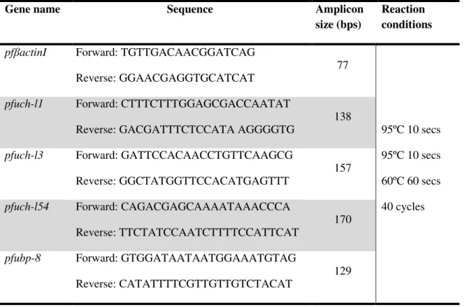
Table 5. Plasmodium falciparum RT-PCR primers designed from mRNA sequence
46
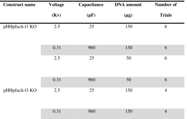
Table 6. Amplification of pfuch-l1and pfuch-l3 PCR products for transfection
49 Table 7. Electroporation settings for transfection of P.falciparum parasites
50 Table 8. Primers designed for recombinant protein production
54 Table 9. PCR reaction components and conditions for the amplification of the gene sequences of pfuch-l, pfuch-l3,pfuch-l54and pfubp-8 55
Table 10. Illustration of Plasmodium falciparum DUBs studied and their respective predictive active site obtained from the database Pfam 59
Table 11. Acute toxicity test for curcumin 67
Table 12. Determination of the in vitro IC50 of artemisinin, chloroquine and
curcumin 77
Table 13. In vivo acute toxicity test of curcumin in Balb/C mice
105 Table14. Differentially expressed proteins in Plasmodium falciparum curcumin
LIST OF ABBREVIATIONS
ACTs Artemisinin combination therapy ATG8 Autophagy related protein 8 ATP Adenosine-5-triphosphate AU Absorbance units
Bti Bacillus thurigiensisvar israeliensis Bs Bacillus sphaericus
BSA Bovine serum albumin CFA Complete freunds adjuvant CP 20S Core protein
CSP Plasmodium falciparum circunsporozoite protein Cy2 Cyanine dye 2
Cy3 Cyanine dye 3 Cy5 Cyanine dye 5
DDT Dichloro-diphenyl-trichloroethane Dhfr Dehydrofolate reductase enzyme Dhps Dehydrofolate pteoroate synthase DMSO Dimethyl sulfoxide
DNA Deoxyribonucleic acid DTT Dithiothreitol
DUBs De-ubiquitylating enzymes E1 Ubiquitin activating enzyme E2 Ubiquitin conjugating enzymes E3 Ubiquitin ligase
pfEBL-1 Plasmodium falciparum erythrocyte-binding ligand EDTA Ethylenediamine tetratacetic acid
ED50 Concentration of drug able to reduce parasitaemia to 50% ER Endoplasmic reticulum
GDP Gross Domestic product
HIV Human immunodeficiency virus HRP Horseradish peroxidase
HUB-1 Homologous to ubiquitin
IC50 Concentration needed to achive 50% inhibition IEF Isoelectric focusing
IFA Incomplete freunds adjuvant IPG Immobalized pH gradrient
IPTG Isopropyl β-D-1thiogalactopyranoside IRS Indoor residual spraying
IS Internal standard ITNs Insecticide treated nets kDa kiloDaltons
KO Knockout
LB Luria broth medium
LD50 Lethal dose to achieve 50% inhibition mRNA messenger RNA
MW Molecular weight NEM N-ethylmaleimide
NEDD8 Neural precursor cell expressed developementally down NI-NTA Nickel nitrilotriacetic acid
OUT Ovarian tumour domain containing protease OD Optical density
PBS Phosphate buffered saline PCR Polymerase chain reaction
pfatp6 Plasmodium falciparum Ca2+ depending SERCA type ATPase protein pfcrt Plasm odium falciparum chloroquine resistance transporter
pfHRD-1 Plasmodium falciparum ubiquitin ligase pfmdr-1 Plasmodium falciparum multidrug resistance 1
pftctp Plasmodium falciparum translationally controlled tumor gene pfUB Ubiquitin gene
pfUBA-1 Plasmodium falciparum ubiquitin activating enzyme 1 pfUBC Plasmodium falciparum uiquitin conjugating enzyme 2 pfuchl-1 Plasmodium falciparum ubiquitin carboxyl hydrolase pfRpn6 Plasmodium falciparum proteosome lid subunit 6 PI Isoelectric point
PMSF Phenylmethylsulfonyl fluoride PVDF Polyvinylidene fluoride RNA Ribonucleic acid RT-PCR Real time PCR
SDS Sodium dodecyl sulphate SUMO Small ubiquitin like modifier Ub Ubiquitin
Ub-AMC Ubiquitin-7-amino-4-methylcoumarin UbLp Ubiquitin like proteins
UBPs Ubiquitin proteases
UCHs Ubiquitin C-terminal hydrolases UN United Nations
UPS Ubiquitin/proteasome system URM-1 Ubiquitin related modifier -1 USD United states/American Dollar WHA World health assembly
Three letter amino acid code
Alanine ala
Asparagine asp
Cysteine cys
Glycine gly
Histidine his
Lysine lys
Serine ser
1.1 An introduction to malaria
1.1.1 A Historical overview on the discovery of malaria
Malaria is one of the oldest human parasitic diseases (Alonso & Tanner, 2013). Historical accounts refer to the existence of malaria back in Chinese documents (The canon of medicine) dating back to 2700 BC (Aikwa, 1971; Cowman, 2006; Cox, 2010). In 400 BC in the Greek era of Hippocrates there were also symptoms of malaria associated with swamps and poor sanitation (Aikwa, 1971). With the advancement of science, in 1886, the French military surgeon Alphonse Louis Laveran was responsible for the observation of
Plasmodium parasites in the blood of soldiers with malaria symptoms working in Constantine, Algeria (Bruce-Chwat, 1982; Cox, 2010). In 1897, the scientist William H Welch named Plasmodium falciparum specie (Coatney,1971) and this name became widely used in the literature. Between 1882 and 1899, the English scientist Sir Ronald Ross allowed mosquitoes to feed on birds infected with Plasmodium relictum and through the dissection of mosquito stomach demonstrated that mosquitoes were responsible for malaria transmission (MacCallum, 1897). Since then it has become clear that malaria is transmitted to humans by the bite of female Anopheles mosquitoes (Coatney, 1971; Baird 2009; Eade et al, 2009).
chloroquine (CQ) began to emerge in the 1950´s in South East Asia and contributed to the lack of success at the first attempt of global eradication (Overgaard and Angstreich, 2007).
1.1.2 An overview on the current malaria dissemination
Today malaria affects mainly populations in Sub-Saharan Africa, South East Asia and certain regions of South America and the Middle East (figure 1). According to the WHO in 2010 there were 219 million cases of malaria and 66000 deaths (WHO report, 2013), 91% of deaths are estimated to have occurred in the African region. The most affected groups are pregnant women, vulnerable HIV/AIDS patients and young children under five years of age who have not developed protective immunity against the most severe form of the disease. Malaria has a huge economic impact on endemic countries (Sachs and Malaney, 2002) and the disease accounts for approximately 22% of childhood deaths and it can decrease gross domestic product (GDP) by 1.3% (Sachs and Malaney, 2002).
The health costs of malaria in high transmission areas accounts for 40% of public expenditure and 50% of hospitals admissions, contributing to the poverty often seen in malaria endemic countries (Chima et al., 2003; Onwujekwe et al., 2010). The 6th United Nations (UN) development goal established in 2000 declares a combat against malaria, tuberculosis and HIV/AIDS. With the establishment of those goals, new mechanisms were introduced to finance the fight against those diseases. The latest WHO report (2013) indicates that funding for malaria control was 100 million USD back in 2000 and has risen since to 1.71 billion USD in 2010 and increased to 1.94 billion USD in 2012 and 1.97 billion USD in 2013 which has contributed to a 51% reduction in the malaria mortality rate in young children under five years of age (WHO, 2013).
1.1.3 The malaria parasite and its life cycle
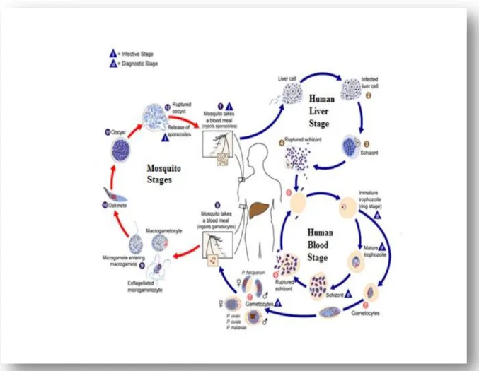
Plasmodium is a eukaryotic organism that belongs to the phylum Apicomplexa (Antinori et al., 2012).There are five species of Plasmodium responsible for human malaria and these are: falciparum, vivax, ovale, malaria and knowlesi. P. knowlesi predominantly infects
Macaca fascicularis by the bite of Anopheles leucosphyrus mosquitoes but it can also infect humans (Rosenberg et al., 1999; Antinori et al., 2012). In general the life cycle of human malaria parasites is divided into two phases: an exogenous sexual phase (sporogony) which occurs in the stomach of the female Anopheles mosquitoes (figure 2) and an endogenous asexual phase (schizogony) which occurs in the vertebrate host (figure 2).
During a blood meal the malaria infected female Anopheles mosquito inoculates sporozoites into the human host (Mackinnon et al., 2004). Sporozoites are injected subcutaneous under the skin and from there, the sporozoite travel and enter the parenchymal cells of the liver, (Rosenberg et al., 1999; Amino et al., 2006). Once the sporozoites have entered the parenchymal cells of the liver, the circulating sporozoites are first taken up by the Kupffer cells and then they enter the hepatocytes through the thrombosponding domain of the circumsporozoite protein (CSP) on the sporozoite and the heparin sulphate proteoglycan receptor on the hepatocyte (Pradel et al., 2002).
Once inside the liver cells, the parasite undergo asexual division and becomes a tissue schizont that contains thousands of merozoites (exo-erythrocytic schizogony) in what is known as the liver stage of the infection (Collins et al., 2005). Plasmodium falciparum can mature within 7 days and each sporozoite produces 40.000 merozoites, in P. vivax and P.
ovale dormant stages of the parasite, called hypnozoites, can persist in the liver causing relapses characterized by the appearance of parasitemia in the blood. Plasmodium
Once the hepatocytes rupture, they release merozoites that can invade the erythrocytes and initiate the intraerythrocytic phase of the infection also known as the blood stage (figure 2). Invasion of erythrocytes by the merozoites involves the interaction between protein ligands on the surface of the merozoite such as the merozoite surface protein (MSP-1) or the
Plasmodium falciparum erythrocyte binding ligand-1 (EBL-1) to the red blood cell receptors such as the glycophorin family of receptors (Mayer et al., 2009). Once inside the erythrocyte, trophozoite maturation occurs over a period of 24-72 hours depending on the species (Collins et al., 2005). The surface area of the young trophozoite (figure 2) begins to enlarge giving rise to a mature trophozoite (Collins et al., 2005; Antinori et al., 2012). Further mitotic divisions allow the parasite to mature into a schizont. The mature schizont can contain between 12-16 merozoites, the blood cell ruptures releasing those merozoites which invade new red blood cells maintaining the asexual life cycle. This phase of the infection is associated with malaria symptoms which is characterized by rapid rise of temperature, skin vasodilation and headaches (Collins et al, 2005; Cowman et al., 2006).
Within the erythrocyte, some parasites undergoe gametocytogenesis producing male (microgametocyte) or female (macrogametocytes) gametocytes (figure 2). If the female mosquitoes bite a malaria infected individual, gametocytes will be ingested with the blood meal and the recombination can occur in the mosquito stomach (sporogonic cycle) (Collins
et al., 2005). Once ingested by a mosquito, malaria parasites can develop during a period that may range between 10 to 21 days depending on the parasite species and environmental conditions such as temperature. In the mosquito stomach, gametocytogenesis will lead to the production of a zygote (figure 2), which will further develop into elongated ookinetes which invade the midgut wall of the mosquito, developing into large oocystis. The oocysts will develop and after their maturation the rupture occurs and the sporozoites are released (figure 2). The sporozoites make their way into the mosquitoes salivary glands (Collins et
1.2 Malaria control measures
1.2.1. Vector control measures
1.2.1.1. Chemical vector control measures
Vector control can be used in many levels in order to disrupt the parasite´s life cycle and block transmission. In many countries where malaria is endemic IRS has now become an important tool in the combat against malaria. The aim of IRS is to kill female mosquitoes with endophilic behavior, resting inside the house, thereby blocking transmission of disease (WHO report, 2013). Usually IRS is applied three times per year in areas of moderate and high transmission and a wide range of insecticides is available such as DDT, malathion, fenitrophion, alpha-cypermethrin just to name a few. However resistance to insecticides has already been reported in 27 countries (Abilio et al., 2011) and threatens the goals that have been achieved thus far. Another anti-vectorial measure is the use of ITNs impregnated with insecticides (WHO report,2013) and according to the WHO report, between 2004-2010, the number of ITNs rose from 5.6 million to 145 million in Sub-Saharan Africa contributing to the drop in the number of malaria cases (WHO report, 2013).
1.2.1.2. Non chemical vector control measures
Non chemical control measures are also seen as an alternative approach to insecticide use. Microorganisms such as Bacillus thurigiensisvar israeliensis (Bti) and Bacillus sphaericus (Bs)biolarvicidecan be placed in mosquitoe breeding sites (Lingenfelser et al., 2010), they have shown activity against Anopheles larvae without affecting the vertebrate host. The use of these microrganisms may help reduce the constant exposure of mosquitoes to insecticides which tends to exacerbate insecticide resistance (Mwuangangi et al, 2011). Predator fish such as Tilapia guineeensis have also shown efficacy in the removal of late stage Anopheles larvae in the Gambia Republic (Louca et al., 2009) reducing once again the excessive use of insecticides.
1.2.2. Malaria Vaccines
2009). This protein is involved in the adhesion of the sporozoite to the hepatocyte and during the liver stage of the disease. Anti-CS antibodies have been shown to inhibit parasite invasion and are also associated with a reduced risk of clinical malaria (Plassmeyer et al., 2009). The vaccine induced anti-CS responses and the fact that CS is the predominant surface antigen of sporozoites, makes CS a very good antigen for use in pre-erythrocytic vaccines (Audran et al., 2005; Schwartz et al., 2012). At the present time, the most promising vaccine is the vaccine candidate RTS,S /AS01E (which contains part of the CSP protein of P. falciparum fused to hepatitis B antigens and the adjuvant ASO2A). RTS,S/AS01E is the most advanced malaria vaccine candidate produced thus far (Enosse
et al., 2006) and has shown 50% efficacy against clinical malarial cases in children ages between 5-17 months after administration of three doses, in a study carried out in Mozambique (Enosse et al., 2006; Alonso, 2012). Trials are now being extended to other African countries to assess the efficacy of the vaccine.
1.2.3. Antimalarial drugs
1.2.3.1.Chloroquine
Chloroquine is a blood schizonticide and acts on the parasite food vacuole (Fidock et al., 2000; Daily, 2006; Aguiar et al., 2012). As the parasite digests hemoglobin, large amount of heme is formed as a byproduct, which is toxic for the parasite. The parasite detoxifies this product in its food vacuole by forming an inert crystal called hemozoin (Fidock et al., 2000; Daily, 2006; Aguiar et al., 2012). CQ acts by inhibiting the detoxification process, resulting in accumulation of millimolar levels of heme in the digestive vacuole of the parasite, causing their death. The heme-chloroquine complex may permeabilize membranes, interfere with free radical detoxification and block protein synthesis (Fidock et
reach levels required for inhibition of hemozoin formation (Fidock et al., 2000; Plowe, 2003; Farooq et al., 2004). Transfection assays (Fidock et al., 2000) demonstrated that the mutation in the (pfcrt) gene referred before was sufficient to induce a chloroquine resistance phenotype. Chloroquine resistance emerged first in South East Asia and it is believed that (CQ) resistance is the result of massive CQ pressure (Fidock et al., 2000; Farooq et al., 2004). CQ was widely used as the first line of antimalarial therapy for more than 50 years, because it was cheap to produce and had a good safety profile (Fidock et al., 2000). However due to the spread of chloroquine resistance worldwide (figure 3) CQ in many countries is no longer used in the clinic. Derivatives of quinine such as amodiaquine and mefloquine have also been synthesized for the treatment of P. falciparum infections. It is thought that the mode of action of amodiaquine and mefloquine is similar to chloroquine (Price et al., 2004). Resistance to mefloquine (figure 3) has been associated with an increase in the expression of the pfmdr-1 gene or increase in gene copy number (Price et
al., 2004; Daily et al., 2006).
1.2.3.2 Sulphadoxine/Pyrimethamine
Antifolate drugs, such as sulphadoxine/pyrimethamine act through sequential inhibition of two key enzymes which are dehydrofolate reductase (Dhfr) and Dehydropteroate synthase
1.2.3.3. Atovaquone/Proguanil
Atovaquone is a competitive inhibitor of the Quinol oxidation (Qo) site of the mitochondrial cytochrome b complex (Baggish and Hill, 2002). Atovaquone acts by inhibiting the parasite´s mitochondrial electron transport at the cytochrome b complex (Kroodsood et al., 2007). Although resistance to atovaquone develops very rapidly when used alone, when combined with proguanil, which is converted in the liver to cycloguanil (the active compound) which is an inhibitor of Dhfr enzyme, enhances the activity of atovaquone (Baggish and Hill, 2002). The combination is commercially known as
“malarone”. Atovaquone is expensive compared to chloroquine and mefloquine and therefore it is used mainly for prophylactic purposes for travelers visiting endemic areas. However, resistance to malarone has also emerged (Baggish and Hill, 2002) and it is conferred by single point mutations tyrosine (tyr) to serine (ser) at codon 268 in the cytochrome-b gene (Baggish and Hill, 2002).
1.2.3.4. Antibiotics
1.2.3.5. Artemisinin and its derivatives
Artemisinin is an active compound derived from the Chinese plant Artemisia annua (Klayman et al., 1985). At present artemisinin and its derivatives are the best antimalarial drugs available for the treatment of malaria. Artemisinin is a sequisterpene containing a peroxide bridge (Meshnick et al., 1996) this structure is unique to this compound and it is vital for its antimalarial activity (Woodrow et al., 2005; O´Neill et al., 2010). Evidence suggests that artemisinin´s mode of action is the result of interaction of artemisinin´s endoperoxide bridge with heme group present in the parasite´s digestive vacuole forming highly reactive species which leads to destruction of the parasite´s membranes and lysis of infected erythrocytes (Ellis et al., 1985; Meshnick et al., 1996).
The only disadvantage shown by artemisinin is that it has a short half life (Krishna et al., 2004). Now, several derivatives of artemisinin with better pharmacokinetic profile have been developed, namely artesunate, artemether, arteether, dihydroartemisinin. Those derivatives have between two to eight hours, meaning that not all parasites will be eliminated within that time frame thus resulting in a high risk of recrudescence (Krishna et
al., 2004). In order to avoid that, new policies regarding artemisinin in the treatment of malaria were endorsed by the World Health Assembly (WHA) in 2007, where the use of artemisinin alone was discouraged and current treatment of malaria is based on the association of artemisinin or its derivatives with other traditional antimalarial drugs such mefloquine, amodiaquine and others forming what is now known as artemisinin combination therapy (ACT).
According to the WHO report 2013, resistance to ACTs has been confirmed in Cambodia although it is not clear what are the mechanisms behind ACTs drug resistance (Wang et
al., 2011). It is thought that ACT resistance may be attributed to the fact that some of the partner drugs used in the ACT combination such as mefloquine is no longer eficaccious (figure 3). It is not clear which genes are potentially involved in artemisinin drug resistance. However, potential candidate genes were identified and sequenced in field isolates. Those were: P. falciparum Ca2+ depending SERCA type ATPase (pfATP6) (Valderramso et al., 2010), pfcrt gene (Fidock et al., 2000) pfmdr1 gene (Price et al., 1999), translationally controlled tumor gene (pftctp) (Eckstein-Ludwig et al., 2003) and the
been sequenced for mutations related to artemisinin resistance. However, so far no mutations were identified in those candidate genes, which can be linked to drug resistance to ACTs (Wang et al., 2011; Zakeri et al., 2012). The reality is that resistance to artemisinin and its derivatives has emerged and the failure to develop new antimalarials that preferentially act on new targets would only contribute to the spread of drug resistance and would jeopardize all the efforts that have been made thus far to control the disease.
1.3
Plasmodium
genome and its potential drug targets
1.3.1 Identification of potential drug targets
Sequencing of the Plasmodium genome was a big land mark in the history of malaria (Gardner et al., 2002) and revealed 5.403 nuclear genes identified, but only 1.800 genes encode proteins with known function (Gardner et al., 2002) which means that the
Plasmodium parasite may have interesting potential drug targets which must be identified and validated especially now that resistance to ACTs has become evident (Imwong et al., 2010). Most of the proteins with known or partially known function are involved in post-translational modifications (Chung et al., 2009). Plasmodium drug targets, have been identified by looking at proteins in the human host which are drug targets in cancer therapy or looking at proteases which are drug targets in other microorganisms (bacteria, virus) and then identifying whether those targets are also present in the Plasmodium genome (Fidock
et al., 2004).
Other approaches include, the identification of inhibitors against human phosphorylation/dephosphorylation, ubiquitin/proteasome (UPS), methylation, acetylation pathways (Brumlik et al., 2011; Fidock et al., 2004; Prudhomme et al., 2008; Sumanadasa
this system is responsible for most of the protein regulation inside eukaryotic cells (Aminake et al., 2012).Therefore deficiencies in the UPS can lead to the development or progression of metabolic, neurodegenerative and oncogenic diseases (Aminake et al., 2012), which has prompted the development of specific inhibitors against those enzymes involved in the UPS.Since the UPS or its components are present in many parasitic protozoa responsible for human diseases such as Plasmodium spp, Trypanosomes spp,
Leishmania spp, Giardia spp, Cryptosporidum spp and Theileria spp (Ponder and Bogyo, 2007) there is a huge interest in the characterization and validation of the UPS as a potential drug target in parasitic protozoa. Development of drugs against components of the UPS in one parasite may be efficient in the killing of another parasite making it easier to treat diseases caused by protozoa and reducing the costs of drug development. In the next section the components/enzymes involved in the UPS in Plasmodim falciparum shall be discussed in detail.
1.3.2. Discovery of the ubiquitin molecule in Plasmodium genome
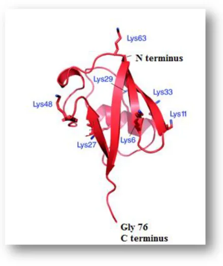
The ubiquitin molecule (Ub) was discovered in 1970 and its name reflects the fact that is a ubiquitous molecule meaning that it is found in various organelles inside the cell (Goldstein et al., 1975) and it is a key component in the UPS. Addition of this molecule to target proteins is called ubiquitylation (Horrocks and Newbold, 2000). The Ub molecule has special features such as the C terminal glycine (gly) 76 residue and also has on its sequence seven lys residues (lys6, lys11, lys29, lys33, lys48 and lys63) which play a crucial role in the activity of this molecule (figure 4).
The Plasmodium genome encodes an ubiquitin gene, whose official symbol is pfUB (Horrocks and Newbold, 2000; Ponts et al., 2008) found on chromosome 12. Gene expression studies in P.falciparum confirmed that pfUB is expressed in all life cycle stages of the parasite especially the late trophozoite stage (Horrocks and Newbold, 2000; Le Roch
et al., 2003) which means that ubiquitylation is an ongoing process which appears to be vital for the parasite (Ponts et al., 2008). PfUB was cloned and the protein was expressed and showed to be an 8.5 Kda protein on SDS-PAGE and its activity confirmed by ubiquitylation assays (Horrocks and Newbold, 2000). The human Ub sequence shares 98% similarity with Plasmodium pfUB and yeast Ub sequence meaning that Ub is very much conserved amongst eukaryotic organisms.
The Plasmodium genome also has ubiquitin like proteins/ubiquitin like modifiers (UbLps) these are: SUMO, NEDD8, HUB-1, URM1 (Ponts et al., 2011) which appear to be expressed in all stages of the Plasmodium life cycle. These molecules share similar tertiary structure with Ub and attachment of UbLps occurs in a similar mechanism as ubiquitin as explained below. PfSUMO an ubiquitin like molecule has also been characterized (table 1) and it was 40% identical to human SUMO-1 and found mainly in the nucleus and the cytoplasm of the parasite Plasmodium falciparum (Issar et al., 2008). Proteins tagged with (UbLPs) usually function in regulatory activities rather than being tagged for degradation (Frickel et al., 2007; Aminake et al., 2012).
Inhibitors of both Ub and UbLPs (SUMO) have been developed (table 1) and are now being tested for the treatment of neurological, microbial diseases and cancer (Edelmann et
Table 1. Characterization of ubiquitin (Ub) and ubiquitin like proteins (UbLps) in Plasmodium falciparum.
Component of the UPS
Putative biological role Biological characterization
Inhibitors available References
Ubiquitin Post translational modification
pfUB was characterized involved in ubiquitylation of proteins
Synthetic compound
Ubiquitin aldehyde and ubiquitin vinyl sulfone
Hershko and Rose 1987; Horrocks and Newbold, 2000
SUMO Post translational modification
pfSUMO was characterized as an ubiquitin like molecule
Anacardic acid isolated from cashew nut plant Anacardium occidentale
Ginkgolic acid isolated from Ginkgo biloba leaves
Issar et al., 2008 Fukuda et al.,2009
Nedd8 Post translational modification
Not available
URM-1
and
HUB-1
Post translational modification
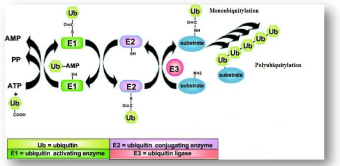
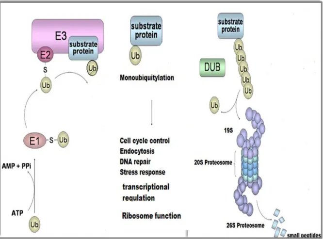
1.3.4 Ubiquitin Ligases
Attachment of ubiquitin molecules (figure 4) to target proteins is catalyzed by the action of ubiquitin activating enzymes also known as ubiquitin carrier protein (E1), Ubiquitin conjugating enzyme (E2) and ubiquitin ligase (E3) altogether known as ubiquitin ligases (Ponder and Bogyo, 2007; Ponts et al., 2008). The ubiquitylation cascade initiates when (E1) adenylates the ubiquitin molecule in an ATP dependent reaction (figure 5). Then ubiquitin is transferred to the active site cysteine (cys) residue on the E1 enzyme forming an E1-Ub thiolester complex, thereby activating the Ub molecule. The next step requires the action of another enzyme known as Ubiquitin conjugating enzymes (E2) (Ciechanover
et al., 2000; Glickman and Ciechanover, 2002).
E2 is responsible for the transfer of the activated Ub molecule from E1 via a high energy thiolester intermediate forming an E2-Ub complex at the active site cys residue of the enzyme. The E2-ubiquitin complex is then transferred to the active site cysteine residue on the E3 enzyme where the substrate protein binds directly to the E3 ligase (figure 5) via NH2 terminal residue (Glickman and Ciechanover, 2002; Ponts et al., 2008). Hence the E3 ligases are responsible for the last step in the reaction cascade and the E3-ubiquitin complex is transferred to the lysine (lys) residue on the target protein via an isopeptide bond which is formed between the glycine (gly) 76 amino acid (figure 4) on the Ub molecule and the lys residue on the target protein (Ciechanover et al., 2000; Glickman and Ciechanover, 2002).
1.3.5 Ubiquitin Ligases in the Plasmodium genome
In Plasmodium falciparum genome, at least 8 putative E1 genes encoding enzymes have been identified. At least 14 putative genes encoding E2 enzymes (Ponts et al., 2008) have also been identified and were detected at the trophozoite and schizont stages of the parasite. Finally 54 putative E3 ligases have been identified and were detected at the ring, trophozoite and schizont stages of the parasite (Ponts et al., 2011). Making a total of 76 putative genes encoding ubiquitin ligases. The E3 family of enzymes is very diverse, the large number of E3 is due to the fact that there are different domain families of E3 ligases (Ring finger domain E3s, HECT domain E3s and U-box domain E3 ligases).
The Ring finger family represents the largest group of enzymes in eukaryotes (Ponts et al., 2008) and they contain a cystein/histadine/zinc domain involved in protein-protein interaction. A total of 68 putative ub ligases have also been identified in Plasmodium
vivax, 62 putative ubiquitin ligases have been identified in Plasmodium yoelli but so far no biochemical characterization has been done (Ponts et al., 2008). Of the predictive 76 putative Ub ligases identified in the Plasmodium falciparum genome only three ubiquitin ligases have so far been characterized (table 2): The E1 Plasmodium falciparumpfUBA1, the E2 Plasmodium falciparum pfUBC, the E3 enzyme Plasmodium falciparum pfHRD1, which are involved in ubiquitylation of proteins in the endoplasmic reticulum (ER) (Chung
et al., 2012) and its ubiquitylating enzyme activity was confirmed by ubiquitylation assays. Several inhibitors that were discovered in the field of cancer research are now being tested for their antimalarial activity (table 2) (Chung et al., 2012).
Table 2. Characterization of ubiquitin ligases in Plasmodium falciparum.
Component
of the UPS
Putative biological
role
Biological characterization Inhibitors available References
Ubiquitin activating enzymes
(E1)
Activation of ubiquitin
pfUBA-1 characterized as an E1 and found in the ER
Synthetic compounds, Benzothiazole derivatives and PYR-41
Guedat and Colland, 2007; Yang et al., 2007
Ubiquitin conjugating enzymes
(E2)
Conjugation of ubiquitin
pfUBC characterized as an E2 found in the ER
Leucettamol A Isolated from marine sponge
Leucetta aff.microraphis
Edelmann et al., 2011
Ubiquitin
ligase (E3)
Ligation of ubiquitin
pfHRD-1 characterized as an E3 found in the ER
Synthetic compound Eeyarestatin*
1.3.6 The Proteosome
The proteosome is also known as the 26S proteasome, it is a complex with many subunits (figure 6) involved in the regulated degradation of ubiquitylated proteins (Ciechanover et
1.3.7 The proteasome in the Plasmodium genome
In P. falciparum genome, 14 putative proteins, homologous to the yeast 20S CP subunit of the proteasome, were identified (Mordmuller et al., 2006). 20S CP subunits were shown to be present in the cytoplasm and nucleus of blood stage Plasmodium parasites, particularly in trophozoites and schizonts where there is a peak of ubiquitylated proteins (Aminake et
al., 2012; Ponts et al., 2011) and the 26S CP subunit is expressed at the trophozoite and schizont stages of the parasite´s life cycle (Kreidenweiss et al., 2008).
Another component of the proteasome of P. falciparum is the protein RPn6 which is found in the lid of the protesome, this protein was characterized in P. falciparum and found in the cytosol of the parasite and biochemical assays indicated that this protein is an important part of the proteosome specially for the degradation of ubiquitylated proteins (Muralidharan et al., 2011). Thus, confirming the existence of an active UPS in the malaria parasite. Research carried out in cancer in the last decade has identified many proteasome inhibitors with antimalarial activity marked with an asterisk in the (table 3).
The proteasome inhibitor epoxomicin for example has antimalarial activity against chloroquine sensitive strains with an IC50 of 6.8 nM and it also has activity against field isolates from Gabon with an IC50 of 8.5 nM (Kredeinweiss et al., 2008). Even though many proteasome inhibitors with antimalarial activity have been developed, proteosome inhibitors are also known to be very toxic (Reynolds et al., 2007), hence it is thought that targeting individual enzymes involved in the UPS may be a more viable alternative chemotherapy for both infectious and non-infectious diseases.
Another proteasome inhibitor used in the treatment of multiple myeloma known as Bortezomib (table 3) is a well known proteosome inhibitor able to inhibit the intraerythrocytic developmental stages of P. falciparum (Reynolds et al., 2007). Salinosporomide A is a proteasome inhibitor, now in clinical trials phase I for the treatment of multiple myeloma and has antimalarial activity against P. falciparum and P.
Table 3. Characterization of the proteosome in Plasmodium falciparum.
Component of the
proteosome Putative biological
Role
Biological characterization Inhibitors
Available
References
20S proteasome subunit
and Rpn6 subunit
Protein degradation Found to be expressed in the trophozoite and schizont stages and localized in the cytosol
Lactacystin* isolated from Streptomyces
Synthetic compound bortezomib*
Gantt et al., 1998 Reynolds et al 2007
Salinosporomide A* isolated from marine bacteria
Salinospora tropica
Prudhomme et al., 2008
Synthetic compounds MG132*and Epoxomicin* isolated from Actinomycetes
Kreidenweiss et al., 2008;Cszesny et al.,2009
1.3.8 De-ubiquitylating enzymes (DUBs)
Protein ubiquitylation is a reversible process; the removal of ubiquitin molecules is carried out by ubiquitylating enzymes also found in the literature as ubiquitinases or ubiquitinating enzymes, but in the present study they shall be referred to as de-ubiquitylating enzymes (DUBs) (Mullaly and FitzPatrick 2002; Amerik and Hochstrasse, 2004; Nijman et al., 2005), which are responsible for the generation of free ubiquitin molecules (figure 6) and the disassembly of mono or polyubiquitin chains on substrate proteins.
DUBs are classified as proteases (Nijman et al., 2005) depending on their mechanism of catalysis they have been divided into: cysteine proteases and zinc dependent mettalloproteases which can be further subdivided into distinct subfamilies (Nijman et al., 2005; Mukhopadhyay and Riezman, 2007) these are: The Ubiquitin C terminal Hydrolases (UCHs) (figure 7), the Ubiquitin proteases (UBPs) or USPs (Ubiquitin specific proteases) from now on referred to as UBPs. The Machado Joseph Disease protein domain proteases (MJDs). The Outbains (OTUs), JAMM (motif mettallo proteases) which are classified as zinc dependent proteases. In addition to this major group of DUBs, there is also three distinct families of de-ubiquitylating like enzymes (DUBLs) these are the SUMO specific proteases, the autophagins proteases and the WLM (weak suppressor mettalloproteases) family of zinc dependent proteases (Nijman et al., 2005; Ponder and Bogyo, 2007) which will not be discussed further as they are beyond the scope of this project and in this project the focus will be mainly on UCHs and UBPs.
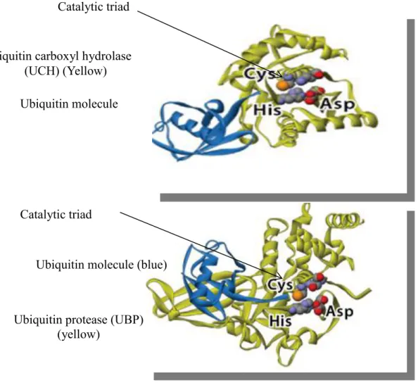
In general Dubs have in their catalytic site three key amino acids (aa) cysteine (cys), histidine (his) (figure 7) and an aspartate residue (asp) known as the catalytic triad (Nijman
Figure 7. A general structure of the catalytic domain of ubiquitin carboxyl hydrolase (UCH) and ubiquitin protease (UBP). DUBs are highlighted in yellow and interacting with the ubiquitin molecule highlighted in blue. In general DUBs have in their catalytic site three key amino acids cysteine (cys), histidine (his) and an aspartate residue (asp) known as the catalytic triad. De-ubiquitylation occurs when the cysteine residue launches a nucleophilic attack on the isopeptide bond that is between the ubiquitin C terminal and the lysine (lys) residue on the target protein. The reaction results in the release of the target protein and free ubiquitin molecules. Adapted and modified from (Nijman et al., 2005).
Ubiquitin molecule (blue)
Ubiquitin carboxyl hydrolase (UCH) (Yellow)
Catalytic triad
Ubiquitin molecule (blue)
Ubiquitin protease (UBP) (yellow)
(UBP)
1.3.9 De-ubiquitylating (DUBs) enzymes in the Plasmodium genome
An in silico study has shown that the Plasmodium genome encodes at least 40 putative DUBs whereas the human genome contains approximately 95 putative DUBs (Nijman et
al., 2005; Ponder and Bogyo, 2007; Ponts et al., 2008; Wilkinson, 2009). In spite of the large number of DUBs in the Plasmodium genome, very few proteins have been characterized. The first Plasmodium DUB to be characterized was Plasmodium falciparum
ubiquitin carboxyl hydrolase 54 (Pfuch-l54) (Artavanis-Tsakonas et al., 2006). The crystal structure of this enzyme was determined and it was found that Pfuch-l54 protein has 54 KDa and has moderate sequence identity with human ubiquitin carboxyl hydrolase 1 (huch-l1) and (huch-l3) but the active site residues were conserved amongst those proteins (Artavanis-Tsakonas et al., 2006).
No biochemical assays, gene knockout assays and localization studies were carried out in order to clarify the role and the location of this protein in Plasmodium´s life cycle. The second DUB whose crystal structure was determined was Plasmodium falciparum
ubiquitin carboxyl hydrolase-3 (Pfuch-l3)(table 4), this protein is 30% identical to huch-l3 (Artavanis-Tsakonas et al., 2006).The recombinant proteinappeared on the SDS-PAGE as a 30-32Kds band but in the literature is known as Pfuch-l3 (Artavanis-Tsakonas et al., 2011) and site directed mutagenesis assays showed that Pfuch-l3 gene product might be essential for parasite survival as substitution of a cys residue by an alanine (ala) in the active site resulted in the death of mutant parasites (Artavanis-Tsakonas et al., 2011). Furthermore, there is only moderate identity between the human and PlasmodiumPfuch-l3 which means that these enzymes may be selectively targeted in malaria chemotherapy without damaging the host´s enzyme (Artavanis-Tsakonas et al., 2011) generating an interest in the characterization of these proteins as future drug targets.
The genome of Plasmodium berghei has about 24 putative genes encoding DUBs,
against important parasites responsible for human diseases.In order to develop new drugs several parameters must be taken into account such as: drug efficacy, pharmacology, general toxicity and potential side effects.Developing anti-malarial drugs from scratch is time consuming and often results is drug development projects being abandoned halfway due to the lack of return on investments (Fidock et al., 2004; Chung et al., 2009).
Recent approaches to speed up the discovery and development of new compounds with antimalarial activity involve screening of compounds which have shown therapeutical potential in the treatment of other diseases, but also display antimalarial activity. The natural compound curcumin is a polyphenolic compound with cancer, anti-inflammatory, anti-viral and antimalarial activity. Curcumin is widely used in traditional Indian medicine in the treatment of cancer. Now several studies have shown that curcumin is active against coxsackie viral infections, prevents mycocardial infarction, rheumatoid arthiritis, multiple sclerosis and Alzheimer disease (Anand et al., 2007; Mimche et al., 2011).There is a growing body of evidence indicating that curcumin has potent antimalarial activity both in vivo and in vitro (Mullaly and Fitzpatrick et al., 2002, Nandakumar et al., 2006, Martinelli et al., 2008).
Curcumin has proved to be potent against other parasitic organisms including: Schistosoma
Table 4. Characterization of de-ubiquitylating enzymes (DUBs) and de-ubiquitylating like enzymes (DUBLs) in Plasmodium falciparum.
Component of the UPS Putative biological Role Biological characterization Inhibitors available References
DUBs & DUBLs
Ubiquitin carboxyl hydrolase
(UCH)
de-ubiquitylation Pfuch-l3 & Pfuch-l54 characterized as DUBs
Synthetic compounds Cyclopentenone and DBA
Guedat and Colland, 2007; Artavanis-Tsakonas et al., 2011
Ubiquitin proteases
(UBP)
de-ubiquitylation Not available Curcumin* isolated from the plant Curcuma longa
Shikoccin isolated from the plant Rabdosia shikokiana occidentalis
Mullaly and FitzPatrick 2002; Reddy et al., 2005; Nandakumar et al., 2006
Machado Joseph disease
(MJD)
de-ubiquitylation Not available
JAMM motif metalloprotease
(JAMM)
SUMO specific proteases
(SENPs)
de-ubiquitylation pfSENP-1 and pfSENP-2 characterized and SUMO cleavage activity confirmed
Synthetic compound
JCP-666*
Ponder et al.,2011
Ovarian tumour proteases (OTU)
de-ubiquitylation Not available
Given the fact that antimalarial drug resistance is emerging at a much faster rate then antimalarial drug development, the enzymes involve in the UPS and its inhibitors represent a promising avenue in antimalarial chemotherapy. In this project the main focus will be on de-ubiquitylating enzymes (DUBs) as previous study carried out by our laboratory identified a mutation in a gene encoding a ubiquitin carboxyl hydrolase-1 enzyme in Plasmodium chabaudi (pcuch-l1) strains resistant to artemisinin and artesunate (Afonso et al., 2006; Hunt et al., 2007). In light of the previous research that was carried out in our laboratory, there is now a major interest in characterizing DUBs not only because of their possible involvement in drug resistance (Hunt et al., 2007) but also because of their involvement in the development and the progression of infectious and non infectious diseases (Le Negrate et al., 2008; Luise et al., 2011).
Given the large number of DUBs in the Plasmodium genome (section 1.1.3.9) four of them were identified in the Plasmodium genome using the PlasmoDB and the Protein Data Bank (APPENDIX A and APPENDIX B and APPENDIX I) to be characterized in this project, these are: Plasmodium falciparum ubiquitin carboxyl hydrolase-1 (Pfuch-l1) Plasmodium falciparum ubiquitin carboxyl hydrolase-3 (Pfuch-l3)
Plasmodium falciparum Ubiquitin carboxyl hydrolase 54 (Pfuch-l54) and Plasmodium falciparum Ubiquitin protesase 8 (Pfubp-8).Those genes were chosen based on previous work published by others and their relevance in other biological systems.The human homologue of Pfuch-l1 has been implicated in kidney carcinomas (Luise et al., 2011) and point mutations in the human gene huch-l1 are associated with Parkinson´s disease (Liu et al., 2002). This gene also appears to be mutated in Plasmodium chabaudi parasites resistant to artesunate (Hunt et al., 2007).The protein sequence of Pfuch-l1 was used to interrogate the protein data bank (PDB) the protein query indicated that human ubiquitin carboxyl hydrolase 8 (huch-l8 ) is the human homologue of Pfuch-l1.