Study of the embryofeto-toxicity
of Crown-of-Thorns
(Euphorbia milii)
latex, a natural molluscicide
Laboratório de Toxicologia Ambiental, Departamento de Ciências Biológicas, Escola Nacional de Saúde Pública, FIOCRUZ, Rio de Janeiro, RJ, Brasil C.A.M. Souza,
R.R. de-Carvalho, S.N. Kuriyama, I.B. Araujo, R.P. Rodrigues, R.S. Vollmer, E.N. Alves and F.J.R. Paumgartten
Abstract
The crude latex of Crown-of-Thorns (Euphorbia milii var. hislopii) is a potent plant molluscicide and a promising alternative to the synthetic molluscicides used in schistosomiasis control. The present study was undertaken to investigate the embryofeto-toxic potential of E. milii
latex. The study is part of a comprehensive safety evaluation of this plant molluscicide. Lyophilized latex (0, 125, 250 and 500 mg/kg body weight) in corn oil was given by gavage to Wistar rats (N = 100) from days 6 to 15 of pregnancy and cesarean sections were performed on day 21 of pregnancy. The numbers of implantation sites, living and dead fetuses, resorptions and corpora lutea were recorded. Fetuses were weighed, examined for external malformations, and fixed for visceral examination, or cleared and stained with Alizarin red S for skeleton evaluation. A reduction of body weight minus uterine weight at term indicated that E. milii latex was maternally toxic over the dose range tested. No latex-induced embryolethality was noted at the lowest dose (125 mg/kg) but the resorption rate was markedly in-creased at 250 mg/kg (62.5%) and 500 mg/kg (93.4%). A higher frequency of fetuses showing signs of delayed ossification (control: 17.4%; 125 mg/kg: 27.4% and 250 mg/kg: 62.8%; P<0.05 vs control) indicated that fetal growth was retarded at doses ≥125 mg latex/kg body weight. No increase in the proportion of fetuses with skeletal anomalies was observed at the lowest dose but the incidence of minor skeletal malformations was higher at 250 mg/kg body weight (control: 13.7%; 125 mg/kg: 14.8%; 250 mg/kg: 45.7%; P<0.05 vs control). Since a higher frequency of minor malformations was noted only at very high doses of latex which are embryolethal and maternally toxic, it is reasonable to conclude that this plant molluscicide poses no teratogenic hazard or, at least, that this possibility is of a considerably low order of magnitude.
Correspondence
F.J.R. Paumgartten Laboratório de Toxicologia Ambiental
Departamento de Ciências Biológicas
ENSP, Fundação Oswaldo Cruz Av. Brasil, 4365
21045-900 Rio de Janeiro, RJ Brasil
Research supported by CNPq and PAPES-FIOCRUZ. F.J.R. Paumgartten and S.N. Kuriyama are recipients of CNPq fellowships.
Received March 13, 1997 Accepted August 25, 1997
Key words
•Euphorbiaceae
•Embryofeto-toxicity
•Plant molluscicides
•Euphorbia milii
•Reproductive toxicity
Introduction
In addition to treating the affected indi-viduals, one of the most effective and rapid means of reducing schistosomiasis transmis-sion is snail control with molluscicides. De-sirable attributes of a molluscicide to be used in public health programs are a high activity and specificity for snails, a low tox-icity to mammalian species, stability under different environmental and storage condi-tions, safety to the applicator, and low cost (1). None of the synthetic molluscicides cur-rently available meet all these requirements and only one, niclosamide (Bayluscide®), is
recommended by the WHO for large-scale use in schistosomiasis control (1). Because developmental costs are high and the market is confined to the developing countries where the disease is endemic, no new molluscicide has been developed by the industry in recent years. However, considerable efforts have been made to find a plant molluscicide that would be at one time highly active, cost-effective, safe, easy to apply and locally available in the endemic areas.
To date hundreds of botanical species have been screened for molluscicidal activ-ity but the application of crude plant material to snail habitats, with very few exceptions (e.g., dried leaves of Ambrosia maritima and unripe berries of Phytolacca dodecandra), has been unsuccessful (2). Within this con-text, the crude latex of “Crown-of-Thorns” (Euphorbia milii var. hislopii Des Moul. ex Boiss, syn. E. splendens) seems to be one of the most promising plant molluscicides tested so far. It is active against the snails which are intermediate hosts of S. mansoni in the labo-ratory at concentrations as low as 0.5 ppm (3) and under field conditions at concentra-tions between 5 and 12 ppm (4,5). Besides its remarkable potency, E. milii also presents some very interesting characteristics for a plant molluscicide: Crown-of-Thorns is a cosmopolitan plant easily cultivable in en-demic areas, it yields a rather large amount
of latex throughout the year and no marked geographic or seasonal variation in the mol-luscicidal activity of latex has been found (6,7). An ecotoxicological study has also shown that latex is less harmful to non-target freshwater organisms than niclosamide (8).
The present study was carried out to in-vestigate the teratogenic or embryofeto-toxic potential of E. milii latex in rats. The study is part of a more comprehensive toxicological evaluation of the health risks posed by this plant molluscicide.
Material and Methods
Animals
Male and virgin female Wistar rats from the Oswaldo Cruz Foundation Central Ani-mal House breeding stock were used. The animals were housed in standard plastic cages with stainless steel cover lids and wood shav-ings as bedding, and kept under controlled temperature (23 ± 1oC), humidity
(approxi-mately 70%), and dark-light cycle (lights on from 10:00 to 22:00 h). A pelleted diet (Nuvital®, Nuvilab Ltd., Curitiba, PR,
Bra-zil) and tap water were available ad libitum.
Mating procedure
Mating was performed by transferring two females to the cage of one male for 2 h (8:00 to 10:00 h). Copulation was confirmed by the presence of sperm in the vaginal smear and the day when spermatozoa were found was designated as day 0 of pregnancy.
Plant material
longitudi-nal incision was made in the plant stem and the latex was collected into glass tubes which were then sealed, wrapped in aluminum foil, and immediately transported to the labora-tory where it was lyophilized. Lyophilized latex was protected from light and stored in the refrigerator until further use. Under these storage conditions, E. milii latex keeps its molluscicidal potency for at least 18 months (7).
Treatment
The lyophilized latex was suspended in corn oil (Mazola®) and administered by
ga-vage to rats (N = 100) once a day from day 6 to day 15 of pregnancy. Three groups of pregnant rats were treated with different doses of lyophilized latex (125, 250 and 500 mg/kg body weight) and a control group received only corn oil (3.75 g/kg body weight). All rats were weighed on days 0, 6 to 15, and 21 of pregnancy.
Cesarean section
On day 21 of pregnancy the rats were anesthetized by ethyl ether inhalation and killed by decapitation. The gravid uterus was weighed with its contents. Living and dead fetuses, resorptions and corpora lutea were recorded. The number of implantation sites was determined by the method of Salewski (9). The fetuses were weighed, numbered with a marker pen, examined for external malformations and fixed in 5% formalin so-lution. One-third of the fetuses of each litter, selected at random, were evaluated for vis-ceral malformations using a microsectioning technique adapted from Sterz (10). Heart, lungs, thymus, spleen, liver and kidneys of fetuses which were microdissected were also weighed. The remaining fetuses were cleared with KOH, stained with Alizarin red S (11) and examined for skeletal abnormali-ties.
Statistical analysis
Data were analyzed by one-way analysis of variance or, alternatively, by the Kruskal-Wallis test whenever the data did not fit a normal distribution. Differences between groups were tested by the two-sided Student t-test or Mann-Whitney U-test. Proportions were analyzed by the chi-square test or, al-ternatively, by the Fisher exact test. Statisti-cal evaluation was performed using a MINITAB program (MTB, University of Pennsylvania, 1984), and differences were considered to be statistically significant at P<0.05.
Results
A higher proportion of resorptions per implantation site as well as an increase in the mean number of resorptions per litter were observed in the groups treated with the two highest doses of E. milii latex (Table 2). A
parallel decrease in the number of live fe-tuses per litter was also noted (Table 2). The results therefore demonstrated that E. milii latex, at doses equal to or higher than 250 mg/kg, induced a high frequency of
post-Table 1 - Maternal weight gain of rats treated orally with Euphorbia milii latex (0, 125, 250 and 500 mg/kg, po) on days 6 to 15 of pregnancy.
+Two rats treated with 500 mg/kg and one treated with 250 mg/kg died and were not included. % of pregnant
females was analyzed by the chi-square test. Maternal weight gain was analyzed by the Kruskal-Wallis test followed by the Mann-Whitney U-test. All other parameters were analyzed by one-way analysis of variance and the Student t-test. Data are reported as mean ± SD. *P<0.05 compared to control.
Treatment E. milii latex (mg/kg body weight)
0 125 250 500
Treated females 29 24 28+ 16+
Pregnant females 19 21 22 13
Pregnant/treated females (%) 65.5 87.5 78.6 81.2
Maternal weight (g)
Day 0 236 ± 16 230 ± 22 229 ± 11 227 ± 18
Day 21 341 ± 32 326 ± 35 288 ± 25* 250 ± 20*
Gravid uterus weight (g) 67.0 ± 18.1 63.8 ± 23.1 30.6 ± 19.9* 4.6 ± 8.1*
Maternal weight gain (g)
Days 6-11 9.8 ± 4.1 -9.8 ± 4.8* -23.8 ± 8.7* -31.1 ± 6.7*
Days 6-15 26.0 ± 7.4 0.5 ± 10.0* -20.7 ± 13.7* -33.3 ± 7.2*
Days 0-21 104.6 ± 23.5 85.1 ± 29.9 52.0 ± 28.2* 17.6 ± 17.4*
Days 0-21 (minus uterus weight) 37.7 ± 16.6 21.4 ± 16.4* 19.9 ± 11.7* 13.0 ± 14.6*
Table 2 - Effects of treatment with E. milii latex (0, 125, 250 and 500 mg/kg, po) on days 6 to 15 of pregnancy on parameters assessed at the time of cesarean section of rats.
Percent resorptions was analyzed by the chi-square test. Numbers of corpora lutea, implantation sites and live fetuses per litter were analyzed by the Kruskal-Wallis test followed by the Mann-Whitney U-test. All other parameters were analyzed by one-way analysis of variance and the Student t-test. Data are reported as mean ± SD. *P<0.05 compared to control.
Treatment E. milii latex (mg/kg body weight)
0 125 250 500
Corpora lutea 11.9 ± 1.8 11.1 ± 2.3 10.4 ± 2.8 11.0 ± 1.4
Implantation sites 12.3 ± 2.3 11.8 ± 13.7 11.4 ± 3.1 11.0 ± 2.8
Resorptions 33 37 140 156
Resorptions/implantations (%) 13.9 17.7 62.5* 93.4*
Resorptions per litter 1.8 ± 1.9 1.8 ± 1.6 6.4 ± 3.4* 12.0 ± 3.2*
Live fetuses 200 210 104 7
Live fetuses per litter 10.5 ± 2.6 10.0 ± 4.1 4.7 ± 4.2* 0.4 ± 12.1*
Fetal weight (g)
Individual 4.82 ± 0.44 4.66 ± 0.51* 4.41 ± 0.71* 3.44 ± 0.66*
implantation losses.
A dose-dependent reduction of fetal body weight (Table 2) and a higher proportion of fetuses exhibiting signs of delayed ossifica-tion (Table 3) indicated that E. milii latex caused fetal growth retardation at doses ≥ 125 mg/kg body weight. Owing to the ex-tremely high resorption rate (93.4%) and to the very small number of living fetuses ob-tained, frequencies of fetal anomalies at the highest dose (500 mg/kg) were not included in the statistical evaluation of data and thus are not shown in Tables 3, 4, and 5.
Edema, paleness, tail abnormalities (kinky tails and tails with a bent end), gastroschisis, and irregular positioning of hindpaws were occasionally observed in latex-exposed tuses (Table 4). A higher proportion of fe-tuses with an abnormally shaped thymus (not related to the dose), as well as a dose-dependent increase in the frequency of fe-tuses with an accessory lobe (seventh) in the liver, were the only treatment-related find-ings revealed by visceral examination (Table 4). No statistically significant (P>0.05, ANOVA) alterations in fetal organ weight (mg; mean ± SD) were found after exposure to E. milii latex from days 6 to 15 of preg-nancy: liver (control: 364 ± 47; 125 mg/kg: 356 ± 48; 250 mg/kg: 352 ± 62), spleen (control: 6.4 ± 1.9; 125 mg/kg: 6.7 ± 2.0; 250 mg/kg: 6.6 ± 2.6), thymus (control: 7.7 ± 1.8; 125 mg/kg: 8.2 ± 1.5; 250 mg/kg: 7.7 ± 1.8), lungs (control: 131.0 ± 21.0; 125 mg/kg: 130.0 ± 15.5; 250 mg/kg: 126.0 ± 18.9), heart (control: 31.9 ± 5.4; 125 mg/kg: 31.0 ± 7.2; 250 mg/kg: 30.9 ± 7.2) and kidneys (left, control: 11.6 ± 2.3; 125 mg/kg: 11.1 ± 1.7; 250 mg/kg: 12.1 ± 2.3; right, control: 12.3 ± 2.3; 125 mg/kg: 11.8 ± 1.9; 250 mg/kg: 12.6 ± 2.6).
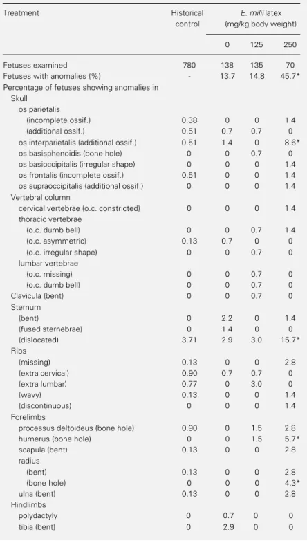
The occurrence of gross structural skel-etal abnormalities in fetuses exposed to E. milii latex from pregnancy day 6 to day 15 is shown in Table 5. No increase in the propor-tion of skeletal malformapropor-tions was noted at the lowest dose tested, but the frequency of
fetuses showing one or more abnormalities was higher at 250 mg latex/kg body weight. The overall increase in the occurrence of skeletal anomalies seemed to have resulted, to a large extent, from higher incidences of dislocated sternum (non-aligned sternebrae), additional ossification of the os
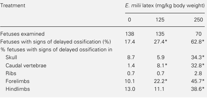
interparieta-Table 3 - Occurrence of signs of delayed ossification in the offspring of rats treated orally with E. milii latex (0, 125 and 250 mg/kg) on pregnancy days 6-15.
Signs of delayed ossification: not ossified (whole bone not stained); poorly ossified (whole bone is poorly ossified), and irregular spongy bones. Data were analyzed by the chi-square test or by the Fisher exact test. *P<0.05 vs control. Because of the high resorption rate at the highest dose tested (500 mg/kg), only six fetuses of this group were available for skeleton examination, and thus they were not included in the table.
Treatment E. milii latex (mg/kg body weight)
0 125 250
Fetuses examined 138 135 70
Fetuses with signs of delayed ossification (%) 17.4 27.4* 62.8*
% fetuses with signs of delayed ossification in
Skull 8.7 5.9 34.3*
Caudal vertebrae 1.4 8.1* 32.8*
Ribs 0.7 0.7 2.8
Forelimbs 10.1 22.2* 45.7*
Hindlimbs 13.0 11.1 38.6*
Table 4 - Occurrence of externally visible and visceral abnormalities in fetuses of rats treated orally with E. milii latex (0, 125 and 250 mg/kg body weight) on days 6 to 15 of pregnancy.
Data were analyzed by the chi-square test or by the Fisher exact test. *P<0.05 vs control. One fetus exposed to the highest dose (500 mg/kg) was not included in the table because it was the only one available for visceral examination in this group.
Treatment E. milii latex (mg/kg body weight)
0 125 250
External examination (number of fetuses) 200 210 104
Gastroschisis (%) 0 0 1 (1.0)
Edema (%) 0 0 2 (1.9)
Paleness (%) 0 0 2 (1.9)
Tail anomalies
Bent tip (%) 0 8 (3.8) 1 (1.0)
Kinky (%) 0 2 (1.0) 1 (1.0)
Visceral examination (number of fetuses) 62 62 28
Abnormal shape of thymus (%) 1 (1.6) 6 (9.7)* 2 (7.1)*
lis, hole in the humerus and hole in the radius.
Discussion
The maternal deaths as well as the de-crease in overall weight gain during preg-nancy, still apparent after subtraction of gravid uterus weight, clearly indicated that E. milii latex was maternally toxic over the dose range tested. These maternally toxic doses of latex also proved to be embryofeto-toxic as revealed by the three outcomes evalu-ated: embryolethality, prenatal growth retar-dation and fetal malformations.
No post-implantation loss was noted at the lowest dose tested (125 mg/kg). None-theless, a sharp dose-related increase in the resorption rate, as well as a parallel decrease in the number of living fetuses, showed that embryolethality occurred at doses ≥250 mg latex/kg.
Signs of prenatal growth retardation were observed in rats exposed to doses ≥125 mg latex/kg. A dose-dependent increase in the frequency of fetuses exhibiting signs of de-layed ossification indicated that E. milii latex caused retardation of fetal skeleton develop-ment at doses equal to or higher than 125 mg/ kg. Latex-induced reductions in fetal body weight were only slight, and were not sig-nificant when the litter was taken as the statistical unit of analysis in the groups treated with 125 and 250 mg/kg. Thus, in this case, as in previous experiments with other sub-stances (12-14), the proportion of fetuses showing signs of delayed ossification proved to be a more sensitive indicator of prenatal growth retardation than fetal weight at term. No increase in the occurrence of skeletal malformations was produced by E. milii la-tex at the lowest dose tested (125 mg/kg) but a higher frequency of skeletal anomalies was observed at doses as high as 250 mg/kg. It is noteworthy that the E. milii latex-induced increase in skeletal malformations was mainly due to sternum dislocation
(non-Table 5 - Occurrence of skeletal anomalies in fetuses of rats treated orally with E. milii latex (0, 125 and 250 mg/kg body weight) on days 6 to 15 of pregnancy.
Data were analyzed by the chi-square test or, alternatively, by the Fisher exact test. *P<0.05 compared to control. The highest dose group was not included in the table because only six fetuses exposed to 500 mg/kg were examined. Historical control: data from all vehicle-control fetuses (distilled water or corn oil) of Wistar rats from the FIOCRUZ breeding stock evaluated in our laboratory. o.c., Ossification center.
Treatment Historical E. milii latex
control (mg/kg body weight)
0 125 250
Fetuses examined 780 138 135 70
Fetuses with anomalies (%) - 13.7 14.8 45.7*
Percentage of fetuses showing anomalies in Skull
os parietalis
(incomplete ossif.) 0.38 0 0 1.4
(additional ossif.) 0.51 0.7 0.7 0
os interparietalis (additional ossif.) 0.51 1.4 0 8.6*
os basisphenoidis (bone hole) 0 0 0.7 0
os basioccipitalis (irregular shape) 0 0 0 1.4
os frontalis (incomplete ossif.) 0.51 0 0 1.4
os supraoccipitalis (additional ossif.) 0 0 0 1.4
Vertebral column
cervical vertebrae (o.c. constricted) 0 0 0 1.4
thoracic vertebrae
(o.c. dumb bell) 0 0 0.7 1.4
(o.c. asymmetric) 0.13 0.7 0 0
(o.c. irregular shape) 0 0 0.7 0
lumbar vertebrae
(o.c. missing) 0 0 0.7 0
(o.c. dumb bell) 0 0 0.7 0
Clavicula (bent) 0 0 0.7 0
Sternum
(bent) 0 2.2 0 1.4
(fused sternebrae) 0 1.4 0 0
(dislocated) 3.71 2.9 3.0 15.7*
Ribs
(missing) 0.13 0 0 2.8
(extra cervical) 0.90 0.7 0.7 0
(extra lumbar) 0.77 0 3.0 0
(wavy) 0.13 0 0 1.4
(discontinuous) 0 0 0 1.4
Forelimbs
processus deltoideus (bone hole) 0.90 0 1.5 2.8
humerus (bone hole) 0 0 1.5 5.7*
scapula (bent) 0.13 0 0 2.8
radius
(bent) 0.13 0 0 2.8
(bone hole) 0 0 0 4.3*
ulna (bent) 0.13 0 0 2.8
Hindlimbs
polydactyly 0 0.7 0 0
aligned sternebrae), the spontaneous fre-quency of which was high (3.71%) in our rat strain. The second most frequent latex-in-duced skeletal anomaly, an additional ossifi-cation of the os interparietalis, was also found in 1.4% of fetuses exposed only to the ve-hicle (corn oil). The soft tissue and organ variations, such as abnormal shape of the thymus and accessory lobe in the liver, were also observed in vehicle-treated control rats. Thus, structural changes, the frequency of which was increased by E. milii latex, were also found in control fetuses and can be classified either as variations or minor mal-formations.
Dose-response relationships indicate that skeletal malformations were produced by E. milii latex only at doses (≥250 mg/kg) at which a marked embryolethality also oc-curred, and that prenatal growth retardation, already noted at the lowest dose tested (125 mg/kg), preceded both embryolethality and malformations. As pointed out by Manson and Kang (15), this dose-response pattern suggests that embryolethality and fetal skel-etal malformations are different degrees of manifestations of the same primary insult to the embryo.
It is generally accepted that prenatal growth retardation and an increased resorp-tion rate can be secondary to substance-induced maternal toxicity (15). The role of maternal toxicity in causing fetal malforma-tions, however, is still a matter of contro-versy. Khera (16,17) reviewed the published data and examined the relationship between maternal toxicity, malformations and em-bryotoxicity. According to Khera (16), in the mouse, even malformations as severe as neu-ral tube defects, fused or missing ribs, and fused or scrambled sternebrae could be caused by maternal toxicity. On the other hand, in rats and rabbits, Khera (17) found that maternal toxicity was associated with gross structural anomalies such as fused, supernumerary, missing or wavy ribs; fused, missing or split vertebrae, and fused,
miss-ing or non-aligned sternebrae. Although most authors do not agree with Khera’s conclu-sion that major malformations (e.g., exen-cephaly and open eyes) can be secondary to maternal toxicity, it is generally accepted that some variations and reversible minor structural anomalies (e.g., extra or wavy ribs) could result from maternal toxic effects (15,18). Accordingly, it is believed that an increased frequency of variations and minor malformations found only at maternally toxic doses does not necessarily reflect the terato-genic potential of the test substance.
The embryofeto-toxic effects (prenatal growth retardation, embryolethality and higher frequencies of variations and minor malformations) observed in the present study seemed to be a consequence of E. milii latex-induced maternal toxicity.
Embryolethality and a higher frequency of minor skeletal malformations were ob-served only at doses (≥250 mg latex/kg) overtly toxic to the maternal organism. The same holds true for the E. milii-induced fetal growth retardation. This means that the em-bryo was not more susceptible to the toxic effects of latex than was the mother. Under such circumstances, it cannot be ascertained whether embryotoxicity resulted from a di-rect adverse effect on the embryo of doses coincidently toxic to the mother, or from a substance-induced disturbance of maternal metabolism or homeostasis. In any case, no increase in major gross structural anomalies was noted even at embryolethal and mater-nally toxic doses of latex, and thus it seems fair to conclude that this plant molluscicide poses no teratogenic hazard or, at least, that this possibility is of a considerably low order of magnitude.
latex at 250 mg/kg, a maternally toxic dose at which a high proportion of embryonic deaths also occurred. Data also suggest that the no-observed-adverse-effect level (NOAEL) for embryofeto-toxicity is lower than 125 mg lyophilized latex/kg body weight per day, and that a lowest-observed-effect level (LOEL) for gross structural malformations
and embryolethality can be set at 250 mg latex/kg body weight per day by the oral route. Since E. milii latex has proved to be a potent plant molluscicide (3-5), unintentional human exposure to levels comparable to this LOEL is very unlikely to occur if the crude latex is to be used in snail control.
References
1. WHO (1993). The control of schistoso-miasis: second report of the WHO Expert Committee. WHO Technical Report Se-ries, 830. World Health Organization, Geneva.
2. Kloos H & McCullough FS (1987). Plants with recognized molluscicidal activity. In: Mott KE (Editor), Plant Molluscicides. UNDP/World Bank/WHO, John Wiley & Sons, New York, 45-108.
3. Vasconcellos MC & Schall VT (1986). La-tex of “Coroa de Cristo” (Euphorbia splen-dens): an effective molluscicide. Memó-rias do Instituto Oswaldo Cruz, 81: 475-476.
4. Baptista DF, Vasconcellos MC, Lopes FE, Silva IP & Schall VT (1992). Evaluation of the molluscicidal property of Euphorbia splendens var. hislopii (NEB) (Euphorbia-ceae). 2. Investigation in lotic habitat. Me-mórias do Instituto Oswaldo Cruz, 87: 549-553.
5. Mendes NM, Baptista DF, Vasconcellos MC & Schall VT (1992). Evaluation of the molluscicidal property of Euphorbia splen-dens var. hislopii (NEB) (Euphorbiaceae). 1. Experimental test in lentic habitat. Me-mórias do Instituto Oswaldo Cruz, 87: 21-23.
6. Baptista DF, Vasconcellos MC, Lopes FE, Silva IP & Schall VT (1994). Perspectives of using Euphorbia splendens as a mol-luscicide in schistosomiasis control pro-grams. Southeast Asian Journal of Tropi-cal Medicine and Public Health, 25: 419-424.
7. Schall VT, Vasconcellos MC, Villaça-Coelho AL, Ferreira-Lopes FE & Silva IP (1992). Evaluation of temporal, seasonal and geographic stability of the mollusci-cidal property of Euphorbia splendens la-tex. Revista do Instituto de Medicina Tropical de São Paulo, 43: 183-191. 8. Oliveira-Filho E, Otto SS & Paumgartten
FJR (1995). Ecotoxicity of the mollusci-cidal latex of Crown-of-Thorns (Euphorbia milii var. hislopii). Proceedings of the V International Symposium on Schistoso-miasis. September, 10-13, Salvador, BA, Brazil, 77 (Abstract).
9. Salewski E (1964). Färbemethoden zum makroskopischen Nachweis von Implan-tationsstellen am Uterus der Ratte. Naunyn-Schmiedeberg’s Archiv für Experimentelle Pathologie und Pharma-kologie, 247: 367.
10. Sterz H (1977). Routine examination of rat and rabbit fetuses for malformations of internal organs: combination of Barrow’s and Wilson’s methods. In: Neubert D, Merker HJ & Kwasigroch TE (Editors), Methods in Prenatal Toxicology. Georg Thieme Verlag, Stuttgart, 113-122. 11. Dawson AB (1926). A note on the staining
of the skeleton of cleared specimens with Alizarin red S. Stain Technology, 1: 123-124.
12. Delgado IF, Carvalho RR, Nogueira ACMA, Mattos AP, Figueiredo LH, Oliveira SHP, Chahoud I & Paumgartten FJR (1993). Study on embryo-foetotoxicity of ß-myrcene in the rat. Food and Chemi-cal Toxicology, 31: 31-35.
13. Paumgartten FJR, Souza CAM, Carvalho RR & Chahoud I (1995). Embryotoxic ef-fects of misoprostol in the mouse. Brazil-ian Journal of Medical and Biological Re-search, 28: 355-361.
14. Araujo IB, Souza CAM, De-Carvalho RR, Kuriyama SN, Rodrigues RP, Vollmer RS, Alves EN & Paumgartten FJR (1996). Study of the embryofoetotoxicity of α -terpinene in the rat. Food and Chemical Toxicology, 34: 477-482.
15. Manson JM & Kang YJ (1994). Test meth-ods for assessing female reproductive and developmental toxicology. In: Wallace Hayes A (Editor), Principles and Methods of Toxicology. 3rd edn. Raven Press, New York, 989-1037.
16. Khera KS (1984). Maternal toxicity - a pos-sible factor in fetal malformations in mice. Teratology, 29: 411-416.
17. Khera KS (1985). Maternal toxicity: a pos-sible etiologic factor in embryo-fetal deaths and fetal malformations of rodent-rabbit species. Teratology, 31: 129-136. 18. Rogers JM & Kavlock RJ (1996).