Volume 2013, Article ID 912037,8pages http://dx.doi.org/10.1155/2013/912037
Clinical Study
Predictors of Cognitive Decline in the Early Stages
of Parkinson’s Disease: A Brief Cognitive Assessment
Longitudinal Study
Paulo Bugalho
1,2and Miguel Viana-Baptista
1,21Neurology Department, Hospital de Egas Moniz, CHLO, Rua da Junqueira 126, 1349-019 Lisbon, Portugal 2CEDOC and Neurology Department, Faculdade de Ciˆencias M´edicas (FCM), Universidade Nova de Lisboa (UNL),
Campo dos M´artires da P´atria, 130, 1169-056 Lisbon, Portugal
Correspondence should be addressed to Paulo Bugalho; [email protected] Received 4 June 2013; Revised 9 September 2013; Accepted 13 September 2013 Academic Editor: P. Martinez Martin
Copyright © 2013 P. Bugalho and M. Viana-Baptista. This is an open access article distributed under the Creative Commons Attribution License, which permits unrestricted use, distribution, and reproduction in any medium, provided the original work is properly cited.
Our objectives were to perform a longitudinal assessment of mental status in early stage Parkinson’s disease (PD) patients, with brief neuropsychological tests, in order to find predictive factors for cognitive decline. Sixty-one, early stage, and nondemented patients were assessed twice, over a 2-year interval, with a global cognitive test (mini-mental state examination (MMSE)) and a frontal function test (frontal assessment battery (FAB)) and motor function scales. Dementia and hallucinations were diagnosed according to the DSM-IV criteria. Cognitive function scores did not decrease significantly, except for FAB lexical fluency score. Four patients presented with dementia at followup. The MMSE score below cut-off, worse gait dysfunction, the nontremor motor subtype, and hallucinations were significantly related to dementia. Rigidity and speech dysfunction were related to dementia and a decrease in FAB scores. We can conclude that decline in the MMSE and FAB scores is small and heterogeneous in the early stages of PD. Scores below cut-off in the MMSE could be helpful to predict dementia. Nontremor motor deficits could be predictive factors for frontal cognitive decline and dementia.
1. Introduction
Parkinson’s disease is a movement disorder, defined by a combination of tremor, rigidity, bradykinesia, and gait dis-turbances [1]. Lately, a constellation of nonmotor symptoms has also been described [2]. Cognitive dysfunction, which can ultimately lead to dementia in a great number of cases [3], is a cause of great incapacity in PD. As therapeutic alternatives develop [4], the need for an early detection of cognitive deficits and for accurate prediction of cognitive outcome increases. Brief cognitive tests could be useful for a rapid screening of patients at higher risk for cognitive decline. They also could be of use for following cognitive decline and predicting cognitive outcome. Noncognitive symptoms at the baseline, like motor dysfunction severity, or specific motor symptoms could also be useful as cognitive outcome predictors. Several studies have defined significant clinical
heterogeneity at disease onset [5,6] which could determine prognosis. In previous work, we found that early stage, nondemented PD patients presented with significantly lower scores in the frontal assessment battery (FAB) and the mini-mental state examination (MMSE), when compared to non-PD aged controls, and that MMSE scores were related to nontremor motor scores [7]. In the present study, our objectives were to perform a longitudinal analysis of this cohort, in order to assess the relation between motor and cognitive performance at baseline and cognitive dysfunction progression.
2. Methods
clinic. Exclusion criteria were the existence of relevant psy-chiatric, medical or other neurological diseases. The early stage PD was defined as disease duration (time in years from appearance of first motor symptoms to study first assessment) up to 5 years and the Hoehn and Yahr [8] (HY) stage from 1 to 2.5, included, at baseline.
Patients were assessed twice, with a two-year interval (𝑡0 and𝑡1), with the same instruments and by the same observer.
2.1. Motor Assessment. Patients were assessed with the unified Parkinson’s disease rating scale (UPDRS) parts II and III [9], after receiving their usual medication and while on
onstate. Separated scores were derived for tremor, rigidity, bradykinesia, speech, and gait/postural stability symptoms, from items 20 and 21, 22, 23 to 27, 18, and 29 to 30, respectively. Patients were split into tremor, intermediate, and postural instability and gait difficulty (PIGD) predominant motor groups, according to the classification system proposed by Jankovic and coworkers [10]. For statistical purposes, this variable was dichotomized in tremor and nontremor (PIGD + intermediate). Dopaminergic treatment was calculated as L-dopa equivalent doses (DED) [11].
2.2. Cognitive Function Assessment
2.2.1. MMSE. Global cognitive function was evaluated with the MMSE [12]. The MMSE is a widely used bed-side test, which assesses orientation, verbal memory, language, attention/calculation, and visuoconstructive abilities. While some studies have challenged. MMSE efficacy as a screening instrument in Parkinson’s disease, because it lacks specific tests for executive function assessment [13–15], others have found the MMSE useful to detect cognitive deterioration in the early stage PD [16] and also a significant correlation between the MMSE scores and cortical hypometabolism [17], motor symptoms [18–20], and neuropathological status [21]. The MMSE has been recommended by the movement disorder society task force for level I testing, to assessPD
associated with a decreased global cognitive efficiency and also for detecting impairment in more than one cognitive domain [22] and has been previously used to characterize pd dementia in clinical trials [4,23].
2.2.2. FAB. Frontal function was assessed with the FAB [24]. The frontal assessment battery (FAB) is a rapid screening battery (taking approximately ten minutes), which evaluates several frontal function domains (conceptualization, mental flexibility, motor programming, sensitivity to interference, inhibitory control, and environmental autonomy), and it has been validated for PD [25–27]. Studies have shown high correlation with classical frontal neuropsychological tests [24], significant differences between patients and controls [25,26], and correlation between the FAB performance and perfusion in the medial and dorsolateral frontal cortex [27].
Portuguese validated versions of the tests were used [28,
29]. We determined the number of patients with global (MMSE) and frontal (FAB) cognitive dysfunctions, using published cut-off scores for the Portuguese versions of the
tests [28,29]. Cut-off scores for the MMSE were the following: 22 (0 to 2 years of schooling); 24 (3 to 6 years of schooling); 27 (7 or more years of schooling) (scores below the cut-off scores were considered as signifying global cognitive dysfunction). Patients were considered to have frontal dysfunction if they scored one standard deviation below healthy controls mean score of the same age group, as presented in the validation study of the Portuguese version of the test. As reported by the authors, the normative study involved 122 control subjects (68 women and 54 men) who varied widely in age and education. They were from various regions of Portugal, from urban and nonurban areas. None of the participants had any conditions that could affect the mental state, as assessed by an individual clinical interview.
Given that the instruments were applied by the same observer in two different time points, it is important to verify the test-retest variability of the tests and the their intrarater reliability to make sure that the changes we observe are real and not influenced by the variability of the test itself. The validity and reliability of the FAB were confirmed by several studies [30, 31]. Test-retest reliability (intrarater reliability) was found to be adequate. Siderowf et al. [32] have validated the UPDRS and found high intrarater reliability for the total score of part three and also for the tremor, bradykinesia and rigidity subscales. The construct validity of these subscales had been previously verified by McDermott et al. [33]. Validation studies have also confirmed the intrarater and test-retest reliability of the mini-mental state examination [34]. The study by Aarsland and collaborators [35], performed on the PD patients, suggests that the MMSE is valid to measure the change in cognitive state for periods of at least one year.
Dementia and the presence of hallucinations were diag-nosed according to the DSM-IV-R criteria [36]. Halluci-nations were defined as a sensory perception that has the compelling sense of reality of a true perception but that occurs without external stimulation of the relevant sensory organ. Dementia was defined, after exclusion of other causes, as the progressive development of cognitive deficits causing at least two of the following: memory impairment, language disturbance, apraxia, agnosia, and disturbance in executive functioning. The cognitive deficits cause a significant impair-ment in social or occupational functioning and represent a significant decline from a previous level of functioning. We used the incapacity to independently manage the PD medication as criteria for the significant impact of cognitive dysfunction in daily live activities, as suggested in [22].
2.3. Data Analysis. The incidence of dementia was calculated by dividing new cases of dementia by the number of person-years at risk throughout the observation period. Because it is not possible to know the precise time a subject became demented, it was assumed to occur in the midpoint of the period of observation.
Table 1: Comparison between patients who remained in observation and those lost for follow-up.
Remained in observation (𝑛 = 61) Lost for follow-up (𝑛 = 14) 𝑃
Gender (m) 28 4 0.370
Tremor subtype 34 7 0.977
Hallucinations (𝑛 = 4) 4 1 1.000
FAB<cut-off (𝑛 = 17) 17 9 0.014∗
MMSE<cut-off (𝑛 = 6) 4 2 0.638
Age 71.9±7.53 75.4±4.55 0.099
Age of onset 69.1±7.76 72.7±4.75 0.098
Duration 2.8±1.40 2.7±1.27 0.891
Education 4.4±4.22 4.1±3.46 0.858
HY stage 1.8±0.59 1.8±0.54 0.845
DED 390.0±355.80 487.5±695.49 0.453
UPDRS total 18.6±11.05 15.5±13.92 0.374
Dysarthria 0.6±0.75 0.6±0.75 0.987
Tremor 4.1±3.71 4.1±4.37 0.946
Rigidity 2.3±3.07 1.7±1.20 0.551
Bradykinesia 7.4±6.19 5.8±6.73 0.377
Gait/posture 1.4±1.14 1.6±1.28 0.609
Values are mean±standard deviation, except for categorical variables.
a measure of change: score at 𝑡1 − score at 𝑡0. Paired proportions (proportion of patients with global cognitive at𝑡0 versus at𝑡1, proportion of patients with frontal dysfunction at
𝑡0versus at𝑡1) were compared by means of McNemar’s tests.
In a second analysis, we studied the relation between cog-nitive tests scores and clinical variables at𝑡0and the presence of cognitive decline at𝑡1. Predictor variables (independent variables) were UPDRS total score, bradykinesia, rigidity, tremor, speech, gait-posture scores, Hoehn and Yahr scores, age, age of onset, diration of disease, education, gender, DED, global cognitive dysfunction, frontal cognitive dysfunction and presence of hallucinations. Outcome variables (depen-dent variables) were dementia at𝑡1, change in the MMSE, and change in the FAB. To test the relation between predictor and outcome variables, the former were dichotomized at the median. Fisher’s exact test was used to test the rela-tion between predictor variables and dementia, and Mann-Whitney tests were used to test the relation between predictor variables and the MMSE and FAB change. Multivariate analysis was not performed, either because there were few positive cases (dementia) or because few variables were found as significant predictors (FAB and MMSE change).
In a third analysis, we aimed at evaluating clinical het-erogeneity in the early stages of Parkinson’s disease and its influence on cognitive decline. A two-step cluster analysis was performed, in which we entered all predictor variables. Differences between the clusters thus formed were tested by means of two-samples𝑡-tests or chi-square tests.
3. Ethics
Patients signed informed consent forms, and the ethics com-mittee of the institution approved the protocol.
4. Results
Of the 75 patients assessed at 𝑡0, 61 were reassessed at 𝑡1. Fourteen were not reassessed: 2 died from causes unrelated to PD; 1 refused reassessment; diagnosis changed in 4, which at follow-up were found not to have PD; 7 patients were out of contact and were lost for follow-up. Dropouts showed a tendency for older age and older age of onset. Frontal dysfunction was significantly more frequent in this group. There were no other significant differences in demographic, motor, or cognitive variables between patients lost for follow-up and the rest of the cohort (Table 1).
At𝑡0, no patient was demented against 4 at𝑡1(incidence of 33.9/1000 per year). The patients with global cognitive impairment were the same at𝑡0and𝑡1(𝑛 = 6). The MMSE scores decreased in 23 patients (mean change−1.91 ± 1.20, range from−1 to−5), increased in 21 patients (mean change
1.81 ± 0.98, range from 1 to 5), and did not change in 17 patients. The frequency of frontal cognitive dysfunction increased from 26 to 32, but the difference did not reach significance (𝑃 = 0.201). The FAB scores decreased in 29 patients (mean change−2.04 ± 1.37, range from−1 to−5), increased in 23 patients (mean change2.87±1.74, range from 1 to 7), and did not change in 9 patients.
There were no significant variations in the MMSE total and subscores (Table 2).
Changes in the FAB were significant for lexical fluency task (lower at𝑡1) (Table 3).
Table 4shows the relation between predictive variables at
𝑡0 and the occurrence of dementia. Higher speech, rigidity,
gait/posture scores, and the presence of hallucinations and of the nontremor phenotype were significantly related to dementia.
Table 2: Mini-mental state examination scores’ variation.
MMSE scores 𝑡0 𝑡1 Mean variation 𝑃
Total score 27.20 (2.544) 27.11 (2.745) −0.098 (4%) 0.678 Orientation 9.59 (0.844) 9.48 (0.924) −0.115 (1%) 0.289
Registration 3.00 3.00 0
Attention and calculation 4.00 (1.252) 4.07 (1.276) 0.066 (2%) 0.636 Recall 2.31 (0.720) 2.39 (0.737) 0.082 (4%) 0.450 Language 7.62 (0.522) 7.67 (0.598) 0.492 (1%) 0.496 Visuoconstructive ability 0.66 (0.479) 0.54 (0.502) −0.115 (17%) 0.090
Values are mean standard deviation or mean variation (percent variation). Negative value means decrease from𝑡0to𝑡1.𝑃value is significance for paired sample student’s𝑡-tests. Registration scores remained constant throughout the study.
Table 3: Frontal assessment battery scores’ variation.
FAB scores 𝑡0 𝑡1 Mean variation 𝑃
Total 11.20 (2.971) 11.30 (3.309) 0.115 (1%) 0.738 Similarities 0.90 (0.810) 0.87 (0.826) −0.033 (4%) 0.766 Lexical fluency 1.93 (0.873) 1.72 (0.968) −0.213 (11%) 0.022∗ Motor series 2.15 (0.946) 2.25 (0.925) 0.0984 (4%) 0.451 Conflicting instructions 1.77 (1.131) 1.92 (1.187) 0.1475 (8%) 0.268 Go-No Go 1.51 (0.868) 1.56 (1.009) 0.0492 (3%) 0.717
Prehension behaviour 3 3 0
Values are mean standard deviation or mean variation (percent variation). Negative value means decrease from𝑡0to𝑡1.𝑃value is significance for paired sample student’s𝑡-tests. Prehension behaviour scores remained constant throughout the study.∗𝑃 < 0.05.
Table 4: Baseline comparisons between the PD patients with and without dementia.
Dementia
Yes (𝑛 = 4) No (𝑛 = 57) 𝑃 Gender (m) 2 (50.0) 26 (45.6) 1.000 Tremor subtype 0 (0) 34 (59.6) 0.034∗ Hallucinations 2 (50.0) 2 (3.5) 0.019∗ FAB 3 (75.0) 14 (40.0) 0.303 MMSE 3 (75.0) 3 (5.3) 0.002∗∗ Age>72 4 (100) 28 (49.1) 0.114 Age of onset>69 4 (100) 29 (50.9) 0.118 Duration>2 2 (50) 29 (50.9) 1.000 Education>3 2 (50) 33 (57.9) 1.000 HY stage>1 4 (100) 36 (63.2) 0.289 DED>395 4 (100) 29 (50.9) 0.118 UPDRS total>16 4 (100) 29 (50.9) 0.118 Speech 4 (100) 25 (43.9) 0.046∗ Tremor>4 2 (50) 17 (29.8) 0.582 Rigidity>1 4 (100) 24 (42.1) 0.039∗ Bradykinesia 4 (100) 26 (45.6) 0.053 Gait/posture>1 4 (100) 25 (43.9) 0.046∗
Values are number of patients (percentage).𝑃value is significance for chi-square statistics.∗𝑃 < 0.05,∗∗𝑃 <= 0.01.
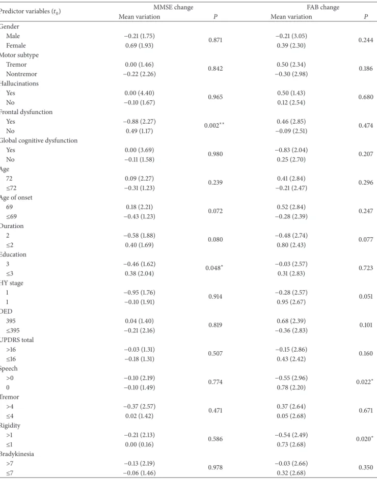
dysfunction and higher education were significantly related to the decreases in the MMSE scores. As chi-square analysis revealed that these two variables were significantly correlated (frontal dysfunction being more frequent in patients with higher education: 57.1% versus 23.1%,𝑃 = 0.010), they were
included as predictor variables in a linear regression analysis model, with the MMSE variation as an outcome variable. This showed that only frontal dysfunction had a significant influence in the MMSE variation (𝑏 = 1.37,𝑃 = 0.003).
Higher rigidity and speech scores were significantly related to the decreases in the FAB scores (Table 5).
Cluster analysis yielded two clusters. Compared to patients in cluster 2 (𝑛 = 35), cluster 1 (𝑛 = 26) patients presented with significantly higher HY stage (𝑃 < 0.00001), UPDRS total score (𝑃 < 0.00001) dysarthria (𝑃 < 0.00001), tremor (𝑃 = 0.019), rigidity (𝑃 = 0.004), bradykinesia (𝑃 < 0.00001), gait and postural instability (𝑃 < 0.00001) scores, and a significantly higher prevalence of the nontremor phenotype (𝑃 < 0.00001). Patients with hallucinations were all included in cluster 1 (𝑃 = 0.029). Patients in cluster 1 showed a more significant decrease in the FAB scores (𝑃 =
0.040). Patients who subsequently developed dementia were all included in cluster 1 (𝑃 = 0.029).
5. Discussion
5.1. Variation of the MMSE and FAB Scores over Time.
Table 5: Baseline comparisons regarding the MMSE and FAB changes from𝑡0to𝑡1.
Predictor variables (𝑡0) MMSE change FAB change
Mean variation 𝑃 Mean variation 𝑃 Gender
Male −0.21 (1.75)
0.871 −0.21 (3.05) 0.244
Female 0.69 (1.93) 0.39 (2.30)
Motor subtype
Tremor 0.00 (1.46)
0.842 0.50 (2.34) 0.186 Nontremor −0.22 (2.26) −0.30 (2.98)
Hallucinations
Yes 0.00 (4.40)
0.965 0.50 (1.43) 0.680
No −0.10 (1.67) 0.12 (2.54)
Frontal dysfunction
Yes −0.88 (2.27)
0.002∗∗ 0.46 (2.85) 0.474
No 0.49 (1.17) −0.09 (2.51)
Global cognitive dysfunction
Yes 0.00 (3.69)
0.980 −0.83 (2.04) 0.207
No −0.11 (1.58) 0.25 (2.70)
Age
72 0.09 (2.27)
0.239 0.41 (2.84) 0.296
≤72 −0.31 (1.23) −0.21 (2.47)
Age of onset
69 0.18 (2.21) 0.072 0.52 (2.84) 0.247
≤69 −0.43 (1.23) −0.28 (2.39)
Duration
2 −0.58 (1.88) 0.080 −0.48 (2.74) 0.077
≤2 0.40 (1.69) 0.80 (2.43)
Education
3 −0.46 (1.62) 0.048∗ −0.03 (2.57) 0.723
≤3 0.38 (2.04) 0.31 (2.83)
HY stage
1 −0.95 (1.76) 0.914 −0.28 (2.57) 0.051
1 −0.10 (1.91) 0.95 (2.67)
DED
395 0.04 (1.40)
0.819 0.68 (2.39) 0.101 ≤395 −0.21 (2.16) −0.36 (2.83)
UPDRS total
>16 −0.03 (1.31)
0.507 −0.15 (2.86) 0.160
≤16 −0.18 (1.31) 0.43 (2.42)
Speech
>0 −0.10 (2.19)
0.774 −0.55 (2.96) 0.022∗
0 −0.10 (1.49) 0.78 (2.20)
Tremor
>4 −0.37 (2.57)
0.471 0.37 (2.64) 0.671
≤4 0.02 (1.42) 0.05 (2.68)
Rigidity
>1 −0.21 (2.13)
0.586 −0.54 (2.49) 0.020∗
≤1 0.00 (0.16) 0.73 (2.68)
Bradykinesia
>7 −0.13 (2.19)
0.978 −0.03 (2.66) 0.350
Table 5: Continued.
Predictor variables (𝑡0) MMSE change FAB change
Mean variation 𝑃 Mean variation 𝑃 Gait/posture
>1 −0.25 (1.70)
0.471 −0.28 (3.03) 0.111
≤1 0.07 (2.00) 0.53 (2.23)
Values are mean standard deviation.𝑃value is significance for Mann-Whitney tests.
annually. More recently, Lessig et al. [40] also found that the MMSE variation was more pronounced after the first 10 years of disease. A third of the patients presented a decline in the MMSE total scores. Improvement in other patients, however, compensated these changes, resulting in almost null mean variation over time. Although the short time interval between evaluations may have promoted a learning effect, contributing to an improvement on follow-up assessment, the different pattern of cognitive change could argue in favor of a heterogeneous progression of neuropsychological deficits in the early stages of PD. It also suggests that the MMSE may not be indicated for detecting cognitive change at these stages of disease, which could require more demanding measures of cognitive dysfunction. Frontal dysfunction progression was also heterogeneous. Significant variations occurred only in relation with lexical fluency, suggesting that this could be fit to measure cognitive changes at these stages of disease. Significant progression in lexical fluency deficits was also found by Azuma et al. [41] and Levy et al. [42].
5.2. Predictors of Dementia. Only four patients presented dementia at follow-up (annual incidence 33.9/1000). Inci-dence was lower than in most studies, in which this figure ranged from 30 to 107/1000 [38,43,44]. This could be due to the specific nature of our cohort, which was constituted only by early stage disease patients and also to the relatively short follow-up time. Previous work has shown that dementia incidence increases as the disease progresses, and the lower values are found in studies performed in early stage patients [45]. This is also in accordance with the wide believed notion that dementia is a late finding in PD.
Nontremor motor scores, nontremor motor phenotype, the presence of hallucinations, and scores below cut-off in the MMSE were related to dementia at follow-up. Significant relation between hallucinations and cognitive dysfunction in PD has been found in several studies [46, 47]. Non-demented PD patients with hallucinations show significant atrophy in frontal and occipitotemporal cortical regions whose progression was linked to cognitive deterioration in longitudinal studies [47], meaning that hallucinations could be a sign of early neuropathological changes leading to dementia in the long run. Nontremor motor phenotype was significantly more frequent in patients that subsequently developed dementia, as also found in previous longitudinal studies [48]. Axial motor dysfunction, like gait and speech disturbances, responds poorly to dopaminergic treatment, and some authors have proposed that cholinergic dysfunction could contribute to these symptoms. PD dementia has also been associated with cholinergic rather than dopaminergic
deficits, and some have suggested that dementia and gait dysfunction could have the same physiopathological basis, originating from cholinergic deficits caused by pedunculo-pontine and of nucleus basalis the Meynert degeneration [42]. Previous work has suggested that the MMSE could be less sensitive to mild cognitive impairment, when compared to frontal oriented tests, like the Montreal cognitive assessment scale [13–15], although it is better for tracking cognitive changes over time in PD [32]. Our data suggests that the MMSE, and particularly the cut-off values, determined in the validation study of the Portuguese version of the test, could be useful for predicting PD dementia in early stage patients. The presence of cognitive deficits in patients that eventually develop a state of full-blown dementia suggests that cognitive dysfunction is a progressive disorder in PD, and that demen-tia, as happens with Alzheimer’s disease, is preceded by a state of subtler neuropsychological dysfunction, compatible with the notion of mild cognitive impairment.
Contrary to the MMSE scores, the FAB scores were not useful in predicting dementia. This could be related to the nature of cognitive deficits in PD-related dementia. Previous investigation has shown a significant relation between the progression to dementia and the superimposition of non-frontal deficits in patients with non-frontal deterioration [49]. In this way, a global, non-frontal test could be more helpful to predict dementia than a frontal-oriented one.
5.3. Predictors of Global and Frontal Cognitive Decline. No variable was predictive of a decline in the MMSE, except for the presence of frontal dysfunction. Although our data does not allow for a straightforward explanation for these findings, we could hypothesize that, frontal dysfunction being the first step in PD-related cognitive deterioration, as mentioned above, it could act as a predictor in some patients of widespread decline of cognitive function. On the other hand, some studies [24, 25] have found significant correlations between FAB and MMSE, which could have biased our results.
the frontal type deficits at the early stages of disease are not related to dementia at follow-up but to COMT gene mutations, that is, to dopamine dysfunction. They hypoth-esized that there could be a dissociation between frontal/ dopaminergic related dysfunction, bearing no relation with dementia at follow-up, and MAPT/ageing/posterior type of cognitive deficits, which could be predictive of dementia. The relation between PD-related dementia and cholinergic dysfunction could explain the improvement of cognitive function in PD patients treated with cholinesterase inhibitors 2.
5.4. Cluster Analysis. Cluster analysis showed two groups that differed regarding the presence of hallucinations and the severity of nontremor motor symptoms. The cluster with worse nontremor symptoms and hallucinations showed a steeper progression of frontal dysfunction and a higher prevalence of dementia, which is in accordance with results from the previous analysis. Comparison with other studies is difficult, because cluster analysis depends on the variables that are included. However, Erro and collaborators [5] did find a cluster in which worse axial motor symptoms are correlated with worse outcome. Lewis et al. also found a group with worse nontremor motor symptoms and significant levels of cognitive impairment [6].
The present study has several limitations, which should be taken into account when discussing the data. Because we assessed an early stage cohort, and the follow-up time was relatively short, the ratio of dementia conversation was very low. This prevents multivariate analysis procedures, weakens the statistical analysis, and precludes definite conclusions regarding the predictive factors for dementia in this group. Patients were recruited from a tertiary centre outpatient clinic, and the sample was small, precluding the generaliza-tion of results to an epidemiological level—it does, however, mirror the clinical experience of a Portuguese neurological center and could be informative in that context.
In conclusion, our data suggests that nontremor motor deficits are predictive factors for dementia and frontal type decline in the early stages of Parkinson’s disease. They also suggest that mini-mental state examination cut-off scores could be useful for predicting dementia in these stages.
References
[1] D. J. Gelb, E. Oliver, and S. Gilman, “Diagnostic criteria for Parkinson’s disease,”Archives of Neurology, vol. 56, no. 1, pp. 33– 39, 1999.
[2] K. R. Chaudhuri, D. G. Healy, and A. H. V. Schapira, “Non-motor symptoms of Parkinson’s disease: diagnosis and manage-ment,”The Lancet Neurology, vol. 5, no. 3, pp. 235–245, 2006. [3] M. A. Hely, W. G. J. Reid, M. A. Adena, G. M. Halliday, and J. G.
L. Morris, “The Sydney multicenter study of Parkinson’s disease: the inevitability of dementia at 20 years,”Movement Disorders, vol. 23, no. 6, pp. 837–844, 2008.
[4] M. Emre, D. Aarsland, A. Albanese et al., “Rivastigmine for dementia associated with Parkinson’s disease,”The New England Journal of Medicine, vol. 351, no. 24, pp. 2509–2518, 2004.
[5] R. Erro, C. Vitale, M. Amboni et al., “The heterogeneity of early Parkinson’s disease: a cluster analysis on newly diagnosed untreated patients,”PLoS ONE, vol. 8, no. 8, Article ID e70244, 2013.
[6] S. J. G. Lewis, T. Foltynie, A. D. Blackwell, T. W. Bobbins, A. M. Owen, and R. A. Barker, “Heterogeneity of Parkinson’s disease in the early clinical stages using a data driven approach,”Journal of Neurology, Neurosurgery and Psychiatry, vol. 76, no. 3, pp. 343–348, 2005.
[7] P. Bugalho and J. Vale, “Brief cognitive assessment in the early stages of parkinson disease,” Cognitive and Behavioral Neurology, vol. 24, no. 4, pp. 169–173, 2011.
[8] M. M. Hoehn and M. D. Yahr, “Parkinsonism: onset, progres-sion and mortality,”Neurology, vol. 17, no. 5, pp. 427–442, 1967. [9] S. Fahn, R. L. Elton, and Members of the UPDRS Development
Committee, “Unified Parkinson’s disease rating scale,” inRecent Developments in Parkinson’s Disease, S. Fahn, C. D. Marsden, D. B. Calne, and M. Goldstein, Eds., pp. 153–163, Macmollan Health Care Information, Florham Park, NJ, USA, 1987. [10] J. Jankovic, M. McDermott, J. Carter et al., “Variable expression
of Parkinson’s disease: a base-line analysis of the DATATOP cohort,”Neurology, vol. 40, no. 10, pp. 1529–1534, 1990. [11] S. G. Parkin, R. P. Gregory, R. Scott et al., “Unilateral and
bilateral pallidotomy for idiopathic Parkinson’s disease: a case series of 115 patients,”Movement Disorders, vol. 17, no. 4, pp. 682–692, 2002.
[12] M. Folstein, S. Folstein, and P. McHugh, ““Mini-mental state”. A practical method for grading the cognitive state of patients for the clinician,”Journal of Psychiatric Research, vol. 12, no. 3, pp. 189–198, 1975.
[13] E. Mamikonyan, P. J. Moberg, A. Siderowf et al., “Mild cog-nitive impairment is common in Parkinson’s disease patients with normal mini-mental state examination (MMSE) scores,”
Parkinsonism and Related Disorders, vol. 15, no. 3, pp. 226–231, 2009.
[14] O. Riedel, J. Klotsche, A. Spottke et al., “Cognitive impairment in 873 patients with idiopathic Parkinson’s disease: results from the German Study on epidemiology of Parkinson’s disease with dementia (GEPAD),”Journal of Neurology, vol. 255, no. 2, pp. 255–264, 2008.
[15] S. Nazem, A. D. Siderowf, J. E. Duda et al., “Montreal cognitive assessment performance in patients with Parkinson’s disease with “normal” global cognition according to mini-mental state examination score,”Journal of the American Geriatrics Society, vol. 57, no. 2, pp. 304–308, 2009.
[16] N. Kandiah, K. Narasimhalu, P.-N. Lau, S.-H. Seah, W. L. Au, and L. C. S. Tan, “Cognitive decline in early Parkinson’s disease,”
Movement Disorders, vol. 24, no. 4, pp. 605–608, 2009. [17] I. Liepelt, M. Reimold, W. Maetzler et al., “Cortical
hypome-tabolism assessed by a metabolic ratio in Parkinson’s disease pri-marily reflects cognitive deterioration—[18F]FDG-PET,” Move-ment Disorders, vol. 24, no. 10, pp. 1504–1511, 2009.
[18] L. N. Williams, P. Seignourel, G. P. Crucian et al., “Laterality, region, and type of motor dysfunction correlate with cognitive impairment in Parkinson’s disease,”Movement Disorders, vol. 22, no. 1, pp. 141–145, 2007.
[19] J. Y. Oh, Y.-S. Kim, B. H. Choi, E. H. Sohn, and A. Y. Lee, “Relationship between clinical phenotypes and cognitive impairment in Parkinson’s disease (PD),”Archives of Gerontol-ogy and Geriatrics, vol. 49, no. 3, pp. 351–354, 2009.
A 6-year prospective study,”Journal of Neurology, vol. 256, no. 10, pp. 1655–1662, 2009.
[21] H. Braak, U. R¨ub, and K. del Tredici, “Cognitive decline correlates with neuropathological stage in Parkinson’s disease,”
Journal of the Neurological Sciences, vol. 248, no. 1-2, pp. 255– 258, 2006.
[22] B. Dubois, D. Burn, C. Goetz et al., “Diagnostic procedures for Parkinson’s disease dementia: recommendations from the movement disorder society task force,”Movement Disorders, vol. 22, no. 16, pp. 2314–2324, 2007.
[23] B. Dubois, E. Tolosa, R. Katzenschlager et al., “Donepezil in Parkinson’s disease dementia: a randomized, double-blind efficacy and safety study,”Movement Disorders, vol. 27, no. 10, pp. 1230–1238, 2012.
[24] B. Dubois, A. Slachevsky, I. Litvan, and B. Pillon, “The FAB: a frontal assessment battery at bedside,”Neurology, vol. 55, no. 11, pp. 1621–1626, 2000.
[25] G. Kenangil, D. N. Orken, E. Ur, and H. Forta, “Frontal assess-ment battery in patients with Parkinson disease in a Turkish population,”Cognitive and Behavioral Neurology, vol. 23, no. 1, pp. 26–28, 2010.
[26] R. Takagi, Y. Kajimoto, S. Kamiyoshi, H. Miwa, and T. Kondo, “The frontal assessment battery at bedside (FAB) in patients with Parkinson’s disease,”No To Shinkei, vol. 54, no. 10, pp. 897– 902, 2002.
[27] E. Guedj, G. Allali, C. Goetz et al., “Frontal assessment battery is a marker of dorsolateral and medial frontal functions: a SPECT study in frontotemporal dementia,”Journal of the Neurological Sciences, vol. 273, no. 1-2, pp. 84–87, 2008.
[28] J. Morgado, C. S. Rocha, C. Maruta, M. Guerreiro, and I. P. Martins, “Novos valores Normativos do mini-mental state examination,”Sinapse, vol. 9, no. 2, pp. 10–16, 2009.
[29] C. F. Lima, L. P. Meireles, R. Fonseca, S. L. Castro, and C. Garrett, “The frontal assessment battery (FAB) in Parkinson’s disease and correlations with formal measures of executive functioning,”Journal of Neurology, vol. 255, no. 11, pp. 1756–1761, 2008.
[30] A. Iavarone, B. Ronga, L. Pellegrino et al., “The frontal assess-ment battery (FAB): normative data from an Italian sample and performances of patients with Alzheimer’s disease and frontotemporal dementia,”Functional Neurology, vol. 19, no. 3, pp. 191–195, 2004.
[31] T. H. Kim, Y. Huh, J. Y. Choe et al., “Korean version of frontal assessment battery: psychometric properties and normative data,”Dementia and Geriatric Cognitive Disorders, vol. 29, no. 4, pp. 363–370, 2010.
[32] A. Siderowf, M. McDermott, K. Kieburtz, K. Blindauer, S. Plumb, and I. Shoulson, “Test-retest reliability of the unified Parkinson’s disease rating scale in patients with early Parkinson’s disease: results from a multicenter clinical trial,” Movement Disorders, vol. 17, no. 4, pp. 758–763, 2002.
[33] M. P. McDermott, J. Jankovic, J. Carter et al., “Factors predictive of the need for levodopa therapy in early, untreated Parkinson’s disease. The Parkinson Study Group,”Archives of Neurology, vol. 52, no. 6, pp. 565–570, 1995.
[34] O. Spreen and E. Strauss,A Compendium of Neuropsychological Tests: Administration, Norms and Commentary, Oxford Univer-sity press, New York, NY, USA, 1998.
[35] D. Aarsland, K. Andersen, J. P. Larsen et al., “The rate of cognitive decline in Parkinson disease,”Archives of Neurology, vol. 61, no. 12, pp. 1906–1911, 2004.
[36] American Psychiatric Association, Diagnostic and Statistical Manual of Mental Disorder, American Psychological Associa-tion, Washington, DC, USA, 4th ediAssocia-tion, 1994.
[37] N. Kandiah, K. Narasimhalu, P.-N. Lau, S.-H. Seah, W. L. Au, and L. C. S. Tan, “Cognitive decline in early Parkinson’s disease,”
Movement Disorders, vol. 24, no. 4, pp. 605–608, 2009. [38] D. Aarsland, G. Muniz, and F. Matthews, “Nonlinear decline of
mini-mental state examination in Parkinson’s disease,” Move-ment Disorders, vol. 26, no. 2, pp. 334–337, 2011.
[39] C. H. Williams-Gray, J. R. Evans, A. Goris et al., “The distinct cognitive syndromes of Parkinson’s disease: 5 year follow-up of the CamPaIGN cohort,”Brain, vol. 132, no. 11, pp. 2958–2969, 2009.
[40] S. Lessig, D. Nie, R. Xu et al., “Changes on brief cognitive instru-ments over time in Parkinson’s disease,”Movement Disorders, vol. 27, no. 9, pp. 1125–1128, 2012.
[41] T. Azuma, R. F. Cruz, K. A. Bayles, C. K. Tomoeda, and E. B. Montgomery Jr., “A longitudinal study of neuropsychological change in individuals with Parkinson’s disease,”International Journal of Geriatric Psychiatry, vol. 18, no. 12, pp. 1115–1120, 2003. [42] G. Levy, M.-X. Tang, L. J. Cote et al., “Motor impairment in PD: relationship to incident dementia and age,”Neurology, vol. 55, no. 4, pp. 539–544, 2000.
[43] D. Aarsland, K. Andersen, J. P. Larsen, A. Lolk, H. Nielsen, and P. Kragh-Sørensen, “Risk of dementia in Parkinson’s disease: a community-based, prospective study,”Neurology, vol. 56, no. 6, pp. 730–736, 2001.
[44] C. H. Williams-Gray, T. Foltynie, C. E. G. Brayne, T. W. Robbins, and R. A. Barker, “Evolution of cognitive dysfunction in an incident Parkinson’s disease cohort,”Brain, vol. 130, no. 7, pp. 1787–1798, 2007.
[45] E. Y. Uc, M. P. McDermott, K. S. Marder et al., “Incidence of and risk factors for cognitive impairment in an early parkinson disease clinical trial cohort,”Neurology, vol. 73, no. 18, pp. 1469– 1477, 2009.
[46] G. F´enelon, F. Mahieux, R. Huon, and M. Zi´egler, “Hallucina-tions in Parkinson’s disease. Prevalence, phenomenology and risk factors,”Brain, vol. 123, no. 4, pp. 733–745, 2000.
[47] N. Ibarretxe-Bilbao, B. Ramirez-Ruiz, C. Junque et al., “Dif-ferential progression of brain atrophy in Parkinson’s disease with and without visual hallucinations,”Journal of Neurology, Neurosurgery and Psychiatry, vol. 81, no. 6, pp. 650–657, 2010. [48] G. Alves, J. P. Larsen, M. Emre, T. Wentzel-Larsen, and D.
Aarsland, “Changes in motor subtype and risk for incident dementia in Parkinson’s disease,”Movement Disorders, vol. 21, no. 8, pp. 1123–1130, 2006.