DOI: 10.1590/0004-282X20160014
ARTICLE
Work and power reduced in L-dopa naïve patients
in the early-stages of Parkinson’s disease
Trabalho e potência reduzidos em indivíduos com doença de Parkinson sem L-dopa nos
estágios iniciais
Lidiane Oliveira Lima1, Francisco Cardoso2, Luci Fuscaldi Teixeira-Salmela3, Fátima Rodrigues-de-Paula3
Parkinson’s disease (PD) is a neuro-degenerative disease in which the progressions of the symptoms are associated with progressive loss of strength and power, which leads to deterioration of physical abilities1,2. Studies that employed
measures of peak torque have observed muscular deicits in
individuals at various stages of PD, mainly in the intermedi-ate and advanced phases3,4. Usually, these individuals
dem-onstrated bilateral motor impairments, which are associated
with deicits in balance and gait and may contribute to im -mobility and poorer functional performance5.
Recently, studies have found that in the early stages, individ-uals with PD already demonstrate cognitive decline6, impaired
planning7, altered dynamic postural control8, and functional
losses when compared to those without the disease9. In
ad-dition to these impairments, it is possible that muscle dei -cits may also contribute to the functional losses. However, few studies have investigated muscular performance in individu-als in the early stages of PD. Bridgewater and Sharpe10 showed
decreases in trunk extensor torque during the early-stages of
PD, which could contribute to the lexed posture observed in
the advanced stages. Koller and Kase11 observed decreases in
the maximal isotonic knee strength of individuals in the ear-ly stages of the disease; however, the hip and ankle muscular groups were not assessed. Furthermore, several studies have
1Universidade Federal do Ceará, Departamento de Fisioterapia, Fortaleza CE, Brazil;
2Universidade Federal de Minas Gerais, Departamento de Clínica Médica, Belo Horizonte MG, Brazil;
3Universidade Federal de Minas Gerais, Departamento de Fisioterapia, Belo Horizonte MG, Brazil.
Correspondence: Fátima Rodrigues-de-Paula; Departamento de Fisioterapia da Universidade Federal de Minas Gerais; Avenida Antônio Carlos, 6627; 31270-901 Belo Horizonte MG, Brasil; E-mail: [email protected]
Conflict of interest: There is no conflict of interest to declare.
Received 05 May 2015; Received in final form 29 October 2015; Accepted 30 November 2015. ABSTRACT
Studies which have investigated muscular performance during the initial stages of Parkinson´s disease (PD) without L-dopa treatments were not found. Objective: to assess whether muscular performance, work and power, of the trunk and lower limbs in L-dopa naïve patients in the early stages of PD was lower than those of healthy subjects and to compare muscular performance between the lower limbs.
Method: Ten subjects with PD, Hoehn and Yahr (HY) I-II, L-dopa naïve and 10 subjects in the control group were assessed with the isokinetic dynamometer. Results: ANOVAs revealed that work and power measures of the trunk, hip, knee, and ankle muscular groups were lower in PD compared with the control group (p < 0.05). There were no significant differences in muscular performance between the lower limbs.
Conclusion: The results suggested the use of specific exercises, as rehabilitation strategies, to improve the ability to produce work and power with this population.
Keywords:Parkinson’s disease; muscle strength; Muscle strength dynamometer; rehabilitation; physical therapy speciality.
RESUMO
Estudos que investigaram o desempenho muscular durante os estágios iniciais da doença de Parkinson (DP), sem tratamento com L-dopa não foram encontrados. Objetivo: Avaliar se o desempenho muscular, por meio de medidas de trabalho e potência, do tronco e dos membros inferiores em pacientes sem o uso de L-dopa nas fases iniciais da DP é menor do que o de indivíduos saudáveis e comparar o desempenho muscular entre os membros inferiores. Método: Dez indivíduos com DP, Hoehn and Yahr (HY) I-II, sem L-dopa e 10 indivíduos do grupo controle foram avaliados com o dinamômetro isocinético. Resultados: Medidas de trabalho e potência muscular do tronco, quadril, joelho, tornozelo foram menores no PD em comparação com o grupo controle (p < 0,05) e não houve diferenças significativas no desempenho muscular entre os membros inferiores. Conclusão: O uso de exercícios específicos, como estratégias de reabilitação, pode melhorar a capacidade de produzir trabalho e potência muscular nesta população.
indicated torque deicits with signiicant diferences between the most- and least-afected lower limbs in the advanced stag -es of PD5,12. However, these results are controversial regarding
the distribution of the weaknesses between the lower limbs in the early stages of PD5,13.
Isokinetic analyses of muscular performance in individu-als with PD generally considered only peak torque measures. However, work has been recognized as a more representative measure for the execution of various motor activities, such as gait14. Some studies have suggested that measures of power are
important indicators of muscular performance in PD because they take into account the time to reach peak torque, since
bra-dykinesia is an important deicit observed in these individuals 1,2.
It is important to note that these studies were in patients with L-dopa treatment. However, few studies have been con-ducted about newly diagnosed, L-dopa naïve patients and their muscular performance in relation to the variable of work and power. Felows et al. showed exaggerated grip force levels in the early stages of PD, in patients with no exposure to L-dopa medication. However, the patients developed grip force markedly slower than did the control subjects15.
Given the short- and long-term complications in
per-formance of functional tasks afecting individuals with
Parkinson’s disease, the current research supports the de-livery of rehabilitation interventions early in the disease’s progress. Studies suggested that rehabilitation can maintain mobility and prevent secondary impairments of neuromus-cular systems associated with reduced physical activity5,16.
herefore, the analysis of muscular performance in the early
stages may help to identify earlier, prevent, or delay muscular abnormalities and decline.
Hence, the main purpose of the present study is to evaluate and compare muscular performance by measuring work and power of the trunk and lower limbs, between individuals in the early stages of PD without L-dopa treatments and individuals
without PD. Speciically, the aims were to investigate if there are signiicant diferences between work and power measures
of the trunk, hip, knee, and ankle muscular groups within
indi-viduals between the most and least afected lower limbs. In PD, a nigral dopaminergic deicit results in reduction of
the excitatory drive to the motor cortex and disruption of the cortical activation of the muscle17 and may manifest as
bra-dykinesia and muscle weakness. herefore, if muscular per -formance in the early stages of PD without L-dopa treatment is decreased, it may be treated and loss and disability in the advanced stages may be decreased.
METHOD
Subjects
his was a two-group comparison study, where partici -pants with PD were compared with individuals without PD in
a control group. he study comprised a convenience sample
including 10 individuals without PD recruited in the community and 10 individuals with PD recruited at the Movement Disorder Clinic of the University Hospital during medical appointments. Outcomes were measured by the research assistant who was blinded to recruitment and the aims of the study. Individuals with PD were diagnosed by movement disorder neurologists ac-cording to the United Kingdom Brain Bank criteria.
he PD group was comprised of 10 individuals (eight men
and two women), who were at stages one to two of the
modi-ied Hoehn and Yahr (HY) scale18. hese individuals had nev
-er taken L-dopa (L-dopa naïve) medication; howev-er, they were taking other drugs, such as dopaminergic agonists
and amantadine. Five individuals with PD were classiied in stage 2, two in stage 1, and three in stage 1.5 of the HY modi
-ied scale. Regarding their medications, two subjects did not
use any medications, three were using dopamine agonists,
and ive were taking dopamine agonists plus amantadine. he control group consisted of 10 subjects without PD,
matched by age, gender, physical activity, and body mass
index (BMI). Persons were classiied as physical activity ac -cording to Physical Activity Trends19. Subjects were excluded
if they had other neurological or systemic disorders; histories of trunk, knee, hip, and ankle surgeries; and detectable cogni-tive impairments, as determined by their Mini-Mental State Examination scores < 2420. he study was approved by the in
-stitutional ethical review committee and all participants pro-vided their consent.
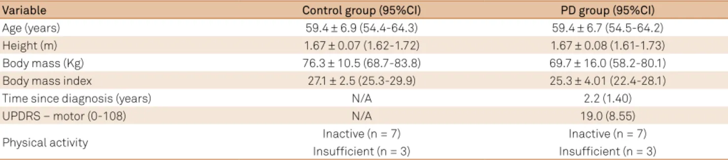
here were no signiicant diferences between the groups
for the variables related to age, height, BMI (Table 1). In both groups, seven subjects were inactive and three patients re-ported some activity during the preceding month but not
enough to be classiied as moderate or vigorous. So, they were classiied as insuicient19.
Procedures
he PD participants were assessed approximately one
hour after taking their usual medications. Initially, demo-graphic and anthropometric data related to age, body mass, physical activity and height, were obtained for character-ization purposes for both groups. Clinical information was also collected regarding their motor capacity levels and
ac-tivity of daily living of the Uniied Parkinson’s Disease Rating
Scale (UPDRS)21.
Outcome measures
he muscular performance of both groups was assessed with the isokinetic Biodex System 3 Pro (Biodex, Shirley, NY).
modes. hese angular speeds were chosen, since they have
commonly been used in previous studies in individuals with PD4,11,12 and are more comprehensive performance indicators.
For the trunk, the evaluated speed was 120°/s.
he range of motion was determined for each joint and
the participants were familiarized with the equipment and procedures, by performing three sub-maximal repetitions
be-fore the evaluations. he tests consisted of ive maximal rep -etitions at 30°/s and 10 rep-etitions at 90°/s, with a rest period of 1.5 minutes between each evaluated speed. For the trunk, the subjects performed the same familiarization procedures
and ive repetitions were assessed at the determined speed. A ive to seven minute rest interval was given between each joint evaluation. hroughout the assessments, the subjects
received verbal encouragement to perform with their
great-est possible eforts. he variables selected for analyses were
work, in Joules and power, in Watts, both normalized by the subjects’ body mass. Work was calculated as torque x
angu-lar displacement and relects the ability to produce and sus -tain torque throughout a determined range of motion. Power
measurement relects how much force the participant can
generate for a given speed of contraction.
Data analyses
Descriptive statistics and tests for normality (Shapiro-Wilk) were carried out for all variables. Multifactorial repeat-ed measure analyses of variance (ANOVA 2x2), followrepeat-ed by planned contrasts were employed to investigate the main
and interaction efects between the PD and control groups
and the sides for the outcome variables of work and power,
with a signiicance level of αcan 0.05. he Mann-Whitney-U test was employed to investigate diferences between the
groups regarding muscular performance of the trunk.
RESULTS
Isokinetic work
Work measures of the trunk, hip, knee, and ankle lexors
and extensors for the PD and control groups are shown in
Table 2. he PD group generated less work than the control
group for the trunk lexion and extension movements and demonstrated signiicant decreases in the work of the hip lexor/extensors, knee lexors, and ankle plantar lexors at all evaluated speeds. For the knee extensors, signiicant difer
-ences were observed only at the speed of 90°/s. No signii
-cant diferences were observed for the ankle dorsilexors for
all evaluated speeds.
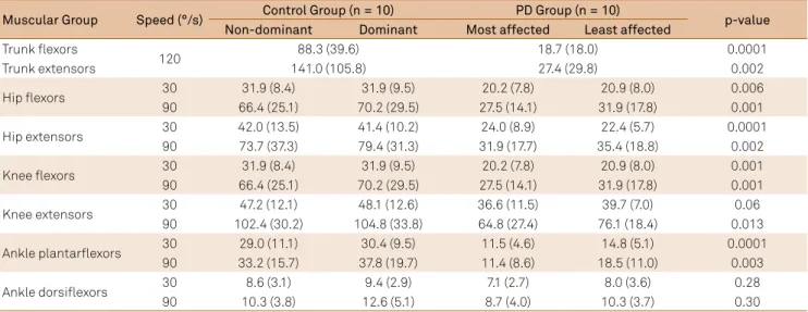
Isokinetic power
Table 3 shows the power values generated by the trunk
and lower limb joints. he PD group also generated less pow
-er than the control group for the trunk lexion and extension movements and demonstrated signiicant decreases in pow
-er of the hip lexor/extensors, knee lexors, and ankle plantar lexors for all evaluated speeds. For the knee extensors, sig
-niicant diferences were observed only at the speed of 90°/s. No signiicant diferences were observed for the ankle dorsi
-lexors for all evaluated speeds.
DISCUSSION
In the present study, L-dopa naïve individuals in the early stages of PD demonstrated poorer muscular performance of the trunk and lower limbs, when compared with those
with-out the disease. hese indings are important, since previous studies have reported torque deicits, mainly, in individuals
in the intermediate and advanced stages of the disease3,4.
he muscular performance variables in the early-stages of
PD have been scarcely investigated and the studies which analyzed these parameters10,11 did not control for the
physi-cal activity and the participants were using L-dopa therapy,
which may have afected their results. he efects of levodopa
in improving muscular strength were previously reported22.
Individuals with PD included in the present study were in
the typical early stages of the disease (HY I to II), did not use
levodopa-based medications, and had physical activity
simi-lar to the control group. he use of well-established inclusion
criteria and the process of matching the groups allowed the
detection of deicits in muscular performance of the trunk
and lower limbs in the early stages of PD.
Table 1. Descriptive statistics and comparisons between the control and PD groups (n = 10) regarding their demographic,
anthropometric, and clinical variables. Means ± standard deviations (95% confidence intervals).
Variable Control group (95%CI) PD group (95%CI)
Age (years) 59.4 ± 6.9 (54.4-64.3) 59.4 ± 6.7 (54.5-64.2)
Height (m) 1.67 ± 0.07 (1.62-1.72) 1.67 ± 0.08 (1.61-1.73)
Body mass (Kg) 76.3 ± 10.5 (68.7-83.8) 69.7 ± 16.0 (58.2-80.1)
Body mass index 27.1 ± 2.5 (25.3-29.9) 25.3 ± 4.01 (22.4-28.1)
Time since diagnosis (years) N/A 2.2 (1.40)
UPDRS – motor (0-108) N/A 19.0 (8.55)
Physical activity Inactive (n = 7) Inactive (n = 7)
Insufficient (n = 3) Insufficient (n = 3)
he PD group demonstrated signiicantly decreased ca -pacities to generate work and power, compared to the control group in all tested speeds. Unfortunately, comparable studies concerning isokinetic work with PD subjects were not found.
Similar to work, measures of power were also signiicantly low -er in the PD group. Measurement of pow-er using isokinetic
dy-namometry relects how much force the participant can gen -erate for a given speed of contraction. It is, therefore, possible to argue that individuals with PD were not able to produce torque through range at that low and high speed.
he PD group demonstrated poorer muscular perfor -mance for all muscular groups of the trunk and hip for all investigated speeds. Bridgewater and Sharpe10 found
sig-niicant decreases in trunk extensor torques and suggested
that there were greater axial strength declines in subjects
with PD in the early stages. However, they did not assess the lower limb muscles. Another study, which analyzed peak torque measures of the trunk and lower limbs, also report-ed poorer performance of the proximal muscles in relation
to the distal ones in individuals mildly afected by PD23. In
the present study, knee extensors at 30°/s and ankle
dorsi-lexors at both speeds demonstrated no signiicant diferenc -es in performance, compared to the control group. However,
these indings could be attributed to the small sample size,
which might have increased the likelihood of type-II errors. Other responsible factors were not directly determined but may be explained by age and disease-associated
impair-ments. he selective atrophy of type II ibres with advancing
age may partly explain poorer performance at high speeds24.
Interestingly, muscles biopsies from persons with PD have
Table 2. Isokinetic work, in Joules/Kg, produced by the trunk and lower limb muscular groups of the control and PD groups. Means (standard deviations).
Muscular Group Speed (°/s) Control group (n = 10) PD group (n = 10) p-value
Non-dominant Dominant Most affected Least affected
Trunk flexors
120 74. 6 (25.4) 20.5 (15.8) 0.0001
Trunk extensors 111.9 (72.2) 27.7 (26.8) 0.002
Hip flexors 30 133.1 (31.4) 145.3 (39.8) 100.1 (26.2) 102.5 (26.0) 0.009
90 111.9 (34.2) 118.7 (37.7) 57.2 (19.6) 64.6 (23.7) 0.001
Hip extensors 30 152.7 (40.3) 156.1 (30.4) 115.7 (43.4) 100.2 (33.0) 0.008
90 125.9 (56.3) 133.8 (55.9) 64.9 (38.2) 69.0 (40.1) 0.009
Knee flexors 30 106.6 (18.9) 107.7 (21.8) 76.6 (25.5) 83.9 (25.9) 0.014
90 85.2 (21.5) 92.5 (19.6) 45.9 (19.2) 50.3 (20.2) 0.0001
Knee extensors 30 192.7 (38.2) 196.2 (36.0) 155.7 (48.6) 167.9 (29.8) 0.064
90 164.4 (37.5) 169.2 (38.8) 114.1 (37.4) 129.0 (28.5) 0.007
Ankle plantarflexors 30 66.3 (17.8) 68.3 (16.6) 37.6 (17.7) 47.0 (21.8) 0.005
90 36.9 (13.7) 41.5 (16.3) 19.8 (9.1) 27.6 (16.3) 0.017
Ankle dorsiflexors 30 21.7 (8.3) 21.6 (4.8) 18.4 (6.7) 17.8 (5.7) 0.18
90 11.8 (1.8) 13.5 (4.6) 10.1 (4.1) 11.8 (3.4) 0.26
p-value: differences between the groups.
Table 3. Isokinetic power, in Watts/Kg produced by the trunk and lower limb muscular groups of the control and PD groups. Means (standard deviations).
Muscular Group Speed (°/s) Control Group (n = 10) PD Group (n = 10) p-value
Non-dominant Dominant Most affected Least affected
Trunk flexors
120 88.3 (39.6) 18.7 (18.0) 0.0001
Trunk extensors 141.0 (105.8) 27.4 (29.8) 0.002
Hip flexors 30 31.9 (8.4) 31.9 (9.5) 20.2 (7.8) 20.9 (8.0) 0.006
90 66.4 (25.1) 70.2 (29.5) 27.5 (14.1) 31.9 (17.8) 0.001
Hip extensors 30 42.0 (13.5) 41.4 (10.2) 24.0 (8.9) 22.4 (5.7) 0.0001
90 73.7 (37.3) 79.4 (31.3) 31.9 (17.7) 35.4 (18.8) 0.002
Knee flexors 30 31.9 (8.4) 31.9 (9.5) 20.2 (7.8) 20.9 (8.0) 0.001
90 66.4 (25.1) 70.2 (29.5) 27.5 (14.1) 31.9 (17.8) 0.001
Knee extensors 30 47.2 (12.1) 48.1 (12.6) 36.6 (11.5) 39.7 (7.0) 0.06
90 102.4 (30.2) 104.8 (33.8) 64.8 (27.4) 76.1 (18.4) 0.013
Ankle plantarflexors 30 29.0 (11.1) 30.4 (9.5) 11.5 (4.6) 14.8 (5.1) 0.0001
90 33.2 (15.7) 37.8 (19.7) 11.4 (8.6) 18.5 (11.0) 0.003
Ankle dorsiflexors 30 8.6 (3.1) 9.4 (2.9) 7.1 (2.7) 8.0 (3.6) 0.28
90 10.3 (3.8) 12.6 (5.1) 8.7 (4.0) 10.3 (3.7) 0.30
shown increased type-I ibres and decreased type-II25. In
ad-dition, patients with PD exibit multiple agonist bursting dur-ing the acceleration phase of movement and increased an-tagonistic movement26. hese abnormal activation patterns
may result in the prolonged planning of the tested
move-ment, contributing to the lack of diferences it a slower rate
in the knee extensors. In the ankle, age induces neural chang-es as decline of 39% in chang-estimated motor unit number in the tibial anterior. However, this can be compensated by
collat-eral reinnervation of muscle ibers and larger size of the re -maining motor untis24. So, this could contribute to a better
ankle performance compared to the hip. Nallegowda et al.27
also observed no diferences in ankle dorsilexor peak torque
values between the groups at 90°/s, 120º/s, and 150º/s speeds
for individuals with mild PD. Future studies aiming to better evaluate the relationships between axial and distal muscles in PD should include a larger sample.
he comparisons between the lower limbs showed sym -metrical performance for both groups, regardless of the
eval-uated speeds. Previous studies demonstrated greater deicits in peak torque of the knee extensor and lexor muscles of the most afected limbs in the early-stages of PD patients during
high12,13 and low12 speeds. However, some participants used
levodopa-based drugs, while others did not take any medica-tions. It is possible that this heterogeneity of medications may have created a tendency for lower limb asymmetries. Pedersen and Oberg28 showed that the observed diferences in peak
torques between the lower limbs of individuals in the early-stages of PD, disappeared after the withdrawal of the
medica-tions. hey reported that the decreases in peak torque were the same for both limbs without medication efects and were not associated with the most afected side. Corroborating
the results of the present study, Nogaki et al.3 also observed
no diferences in knee extension and lexion peak torque val -ues between the lower limb muscular groups at 30°/s, 90º/s,
and 180º/s speeds for individuals with mild PD. he observed
asymmetries occurred at high speeds and with individuals in the advanced stages3. Asymmetrical depletion of dopamine
in the substantia nigra29 is associated with asymmetric
mo-tor features of PD. Frazzita et al. showed that Parkinsonian
pa-tients in stage 3 HY exhibited muscular weakness on the right
side, but not on the left side, as compared to controls30. he
observed absence of asymmetries in muscular performance between the lower limbs in the early-stages suggests that such asymmetries could be particularly found in the intermediate and advanced stages of PD. We hypothesized that the deple-tion of dopamine and some factors such as rigidity, tremor,
bradykinesia and executive deicits that would interfere with
the ability to rapidly generate appropriate torques may not be so exacerbated as in the later stages of PD. Furthermore, it is important to recognize that dynamic postural control during turning is altered even in the early stages of PD. In our study, we decided investigated hip, knee, ankle, and spinal groups of
muscles because these are the major muscle groups that con-trol locomotion and posture.
It has been suggested that isokinetic methods may be
in-luenced by bradykinesia, especially, at high angular speeds3.
Furthermore, this test speed may require a smaller motor
plan-ning time, the processing of which is classically afected in PD7.
hese might represent limitations of the present study. he present indings can be relevant to clinical practice.
From a rehabilitation perspective, it is critical that prevention is the preferred strategy31. Giladi suggested that a person with
movement disorders will develop gait deicit only if all com -pensatory reserves have been depleted31. Prevention
repre-sents strengthening of the cognitive, sensory and motor com-pensatory reserves31. In this case, the aims of the treatment
should be at decreasing disability by preserving and improving
the function of compromised or diseased systems. his may
be act to maintain the motor system in the advanced stages of the disease31. Additionally, according to the physical therapy
international guidelines, one of the goals of physical therapy in the early stages is preserving or improving physical capac-ity (aerobic capaccapac-ity, muscle strength, and joint mobilcapac-ity)5. he
decreased work and power generation observed in the early stages of PD suggest the need for early interventions that im-prove the muscular performance of these individuals. Recently, Corcos et al.32 observed improve in muscular strength and a
7.3 points decrease in UPDRS-III scores, which is a moderate clinically important change, after a 24-month progressive re-sistance exercise program (PRE). PRE progressively increased the resistance over time and each repetition lasted 6-9 sec-onds, that is, at slow speeds32. he authors hypothesized that
PRE may lead to experience-dependent plasticity in the basal ganglia and corticomotor pathways, which could contribute to improving signs and motor performance. On other hand, evidence that resistance training during which the concentric component of the movement is performed as quickly as pos-sible may be critical for gains in power and recovering quickly from a loss of balance in order to avoid falling in people with Parkinson’s disease2. Our indings suggest that the incorpora
-tion of higher speed muscular training sessions within a PRE program will be an optimal training method to integrate the various aspects of the neuromuscular system in early stages of the disease. In summary, this training regime can bring about changes in the motor sign, work and power measures, physi-cal in the early stages of neurodegenerative movement disor-ders maintaining patient mobility and independence in the advanced stages of movement disorders.
References
1. Allen NE, Canning CG, Sherrington C, Fung VSC. Bradykinesia, muscle weakness and reduced muscle power in Parkinson’s disease. Mov Disord. 2009;24(9):1344-51. doi:10.1002/mds.22609
2. Allen NE, Sherrington C, Canning CG, Fung VSC. Reduced muscle power is associated with slower walking velocity and falls in people with Parkinson’s disease. Parkinsonism Relat Disord. 2010;16(4):261-4. doi:10.1016/j.parkreldis.2009.12.011
3. Nogaki H, Kakinuma S, Morimatsu M. Movement velocity dependent muscle strength in Parkinson´s disease. Acta Neurol Scand. 1999;99(3):152-7. doi:10.1111/j.1600-0404.1999.tb07337.x
4. Nogaki H, Kakinuma S, Morimatsu M. Muscle weakness in Parkinson’s disease: a follow-up study. Parkinsonism Relat Disord. 2001;8(1):57-62. doi:10.1016/S1353-8020(01)00002-5
5. Keus SHJ, Munneke M, Graziano M, Paltamaa J, Pelosin E, Domingos J, Brühlmann S, Ramaswamy B, Prins J, Struiksma C, Rochester L, Nieuwboer A, Bloem B. European physiotherapy Guideline for Parkinson’s disease. ParkinsonNet. 2014, the Netherlands. http://parkinsonnet.info/media/14741616/eu_guideline_ parkinson_201412_clinicians.pdf
6. Bugalho P, Viana-Baptista M. Predictors of cognitive decline in the early stages of Parkinson’s disease: a brief cognitive assessment longitudinal study. Parkinson’s Disease; 2013;2013:ID912037. doi:10.1155/2013/912037
7. Trujillo JP, Gerrits NJ, Vriend C, Bendse HW, Heuvel OA, Werf YD. Impaired planning in Parkinson’s disease is reflected by reduced brain activation and connectivity. Hum Brain Mapp. 2015;36(9):3703-15. doi:10.1002/hbm.22873
8. Song J, Sigward S, Fisher B, Salem GJ. Altered dynamic postural control during step turning in persons with early-stage Parkinson’s disease. Parkinson Dis. 2012;2012:ID386962. doi:10.1155/2012/386962
9. Carpinella I, Crenna P, Calabrese E, et al. Locomotor function in the early stage of Parkinson’s disease. IEEE Trans Neural Syst Rehabil Eng. 2007;15(4):543-51. doi:10.1109/TNSRE.2007.908933
10. Bridgewater KJ, Sharpe MH. Trunk muscle performance in early Parkinson’s disease. Phys Ther. 1998; 78(6):566-76.
11. Koller W, Kase S. Muscle strength testing in Parkinson´s disease. Eur Neurol. 1986;25(2):130-3. doi:10.1159/000115998
12. Kakinuma S, Nogaki H, Pramanik B, Morimatsu M. Muscle weakness in Parkinson’s disease: isokinetic study of the lower limbs. Eur Neurol. 1998;39(4):218-22. doi:10.1159/000007937
13. Nogaki H, Fukusako T, Sasabe F, Negoro K, Morimatsu M. Muscle strength in early Parkinson’s Disease. Mov Disord. 1995;10(2):225-6. doi:10.1002/mds.870100218
14. Charteris J. Effects of velocity on upper to lower extremity muscular work and power output ratios of intercollegiate athletes. Br J Sports Med. 1999;33(4):250-4. doi:10.1136/bjsm.33.4.250
15. Fellows SJ, Noth J. Grip force abnormalities in de novo Parkinson’s disease. Mov Disord. 2004;19(5):560-5. doi:10.1002/mds.10710
16. Canning CG. Rehabilitation in Parkinson’s disease: the challenge to provide early and ongoing, evidence-based, patient-centred care. Arq Neuropsiquiatr. 2013;71(12):917-9. doi:10.1590/0004-282X20130228
17. DeLong MR. Primate models of movement disorders of basal ganglia origin. Trends Neurosci. 1990;13(7):281-5. doi:10.1016/0166-2236(90)90110-V
18. Goetz CG, Poewe W, Rascol O, Sampaio C, Stebbins GT, Counsell C et al. Movement Disorder Society Task Force report on the Hoehn and Yahr staging scale: status and recommendations. Mov Disord. 2004;19(9):1020-8. doi:10.1002/mds.20213
19. Centers for Disease Control and Prevention. Physical activity trends: United States (1990-1998). MMWR. 2001;50(9):166-9.
20. Folstein MF, Folstein SE, McHugh PR. “Mini Mental State”: a practical method for grading the cognitive state of patients for the clinican. J Psychiatr Res. 1995;12(3):189-98. doi:10.1016/0022-3956(75)90026-6
21. Fahn S, Elton R. Unified Parkinson’s disease rating scale. In: Fahn S, Marsden CD, Calne DB, Goldstein M, eds. Recent Developments in Parkinson’s Disease, Vol 2. Florham Park, NJ. Macmillan Health Care Information 1987, 153-163, 293-304.
22. Corcos DM, Chen CM, Quinn NP, McAuley J, Rothwell JC. Strength in Parkinson’s disease: relationship to rate of force generation and clinical status. Ann Neurol. 1996;39(1):79-88. doi:10.1002/ana.410390112
23. Inkster LM, Eng JJ, MacIntyre DL, Stoessl AJ. Leg muscle strength is reduced in Parkinson’s disease and relates to the ability to rise from a chair. Mov Disord. 2003;18(2):157-62. doi:10.1002/mds.10299
24. Papegaaij S, Taube W, Baudry S, Otten E, Hortobágyi T. Aging causes a reorganization of cortical and spinal control of posture. Fr.ont Aging Neurosci. 2014;6:28. doi:10.3389/fnagi.2014.00028
25. Rossi B, Siciliano G, Carboncini MC, Manca ML, Massetani R, Viacava P et al. Muscle modifications in Parkinson’s disease: myoelectric manifestations. Electroencephalogr Clin Neurophysiol. 1996;101(3):211-8. doi:10.1016/0924-980X(96)94672-X
26. Moreno Catalá M, Woitalla D, Arampatzis A. Central factors explain muscle weakness in young fallers with Parkinson’s disease. Neurorehabil Neural Repair. 2013;27(8):753-9. doi:10.1177/1545968313491011
27. Nallegowda M, Singh U, Handa G, Khanna M, Wadhwa S, Yada SL et al. Role of sensory input and muscle strength in maintenance of balance, gait, and posture in Parkinson’s disease. Am J Phys Med Rehabil. 2004;83(12):898-908. doi:10.1097/01.PHM.0000146505.18244.43
28. Pedersen SW, Öberg B. Dynamic strength in Parkinson’s disease: Quantitative measurements following withdrawal of medication. Eur Neurol. 1992;33(2):97-102. doi:10.1159/000116913
29. Kempster PA, Gibb WR, Stern GM, Lees AJ. Asymmetry of substantia nigra neuronal loss in Parkinson’s disease and its relevance to the mechanism of levodopa related motor fluctuations. J Neurol Neurosurg Psychiatry. 1989;52(1):72-6. doi:10.1136/jnnp.52.1.72
30. Frazzitta G, Ferrazzoli D, Maestri R, Rovescala R, Guaglio G, Bera R et al. Differences in muscle strength in parkinsonian patients affected on the right and left side. PLoS One. 2015;10(3):E0121251. doi:10.1371/journal.pone.0121251
31. Giladi N. Mobility and exercise in movement disorders. Parkinsonism Relat Disord. 2009;15 Suppl 3:S46-8. doi:10.1016/S1353-8020(09)70033-1