GENOTYPIC AND PHENOTYPIC DETECTION OF CAPSULAR POLYSACCHARIDES IN STAPHYLOCOCCUS AUREUS ISOLATED FROM BOVINE INTRAMAMMARY INFECTIONS IN ARGENTINA
C. Camussone1, P. Rejf 3, N. Pujato2, A. Schwab2, I. Marcipar2, L.F. Calvinho1,3*
1
Estación Experimental Agropecuaria Rafaela, INTA. Ruta 34, Km 227, (2300) Rafaela, Santa Fe, Argentina; 2Facultad de
Bioquímica y Ciencias Biológicas, Universidad Nacional del Litoral, Paraje El Pozo, CC242, (3000) Santa Fe, Argentina; 3
Facultad de Ciencias Veterinarias, Universidad Nacional del Litoral, Rvdo. Padre Kreder 2805, (3080) Esperanza, Santa Fe,
Argentina.
Submitted: February 28, 2011; Returned to authors for corrections: September 19, 2011; Approved: June 07, 2012.
ABSTRACT
Staphylococcus aureus (n=157) isolated from intramammary infections in Argentine dairy areas were
evaluated for presence of cap5 and cap8 loci. Isolates carrying cap5 and cap8 were serotyped using specific
antisera. Sixty four percent of the isolates were genotyped as cap5 or cap8 and 50% of them expressed CP5
or 8.
Key words:Staphylococcus aureus, capsular polysaccharides, bovine mastitis
Staphylococcus aureus capsular polysaccharides have
been shown to confer resistance to phagocytosis by
polymorphonuclear neutrophils (PMN), which are considered
the main mammary gland line of defense against invading
pathogens (5). Conversely, antibodies against CPs have a
protective effect since they can opsonize encapsulated S.
aureus from bovine origin for phagocytic killing by PMN (5).
The existence of 11 CP serotypes has been proposed (13);
however, only four types (CP1, CP2, CP5 and CP8) have been
chemically characterized. Among them, CP5 and CP8 are the
predominant serotypes in S. aureus isolated from human and
bovine infections (13). Distribution of CP serotypes among S.
aureus isolates from bovine milk from different countries
shows variability (6, 15, 23). A study carried out in Argentina
found that only 14% from 195 S. aureus isolates were typeable
by serological methods (21). However, more than 70% of those
isolates belonged to one province and only 9 isolates came
from two provinces that concentrate about 60% of Argentina
dairy production.
Capsular polysaccharides in vitro expression does not
necessarily correlate with expression under in vivo conditions
(11, 13). Therefore, surveys of CP prevalence taking into
account only the in vitro phenotype, could underestimate the
true distribution of virulent CP strains among a bacterial
population. Up to now, the reports of CP prevalence have been
mainly performed by in vitro phenotype analysis and only in
few studies involving S. aureus from bovine origin, a subset of
phenotyped isolates was typed by genetic methodology (21,
23).
Protection afforded by antibodies against CPs is related to
their prevalence and type distribution in the population of
isolates present in different regions (10). Therefore, the latter
information is of paramount importance to estimate the
usefulness of incorporating these components in a vaccine
formulation. The aim of this study was to determine the
prevalence and distribution of capsular genotype and
phenotype of S. aureus isolated from bovine IMI in the four
main dairy provinces of Argentina by genotypic and
phenotypic methods.
One hundred and fifty seven S. aureus isolates were
obtained between 2004 and 2007 from mammary secretion of
cows with clinical or subclinical IMI, including a maximum
number of 3 isolates from the same dairy herd. Isolates were
confirmed to be S. aureus on the basis of conventional
biochemical reactions. Isolates belonged to 83 dairy farms
located in four Argentine provinces that concentrate more than
90% dairy production of the country: Santa Fe (n=91), Buenos
Aires (n=31), Córdoba (n=22) and Entre Ríos (n=13). From
these isolates, 43 were from clinical and 91 from subclinical
IMI; while for the remaining 23 isolates, the clinical origin was
not determined. Clinical IMI was defined as presence of
clinical signs in the mammary quarter (swelling, heat, pain)
and/or changes in the appearance of milk; while subclinical
IMI was defined as absence of clinical signs but somatic cell
counts > 200,000 cells/ml.
Genomic DNA was extracted from each isolate with a
standard phenol-chloroform procedure (14). The presence of
cap5k and cap8I loci was evaluated in all the isolates by
Polymerase Chain Reaction (PCR). PCR was performed using
genomic DNA as a template in a total volume of 25µl
containing: 1x PCR buffer, 2mM MgCl2, 0.25 mM dNTPs
(Genbiotech, Buenos Aires, Argentina), 1U/µl Thermus
aquaticus DNA polymerase (PB-L, Argentina) and 0.2 µM of
the primers Cap5k1 (5’-GTCAAAGATTATGTGATGCTAC
TGAG-3´), Cap5k2 (5´-ACTTCGAATATAAACTTGAATCA
ATGTTATACAG-3´), Cap8k1 (5´-GCCTTATGTTAGGTGA
TAAACC-3´), Cap8k2 (5´-GGAAAAACACTATCATAGCA
GG-3´) (Invitrogen Argentina, Buenos Aires) as described by
Verdier et al. (24). Amplification was carried out on GeneAmp
PCR System (Applied Biosystems, USA) using a program as
follows: an initial 5-min denaturation step at 94°C, followed by
30 cycles of 30 s of denaturation at 94°C, 30 s of annealing at
50°C, and 1 min of extension at 72°C; with a final extension
step at 72°C for 5 min. PCR products were analyzed by
electrophoresis on ethidium bromide-stained 1.5% agarose gels
(Biodynamics, B.A. Argentina). The sizes of the amplicons
were 361 bp for capsular type 5 and 173 bp for capsular type 8.
Bacterial suspensions for preparation of typing sera were
made from cultures of prototype S. aureus strains CP5
(Reynolds) and CP8 (Becker). These strains were isolated in
1979 from blood cultures at Kaiser permanent Hospital, North
Hollywood, California (9) and were a kind gift from Dr. B.
Poutrel (INRA, Nouzilly, France). Bacteria were grown on
Columbia agar (Britania, Buenos Aires) supplemented with
2.5% NaCl, harvested and inactivated following previously
described conditions (8). Two New Zealand white rabbits
weighing 3 kg were immunized with each bacterial prototype
according to the scheme described by Karakawa et al. (8, 12).
Each rabbit serum was absorbed with S. aureus strain 57, to
remove antibodies to noncapsular antigens as previously
described (8), aliquoted and stored at -70°C. CP from prototype
strains 5 and 8 and all isolates typed by genetic method
(n=101) were isolated as described by Fattom et al. (3).
Polysaccharides concentration was determined by
phenol-sulphuric acid method (2), and presence of CP was visualized
by SDS-Page and silver stain. Absence of proteins was verified
by bicinchoninic acid assay (20) and SDS-Page followed by
Coomassie Blue stain. ELISA assays were performed as
follows: 5µg of purified CPs from isolates genotyped as
carrying cap5 and cap8 were used as antigens to sensitize
96-well plates. Plates were blocked with PBS-powdered milk (5%)
and incubated with CP5 or CP8 antisera (1/200), respectively.
Finally, a goat anti-rabbit IgG conjugated to alkaline
peroxidase was used as secondary antibody, and the reaction
was developed with TMB (Zimed). All incubations were
carried out at 37°C, for 60 minutes. Optical Densities (OD)
were measured at 450 nm in an ELISA plate reader (Molecular
distribution of capsular types between provinces and to assess
association between percent distributions of capsular types
with regard to clinical origin of the isolates.
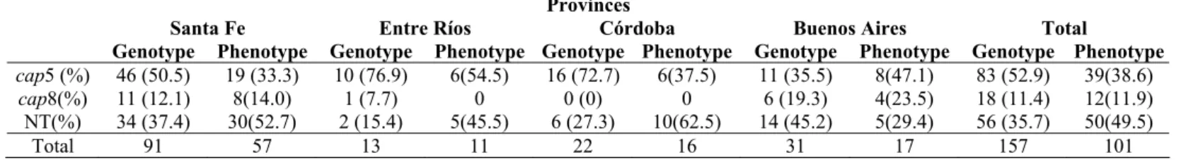
Sixty four percent of the isolates were typeable by PCR
with specific primers for loci cap5 or cap8; being the rest of
the isolates nontypeable (NT). Eighty three (52.87%) isolates
were genotyped as cap5 whereas eighteen (11.4%) as cap8.
None of the isolates positive for either cap5 or cap8 genes was
found to amplify both genes, confirming specificity of PCR
used. Distribution of genotypes among isolates originated in
different geographical areas is shown in Table 1. The prevalent
capsular type among isolates from Córdoba, Santa Fe and Entre
Ríos was cap5. Conversely, the majority of isolates from
Buenos Aires were NT, while type 5 was predominant among
typeable isolates from this province. CP genotype distribution
and percent of NT isolates varied between provinces; however,
differences were not significant (P=0.227). More than 50% of
the isolates from each province could be genotyped by the PCR
methodology. Among isolates from clinical IMI, 31 (72.1%)
were genotyped either as cap5 or cap8, while only 12 (27.9%)
were NT. Among 91 isolates from subclinical IMI, 53 (58.2%)
were typed as cap5 or 8 and 38 (41.8%) were NT (Table 2).
Differences between percentages of typeable isolates according
to the clinical origin were not significant (P=0.12).
Table 1. Distribution of capsular polysaccharide genotypes and phenotypes 5 and 8 among Staphylococcus aureus isolated from
bovine intramammary infections in four Argentinean provinces.
References: NT: nontypeable.
No differences in CP genotype (P = 0.227) or phenotype (P = 0.179) distribution were found between provinces.
Table 2. Distribution of S. aureus isolate types CP5 or CP8, according to mastitis clinical origin.
References: NT: nontypeable.
No statistical association between genotype (P =0.12) or phenotype (P=0.262) and clinical origin of the isolate was not found.
The isolates genotyped as cap5 and cap8 were then tested
with anti-CP5 and anti-CP8, respectively. The results of
serotyping are shown in Table 1. Fifty percent of isolates
genotyped as cap5 and cap8 were shown to produce CPs by
ELISA for detecting either capsular type. Thirty nine isolates
(38.6%) reacted to anti-CP5 serum and 12 (11.9%) to anti-CP8.
From 83 isolates genotyped as cap5, 39 (46.9%) were capable
of expressing CP; while from 18 isolates genotyped as cap8, 12
(66.6%) were capable of expressing CP.
A statistical association between the expression of CP5 or
CP8 capsules and clinical origin of the isolate was not found
(Table 2). From 84 isolates genotyped as cap5 and cap8 for
which clinical status was known, 31 (37%) where from clinical
IMI, and among these isolates 14 (45.2%) and 17 (54.8%) were
serotypeable and non serotypeable, respectively. Among
isolates from subclinical origin, 53 from the 84 isolates were
Provinces
Santa Fe Entre Ríos Córdoba Buenos Aires Total
Genotype Phenotype Genotype Phenotype Genotype Phenotype Genotype Phenotype Genotype Phenotype
cap5 (%) 46 (50.5) 19 (33.3) 10 (76.9) 6(54.5) 16 (72.7) 6(37.5) 11 (35.5) 8(47.1) 83 (52.9) 39(38.6)
cap8(%) 11 (12.1) 8(14.0) 1 (7.7) 0 0 (0) 0 6 (19.3) 4(23.5) 18 (11.4) 12(11.9) NT(%) 34 (37.4) 30(52.7) 2 (15.4) 5(45.5) 6 (27.3) 10(62.5) 14 (45.2) 5(29.4) 56 (35.7) 50(49.5)
Total 91 57 13 11 22 16 31 17 157 101
Clinical Subclinical Genotype Phenotype Genotype Phenotype
Typeable(%) 31(72.1) 14(45.2) 53(58.2) 27(50.9)
NT(%) 12(27.9) 17(54.2) 38(41.8) 26(49.1)
genotyped as cap5 and cap8 (63%), and from these isolates 27
(50.9%) and 26 (49.1) were serotypeable and non serotypeable,
respectively.
Prevalence of isolates expressing CP5 and CP8 in this
study (32.2%) was lower than those observed in most previous
reports of other countries (6, 15, 23). A previous study carried
out in Argentina, including 195 isolates, demonstrated that only
14% could be typed by specific antisera against CP5 or CP8
(21). In the present study, a higher prevalence of isolates
expressing CP5 than previously reported (21) was observed;
however, the percent of isolates expressing CP8 was similar in
both studies. Differences in proportion of isolates expressing
CP5 between studies can be explained mainly by the isolates
geographical origin and the time frame of both studies. While
most isolates from the previous study belonged to Buenos
Aires province, S. aureus isolates included in the present
investigation were obtained from the four main dairy provinces
of Argentina. In addition, in the present study, to avoid bias
produced by clonal dissemination, we included a maximum of
3 isolates per dairy farm to assure bacterial isolate diversity
within each geographical area considered.
The low proportion of phenotype expression with respect
to genotype presence was also reported in bovine mastitis
isolates from Europe and USA (23). Genotype-phenotype
disparity could indicate a restriction in phenotype expression
due to differences between in vivo vs in vitro culture conditions
(16, 17). This implies that conventional phenotypic evaluation
can underestimate isolates ability to express CP in vivo. In
addition, genotype-phenotype disparity could be due to the fact
that some isolates carry cap genes but lack capsule expression
due to mutations within capsule genes (1).
We found no association between genotype or phenotype
and clinical origin of the isolate. In a previous study a
significant association between CP8 expression and mastitis
clinical manifestations was observed only for isolates from
Ireland and Iceland (23). Recent studies have shown an
association between S. aureus genotypes and IMI clinical and
epidemiological features (4, 7); however associations in these
latter cases were established with patterns including several
rather than individual genes (4).
Presence of CP alone is considered to be insufficient to
generate a protective immune response; however, their
inclusion in a multicomponent vaccine would be useful to
improve control of S. aureus IMI (18, 19, 22). In addition,
relevance of CP as candidates for generating protective
responses is underscored by the fact that a commercial vaccine
currently available for S. aureus mastitis control contains
capsulated strains expressing 3 serotypes of CP present among
the population of bovine isolates in USA (10). Sixty four
percent of the isolates evaluated in this study carried cap5 and
cap8 genes, which emphasizes the importance of including
these components for rational design of mastitis vaccines.
ACKNOWLEDGEMENTS
We are grateful to Dr. Marcelo Signorini for technical
assistance and to Dr. Liliana Tirante for providing S. aureus
isolates from Buenos Aires province. This work was supported
by grants from INTA (PNLEC1601) and ANPCyT (PICT
1175).
REFERENCES
1. Cocchiaro, J.L.; Gomez, M.I.; Risley, A.; Solinga, R.; Sordelli, D.O.; Lee, J.C. (2006). Molecular characterization of the capsule locus from non-typeable Staphylococcus aureus. Mol Microbiol. 59, 948-960. 2. Dubois, M.; Gilles, K.A.; Hamilton, J.K.; Rebers, P.A.; Smith, F. (1956).
Colorimetric method for determination of sugars and related substances. Anal Chem. 28, 350–356.
3. Fattom, A.; Schneerson, R.; Szu, S.; Vann, W.; Shiloach, J.; Karakawa, W.; Robbins, J. (1990). Synthesis and immunologic properties in mice of vaccines composed of Staphylococcus aureus type 5 and type 8 capsular polysaccharides conjugated to Pseudomonas aeruginosa exotoxin A. Infect Immun. 58, 2367-2374.
Res Vet Sci. 85, 439-448.
5. Guidry, A.J.; Oliver, S.P.; Squiggins, K.E.; Erbe, E.F.; Dowlen, H.H.; Hambleton, C.N.; Berning, L.M. (1991). Effect of anticapsular antibodies on neutrophil phagocytosis of Staphylococcus aureus. J Dairy Sci. 74, 3360-3369.
6. Guidry, A.; Fattom, A.; Patel, A.; O'Brien, C.; Shepherd, C.; Lohuis, J. (1998). Serotyping scheme for Staphylococcus aureus isolated from cows with mastitis. J Am Vet Med Assoc. 59, 1537-1539.
7. Hensen, S.M.; Pavicić, M.J.; Lohuis, J.A.; de Hoog, J.A.; Poutrel, B. (2000). Location of Staphylococcus aureus within the experimentally infected bovine udder and the expression of capsular polysaccharide type 5 in situ. J Dairy Sci. 83, 1966-1975.
8. Karakawa, W.W.; Fournier, J.M.; Vann, W.F.; Arbeit, R.; Schneerson, R.S.; Robbins, J.B. (1985). Method for the serological typing of the capsular polysaccharides of Staphylococcus aureus. J Clin Microbiol 22, 445-447.
9. Karakawa, W. W.; Vann, W. F. (1982). Capsular polysaccharides of Staphylococcus aureus. Semin. Infect. Dis. 4, 285-293.
10. Ma, J.; Cocchiaro, J.; Lee, J.C. (2004). Evaluation of serotypes of Staphylococcus aureus strains used in the production of a bovine mastitis bacterin. J Dairy Sci. 87, 178–182.
11. Nanra, J.S.; Timofeyeva, Y.; Buitrago, S.M.; Sellman, B.R.; Dilts, D.A.; Fink, P.; Nunez, L.; Hagen, M.; Matsuka, Y.V.; Mininni, T.; Zhu, D.; Pavliak, V.; Green, B.A.; Jansen, K.U.; Anderson, A.S. (2009). Heterogeneous in vivo expression of clumping factor A and capsular polysaccharide by Staphylococcus aureus: implications for vaccine design. Vaccine. 27, 3276-3280.
12. National Research Council (1996) Guide for the Care and Use of Laboratory Animals. Washington, DC: National Academy Press. 13. O'Riordan, K.; Lee, J.C. (2004). Staphylococcus aureus capsular
polysaccharides. Clin Microbiol Rev. 17, 218-234.
14. Pospiech, A.; Neumann, B. (1995). A versatile quick-prep of genomic DNA from gram-positive bacteria. Trends Genet. 11, 217-218.
15. Poutrel, B. ; Boutonnier, A.; Sutra, L. ; Fournier, J.M. (1988). Prevalence
of capsular polysaccharide types 5 and 8 among Staphylococcus aureus isolates from cow, goat, and ewe milk. J Clin Microbiol 26, 38-40. 16. Poutrel, B.; Gilbert, F.B.; Lebrun, M. (1995). Effects of culture
conditions on production of type 5 capsular polysaccharide by human and bovine Staphylococcus aureus strains. Clin Diagn Lab Immunol. 2, 166-171.
17. Poutrel, B.; Rainard, P.; Sarradin, P. (1997). Heterogeneity of cell-associated CP5 expression on Staphylococcus aureus strains demonstrated by flow cytometry. Clin Diagn Lab Immunol. 4, 275-278. 18. Projan, S.J.; Nesin, M.; Dunman, P.M. (2006). Staphylococcal vaccines
and immunotherapy: to dream the impossible dream? Curr Opin Pharmacol. 5, 473-479.
19. Shaffer, A.C.; Lee, J.C. (2008). Vaccination and passive immunization against Staphylococcus aureus. Int J Antimicro Ag. 325, 571-578. 20. Smith, P.K. ; Krohn, R.I.; Hermanson, G.T. ; Mallia, A.K. ; Gartner,
F.H. ; Provenzano, M.D.; Fujimoto, E.K. ; Goeke, N.M.; Olson, B.J.; Klenk, D.C. (1985). Measurement of protein using bicinchoninic acid. Anal Biochem. 150, 76-85.
21. Sordelli, D.O.; Buzzola, F.R.; Gomez, M.I.; Steele-Moore, L.; Berg, D.; Gentilini, E.; Catalano, M.; Reitz, A.J.; Tollersrud, T.; Denamiel, G.; Jeric, P.; Lee, J.C. (2000). Capsule expression by bovine isolates of Staphylococcus aureus from Argentina: genetic and epidemiologic analyses. J Clin Microbiol 38, 846-850.
22. Stranger-Jones, Y.K.; Bae, T.; Schneewind, O. (2006). Vaccine assembly from surface proteins of Staphylococcus aureus. Proc Natl Acad Sci USA. 103, 16942-16947.
23. Tollersrud, T.; Kenny, K.; Reitz, A.J.; Lee, J.C. (2000). Genetic and serologic evaluation of capsule production by bovine mammary isolates of Staphylococcus aureus and other Staphylococcus spp. From Europe and the United States. J Clin Microbiol. 38, 2998-3003.
24. Verdier, I.; Durand, G.; Bes, M.; Taylor, K.; Lina, G.; Vandenesch, F.; Fattom, A.; Etienne, J. (2007). Identification of the capsular polysaccharides in Staphylococcus aureus clinical isolates by PCR and agglutination tests. J Clin Microbiol. 45, 725–729.